(Annals of the Brazilian Academy of Sciences)
Printed version ISSN 0001-3765 / Online version ISSN 1678-2690 www.scielo.br/aabc
Action of Aqueous Extracts of
Phyllanthus niruri
L.
(
Euphorbiaceae)
leaves on Meristematic Root Cells of
Allium cepa
L.
ERASMOVLANE S.B. NEVES1, PAULO MICHEL PINHEIRO FERREIRA2
,
LEONARDO H.G.M. LIMA1 and ANA PAULA PERON1
1Universidade Federal do Piauí (UFPI), Núcleo de Pesquisa Aplicada à Saúde e ao Meio-Ambiente (NUPBSAM),
Laboratório de Citogenética Vegetal e Animal, Campus Senador Helvídio Nunes de Barros (CSHNB), Rua Cícero Duarte, 940, Bairro Junco, 64600-000 Picos, PI, Brasil
2Programa de Pós-Graduação em Ciências Farmacêuticas, Universidade Federal do Piauí,
Centro de Ciências da Saúde, Departamento de Biofísica e Fisiologia, Campus Ministro Petrônio Portela, Avenida Universitária, lado ímpar, Ininga, 64049-550 Teresina, PI, Brasil
Manuscript received on April 29, 2013; accepted for publication on July 17, 2013
ABSTRACT
This study aimed to evaluate the effects of aqueous extracts of dried Phyllanthus niruri L. (stonebreaker) leaves on Allium cepa L. root meristem cells at four concentrations, 0.02 (usual concentration), 0.04, 0.06 and 0.08mg/mL and exposure times of 24 and 48 hours. For each concentration we used a group of ive onion bulbs that were irst embedded in distilled water and then transferred to their respective concentrations. The radicles were collected and ixed in acetic acid (3:1) for 24 hours. The slides were prepared by the crushing technique and stained with 2% acetic orcein. Cells were analyzed throughout the cell cycle, totaling 5000 for each control and exposure time. The calculated mitotic indices were subjected to the Chi-squared statistical analysis (p<0.05). From the results obtained it was observed that all four concentrations tested had signiicant antiproliferative effect on the cell cycle of this test system. We also found the presence of cellular aberrations such as colchicined metaphases, anaphasic and telophasic bridges, and micronuclei in the two exposure times for all concentrations evaluated. Therefore, under the conditions studied the concentrations of aqueous extracts of leaves of P. niruri showed to be cytotoxic and genotoxic.
Key words: medicinal plant, stonebreaker, cell division, cellular aberrations, Allium cepa.
Correspondence to: Ana Paula Peron E-mail: anpapegenpes@hotmail.com
INTRODUCTION
Currently, about 70% of the world population uses
medicinal plants in primary health care (Frescura
et al. 2012). However, many of them have not been
suficiently studied for their potential toxic effects
at the cellular level (Bagatini et al. 2007, Meyer
et al. 2011). These studies are of great importance
for contributing towards standardizing quantities
for safe and effective use of these plants by the
population (Asare et al. 2012).
Phyllanthus niruri
, a species belonging to the
family Euphorbiaceae, subfamily Phyllanthoideae,
originated in India and is widely distributed in
the Americas. This medicinal plant, popularly
known in Brazil as stonebreaker, is classiied as
herbaceous, measuring on average 60 cm tall, with
horizontally ramiied stem. Its lowers,
yellow-green, are minutel, dioecious and inserted in the
leaf axils (Moreira et al. 2013). Its phytochemical
constituents have been well established, particularly
lignans, tannins and lavonoids, the latter two being
present in high concentrations in the leaves (Devi et
al. 2005, Asare et al. 2012).
Aqueous extracts obtained by infusion of
stone-breaker dried leaves are widely used in popular
medicine for the removal of calcium oxalate kidney
stones (Álvarez et al. 2009, Nascimento-Barros
and Albuquerque 2012), as an eupeptic (Asare
et al. 2012), for liver disorders (Murugaiyah and
Chan 2009), in alleviating jaundice and in ighting
bladder and bowel infections (Abdulla et al. 2010).
However, there is a lack of studies in the literature
on its toxic effects at the cellular level.
Bioassays with plants are considered
appro-priate for monitoring of toxic effects of chemical
compounds (USEPA) (Grant 1999, Iganci et al.
2006, Herrero et al. 2012).
Allium cepa
(onion)
is an eficient bioindicator of the cytotoxicity of
aqueous extracts of medicinal plants (Fachinetto et
al. 2007, Sabini et al. 2011). This is due to its kinetic
properties of proliferation and because it processes
large chromosomes in reduced number (2n=16),
which facilitates their analysis (Matsumoto et al.
2006, Caritá and Marin-Morales 2008, Herrero et
al. 2012). This test system is also effective in the
evaluation of mutagenicity of aqueous extracts of
plants with medicinal properties since it enables
the observation of abnormalities of the mitotic
cycle, such as colchicined metaphases, anaphasic
and telophasic bridges, and interphase anomalies,
such as micronuclei and binucleate cells (Leme and
Marin-Morales 2008, Sabini et al. 2011).
Thus, due to the widespread use of
P. niruri
by the population, and the need for further studies
on the action of this plant at a cellular level, and
also considering that the
A. cepa
system is suitable
for assessment of cytotoxicity and mutagenicity of
aqueous extracts of medicinal plants, this study aimed
to evaluate the effect of different concentrations
of aqueous extracts of dried stonebreaker leaves,
obtained by popular use, on
A. cepa
meristematic
root cells in two exposure times.
MATERIALS AND METHODS
This work was developed on the Senador Helvídio
Nunes de Barros Campus of the Universidade
Federal do Piauí (UFPI), municipality of Picos,
state of Piauí.
PLANT COLLECTION
Samples of
P. niruri
were collected in a medicinal
nursery local in the city of Teresina, state of Piauí,
in May of 2012 and identiied by Prof Ms Maria
do Socorro Meireles de Deus, who holds a master
in botany and is a professor at UFPI. The leaves of
these samples were then stored under environmental
conditions for 6 months. Soon after identiication,
a control sample of this species was taken to the
Graziela Barroso Herbarium of UFPI.
PREPARATION OF INFUSIONS
Dry stonebreaker leaves were placed in boiling
water where they remained in infusion for 10
minutes. Subsequently, the aqueous extracts were
iltered and cooled to room temperature. Four
concentrations were established, 0.02; 0.04; 0.06
and 0.08mg/mL, of which 0.02mg/mL is considered
the most common and recommended by the Center
for Drug Information Base of Medicinal and Toxic
Plants (CIMPLAMT 2012).
OBTAINING MERISTEMATIC CELLS FOR CYTOGENETIC ANALYSIS
The bulbs of
Allium cepa
, acquired in the produce
market in the city of Picos - Piauí, were placed to root
in lasks with distilled water, at room temperature
(± 25°C), constantly aerated and with a period of
twelve hours of light and twelve hours of darkness,
until roots with about 1.0cm of length were obtained.
For analysis of each concentration, an experimental
group was stipulated with ive bulbs, according to
Before putting the roots in contact with
their respective concentrations, some roots were
collected and ixed to serve as control (CO) of the
bulb itself. Soon afterwards, the remaining roots
were placed in their respective concentrations, for
24 hours, this procedure denominated as 24-hour
exposure time (ET 24h).
After this time some roots were removed and
ixed. Afterwards, the remaining roots from each bulb
were returned to their respective concentrations where
they remained for 24 hours, which we called 48-hour
exposure time (48h ET). After this period, roots were
again collected and ixed. Exposure times of 24 and
48 h were chosen in order to evaluate the effect of the
concen trations studied in more than one cell cycle.
The ixing of the roots occurred in Carnoy 3:1
(ethanol: acetic acid) at room temperature for 24
hours. For each root collection, an average of three
roots per onion bulb were removed.
PREPARATION AND READING OF THE SLIDES, AND DATA ANALYSIS
The slides, three per bulb on average, were made
following the protocol proposed by Guerra and
Souza (2002). Each slide was stained with two drops
of 2% acetic orcein (Fachinetto and Tedesco 2009)
and examined under an optical microscope at 40X.
For each bulb 1,000 cells were analyzed, totaling
5000 cells for each control and concentration.
During the analysis we observed cells in interphase,
prophase, metaphase, anaphase and telophase. We
calculated the number of cells in interphase and
under division for each control and exposure time
and determined and mitotic index.
We also evaluated the presence of cellular
aberrations such as mitotic cycle anomalies
(colchicined metaphases, anaphasic and telophasic
bridges) interphasic anomalies (micronucleated and
binucleated cells). For this evaluation 1,000 cells
were analyzed for each control and exposure time.
The statistical analysis of all the data was
conducted through the Chi-square (χ
2), with a
level of probability < 0.05, through the statistical
software BioEstat 3.0 (Ayres 2007).
RESULTS AND DISCUSSION
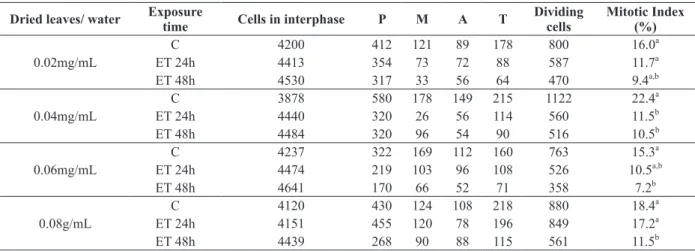
Table I presents the number of cells in interphase and
at different stages of cell division, and the mitotic
Dried leaves/ water Exposure time Cells in interphase P M A T Dividing cells
Mitotic Index (%)
C 4200 412 121 89 178 800 16.0a
0.02mg/mL ET 24h 4413 354 73 72 88 587 11.7a
ET 48h 4530 317 33 56 64 470 9.4a,b
C 3878 580 178 149 215 1122 22.4a
0.04mg/mL ET 24h 4440 320 26 56 114 560 11.5b
ET 48h 4484 320 96 54 90 516 10.5b
C 4237 322 169 112 160 763 15.3a
0.06mg/mL ET 24h 4474 219 103 96 108 526 10.5a,b
ET 48h 4641 170 66 52 71 358 7.2b
C 4120 430 124 108 218 880 18.4a
0.08g/mL ET 24h 4151 455 120 78 196 849 17.2a
ET 48h 4439 268 90 88 115 561 11.5b
TABLE I
Cell cycle analyzes of Allium cepa root tips treated with the infusion
of Phyllanthus niruri leaves at concentrations of 0.02, 0.04, 0.06 and
0.08 mg/mL after 24 h and 48 h. Analysis of 5,000 cells per group.
C – Control; ET – Exposure time, h - hour, d – day.
Means followed by the same letter do not differ signiicantly at the level of 0.05 by the χ2
index values obtained from root meristem cells
of
A. cepa
treated with water and with different
concentrations of
P. niruri
under exposure times of
24 and 48 hours.
The results obtained showed that all four tested
concentrations of
P. niruri
signiicantly inhibited
MI of meristematic root cells of
A. cepa
under
the
two ET when compared to the MI of their respective
controls. As can be seen, at all concentrations,
inhibition of cell division is maintained with the
increase of ET. However, when comparing the MI
between the ET of a same concentration, we found
that they were not statistically signiicant.
Table II presents the number of colchicined
metaphases, bridges in anaphase and telophase,
micronucleated cells, and total cellular aberrations
present in the root meristem cells of
A. cepa
with
water and treated with different concentrations of
P. niruri
in exposure times of 24 and 48 hours.
In both ET evaluated for the four concentrations,
the presence of colchicined metaphases, anaphase
bridges, telophase bridges and micronuclei were
veriied. All concentrations induced a number
of cellular aberrations that differed significantly
from their respective CO, but did not differ
among themselves.
Dried leaves/
water Exposure Time
Number of cells analyzed
Colchicined metaphases
Anaphase and telophase
bridges
Micronucleated
cells Aberrant cells
C 5.000 00 00 00 00a
0.02mg/mL ET 24h 5.000 03 03 21 27b
ET 48h 5.000 06 04 19 29b
C 5.000 00 00 00 00a
‘0.04mg/mL ET 24h 5.000 01 02 20 23b
ET 48h 5.000 04 03 21 28b
C 5.000 0 00 00 00a
0.06mg/mL ET 24h 5.000 04 05 13 22b
ET 48h 5.000 09 08 14 31b
C 5.000 00 00 00 00a
0.08g/mL ET 24h 5.000 03 02 20 25b
ET 48h 5.000 03 03 26 32b,c
TABLE II
Number of cells with colchined metaphases, bridges in anaphase and telophase and micronucleated cells, and total of aberrant cells treated with the infusion of Phyllanthus niruri leaves at concentrations
of 0.02, 0.04, 0.06 and 0.08 mg/mL after 24 h, 48 h and 7 days of exposure.
C – Control; ET – Exposure time, h - hour, d – day.
Means followed by the same letter do not differ signiicantly at the level of 5% by the χ2test.
The cytotoxicity results obtained here
cor-roborate those observed by Barros et al. (2006), who
evaluated the bone marrow of rats provided with
alcoholic extracts of stonebreaker leaves at a dose
of 300mg/ml via gavagem and chronic treatment,
and found that this plant decreased the cell division
rate of marrow cells of these animals, thus being
cytotoxic. However, Asare et al. (2012)
evaluated
the action of alcoholic extracts of
P. niruri
at doses
of 30 and 300mg/ml on peripheral blood of white
mice for ninety days, and found that the extracts
were not cytotoxic nor mutagenic in these animals.
There are no other studies in the scientiic literature
that evaluated cytotoxicity and mutagenicity of
extracts of
P. niruri
in the test systems with normal
cells, i.e those without and previous treatment with
clastogenic drugs or with a cell disorder.
Thus, taking into account the results obtained
in this present work where the aqueous extracts of
the few studies done to date on the toxic effects
of this plant at the cellular level, recognizing that
common sense often considers medicinal plants free
from adverse reactions to the body often leading
to their indiscriminate use, and that the
P. niruri
plant is easily found in medicinal plant nurseries,
herbalists, natural food stores and farmers markets,
it is highly relevant to carry out work similar to
this one with
A. cepa,
with other test-systems,
exposure times and different treatments to thereby
establish, with propriety, what the ideal and safe
concentrations are for the use of this plant.
RESUMO
Este estudo teve por objetivo avaliar a ação de extratos aquosos das folhas secas de Phyllanthus niruri L. (popular quebra-pedras) sobre as células meristemáticas de raízes de
Allium cepa L. em quatro concentrações, 0,02 (concentração
usual), 0,04, 0,06 e 0,08mg/mL, nos tempos de exposição de 24 e 48 horas. Para cada concentração utilizou-se um grupo de cinco bulbos de cebolas, que primeira-mente foram enraizados em água destilada, e em seguida transferidos para as suas respectivas concentrações. As radículas foram coletadas e ixadas em ácido acético (3:1) por 24 horas. As lâminas foram preparadas pela técnica de esmagamento e coradas com orceína acética a 2%. Analisaram-se células em todo ciclo celular, totalizando 5.000 para cada controle e tempo de exposição. Os índices mitóticos calculados foram submetidos à análise estatística do Qui-quadrado (p<0,05). A partir dos resultados obtidos observou-se que as quatro concentrações testadas, tiveram efeito antiproliferativo signiicativo sobre o ciclo celular deste sistema-teste. Também se veriicou a presença de aberrações celulares como metáfases colchicínicas, pontes anáfásicas e telofásicas, e micronúcleos nos dois tempos de exposição avaliados de todas as concentrações. Portanto, nas condições analisadas, as concentrações de extratos aquosos de folhas secas de P. niruri mostraram-se citotóxicas e genotóxicas.
Palavras-chave: planta medicinal, quebra-pedras, divisão
celular, aberrações celulares, Allium cepa.
REFERENCES
ABDULLA MA, AHMED KAA, AL-BAYATY FH AND MASOOD Y. 2010. Gastroprotective effect of Phyllanthus niruri leaf extract against ethanol-induced gastric mucosal injury in rats. J Pharm Pharmacol 4: 226-230.
ÁLVAREZ AL, DIÑEIRO Y, DEL BARRIO G, PICINELLI A, SUÁREZ B AND VALDÉS S. 2009. Bioactivity-guided separation of anti HSV-2 and antioxidant metabolites from the plant
Phyllanthus orbicularis. Planta Med 75: 990-991. ASARE GA, BUGYEI K, SITTIE A, YAHAYA ES, GYAN B, ADJEI S,
ADDO P, WIREDU EK, ADJEI DN AND NYARKO AK. 2012. Genotoxicity, cytotoxicity and toxicological evaluation of whole plant extracts of the medicinal plant Phyllanthus niruri (Phyllanthaceae). Genet Mol 11: 100-111. doi: 10.4238/2012.January.13.3.
AYRES M. 2007.BioEstat 5.0: Aplicações estatísticas nas áreas das ciências biológicas e médicas. Belém: Sociedade Civil Mamirauá, Brasília, CNPq.
BAGATINI MD, SILVA ACF AND TEDESCO SB. 2007. Uso do sistema-teste Allium cepa como bioindicador de genotoxicidade de infusões de plantas medicinais. Revista Bras Farmacogn 18: 509-516.
BARROS ME, LIMA R, MERCURI LP, MATOS JR, SCHOR N AND BOIM MA. 2006. Effect of Extract of Phyllanthus niruri on crystal deposition in experimental urolithiasis. Urol Res 34: 351-357.
CARITÁ R AND MARIN-MORALES MA. 2008. Induction of chromosome aberrations in the Allium cepa test system caused by the exposure of seeds to industrial efluents contaminated with azo dyes. Chemosphere 72: 722-725. CIMPLAMT – 2012. Boletim informativo do Centro de
Infor-mações sobre Medicamentos a base de Plantas medicinais e Tóxicas. Boletim Informativo. São João Del Rei: Universidade Federal de São João Del Rei.
DEVI V, SHANBHAG TV, BAIRY KL AND SHENOY S. 2005. Effect of Phyllanthus niruri on wound healing in rats. Indian J Physiol Pharmacol 49: 487-490.
FACHINETTO JM, BAGATINI MD, DURIGON ACFS AND TEDESCO SB. 2007. Efeito anti-proliferativo das infusões de Achyrocline satureioides DC (Asteraceae) sobre o ciclo de celular de Allium cepa. Revista Bras Farmacogn 17: 49-54.
FACHINETTO JM AND TEDESCO SB. 2009. Atividade anti-proliferativa e mutagênica dos extratos aquosos de
Baccharis trimera (Less.) A. P. de Candolle e Baccharis articulata (Lam.) Pers. (Asteraceae) sobre o sistema teste de Allium cepa. Rev Bras Pl Med 11: 360-367.
FISKESJO G. 1994.Allium Test II: Assessment of a chemicals genotoxic potential by recording aberration in root tips of
Allium cepa L. Environ Toxicol 9: 235-241.
FRESCURA VD, LAUGHINGHOUSE IV AND TEDESCO SB. 2012. Antiproliferative effect of the tree and medicinal species Luehea divaricata on the Allium Cepa cell cycle. Caryologia 65: 27-33.
GUERRA M AND SOUZA MJ.2002. Como observar os cromos-somos: um guia de técnicas em citogenética vegetal, animal e humana. Ribeirão Preto: FUNPEC.
HERRERO O, PEREZ JMM AND FERNÁNDEZ PF. 2012. Toxicological evaluation of three contaminant of emerging concern by use of Allium cepa test. Mut Res 743: 24-34. IGANCI JRV, BROBOWSKI G, HEIDEN GVC, STEIN L AND ROCHA
BHG. 2006. Efeito do extrato aquoso de diferentes espécies de boldo sobre a germinação índice mitótico de
Allium cepa L. Arq Inst Biol 73: 79-82.
LEME DM AND MARIN-MORALES MA. 2008. Chromosome aberration and micronucleus frequencies in Allium cepa
cells exposed to petroleum polluted water – a case study. Mut Res 650: 80-86.
MATSUMOTO ST, MANTOVANI MS, MALAGUTTI MIA, DIAS AL, FONSECA IC AND MARIN-MORALES MA. 2006. Geno-toxicity and mutagenicity of water contamined with tannery, as evaluated by the micronucleus test and comet assay using the ish Oreochromis niloticus and chromosome aberration in onion root-tips. Genet Mol Biol 29: 148-158.
MEYER L, QUADROS KE AND ZENI ALLB. 2011. Etnobotânica na comunidade de Santa Bárbaro Ascurra, Santa Catarina, Brasil. Rev Bras Bioc 10: 258-266.
MOREIRA J, KLEIN-JÚNIOR LC, CECHINEL FILHO V AND DE CAMPOS BUZZI F. 2013. Anti-hyperalgesic activity of corilagin, a tannin isolated from Phyllanthus niruri L. (Euphorbiaceae). J Ethnopharmacol 146: 318-23. doi: 10.1016/j.jep.2012.12.052.
MURUGAIYAH V AND CHAN KL. 2009. Mechanisms of anti-hyperuricemic effect of Phyllanthus niruri and its lignan constituents. J Ethnopharmacol 124: 233-239.
NASCIMENTO-BARROS FR AND ALBUQUERQUE IL. 2012. Substâncias e medicamentos abortivos utilizados por ado-lescentes em unidade secundária de saúde. RBPS 18: 177-184.