Article
0103 - 5053 $6.00+0.00
*e-mail: [email protected]
A New Eremophilane-type Sesquiterpene from the Phytopatogen Fungus
Lasiodiplodia theobromae
(Sphaeropsidaceae)
Fátima M. Nunes,
aMaria da Conceição F. de Oliveira,*
,aÂngela M. C. Arriaga,
aTelma L. G. Lemos,
aManoel Andrade-Neto,
aMarcos C. de Mattos,
aJair Mafezoli,
bFrancisco M. P. Viana,
cViviane M. Ferreira,
cEdson Rodrigues-Filho
dand Antônio G. Ferreira
daDepartamento de Química Orgânica e Inorgânica, Universidade Federal do Ceará, Campus do Pici, CP 6044, 60455-970 Fortaleza-CE, Brazil
bCurso de Farmácia, Universidade de Fortaleza, Av. Washington Soares, 1321, CP 1258, 60811-341 Fortaleza-CE, Brazil
cLaboratório de Fitopatologia, Embrapa – Agroindústria Tropical, Fortaleza-CE, Brazil
d Departamento de Química, Universidade Federal de São Carlos, Rodovia Washington Luiz, km 235, CP 676, 13565-905 São Carlos-SP, Brazil
O fungo fitopatogênico Lasiodiplodia theobromae, isolado de goiaba, foi cultivado em arroz por 32 dias à temperatura ambiente. Extração com CH2Cl2:MeOH (3:7), seguido de fracionamento cromatográfico do extrato forneceu o esteróide ergosterol. Da cultura fúngica em meio de Czapeck por 40 dias à temperatura ambiente, foram isolados a isocumarina cis-4-hidroximeleína e um sesquiterpeno do tipo eremofilano. O sesquiterpeno eremofilano está sendo descrito pela primeira vez na literatura. Este é o primeiro relato do isolamento de um sesquiterpeno eremofilano para o gêneroLasiodiplodia.
The phytopatogenic fungus Lasiodiplodia theobromae, isolated from guava, was cultivated in rice for 32 days at room temperature. Extraction with CH2Cl2:MeOH (3:7), followed by chromatography fractionation of the extract provided ergosterol. From the fungus culture in Czapeck medium for 40 days at room temperature, were isolated isocoumarin cis-4-hydroxymeleine and an eremophilane-type sesquiterpene. The latter compound is being reported for the first time in the literature. Also, this is the first time that an eremophilane sesquiterpene is described for
Lasiodiplodia genus.
Keywords: fungus, Lasiodiplodia theobromae, ergosterol, isocoumarin, eremophilane-type sesquiterpene
Introduction
Microorganisms represent a promising source of biologically active compounds; despite this, only a small portion of the microbial diversity has been chemically investigated. Because of the short life cycle and easy adaptability to external media, fungi can be manipulated for the production of secondary metabolites of biological interest.1 About 1500 secondary metabolites from fungi were reported in the literature from 1993 to 2001, and more than 50% of these compounds showed antibacterial, antifungal
and antitumoral activities.2 Chemical investigation of phytopatogen fungi, especially those associated with serious agricultural problems, was recently begun.
Lasiodiplodia theobromae (Patouillard) Griffon & Maublanc (Sphaeropsidaceae) is a phytopathogen fungus found in more than 280 different genera of host plants from tropical and subtropical regions of the world.3 In Brazil, this fungus is considered a serious problem to agriculture since it is associated with several diseases of tropical fruits.4
L. theobromae is the anamorphous form (asexual state) of
theobromae in the literature, this synonym is falling into disuse.5 The chemical investigation of strains of this fungus is reported in the literature.6-14 Jasmonic acid and thirteen derivatives,6-10 eight hydroxylasiodiplodins,10-12 two cyclohexene derivatives13, 14 and two isocoumarins8, 10 were isolated from L. theobromae.
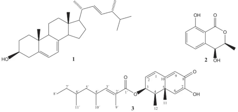
This work reports the isolation of a new eremophilane-type sesquiterpene (3),in addition to the known compounds ergosterol (1) and isocoumarin cis-4-hydroxymelein (2). The presence of an eremophilane-type sesquiterpene in
L. theobromae is being reported for the first time in the literature. The structural elucidation of these compounds was established on the basis of 1D and 2D NMR spectroscopic techniques.
Results and Discussion
Successive chromatography procedures of the CH2Cl2:MeOH (3:7) extract of L. theobromae cultivated in rice provided ergosterol (1). When cultivated in Czapeck broth, this fungus provided isocoumarin 4-hydroxymelein (2) and a new eremophilane-type sesquiterpene (3) after column chromatography of the EtOAc and n-BuOH fractions obtained by partition of the liquid medium (Figure 1).
The structure of compound 1 was established after analysis of its spectroscopic data (IR, 1H and 13C NMR) and comparison with literature data.15 Until now, this is the first report of the isolation of ergosterol (1) from L. theobromae, although the detection of 1 in maize grains has been associated with the presence of this fungus as a contaminant.16 It should be mentioned that TLC analyses of the extracts from the control flasks did not show the presence of compound 1.
Compound2 was identified as the isocoumarin cis -4-hydroxymelein by IR, MS and 1H and 13C NMR techniques
and by comparison with literature data.17 This secondary metabolite was previously isolated from several fungi species, including L. theobromae.10
The molecular formula of compound 3, C23H32O4, was suggested by 1H and 13C NMR. The IR spectrum displayed a broad band at 3299 cm-1characteristic of a hydroxyl group and bands associated with A,B-unsaturated ketone (1643 cm-1) and A,B-unsaturated-ester (1708 cm-1). The analysis of hydrogen broad band decoupled (HBBD) and DEPT 135° 13C NMR spectra revealed the presence of six methyl groups, two methylene carbons, nine methine carbons and six non-hydrogenated carbons, two of which were associated with carbonyl groups, characteristic of anA,B-unsaturated ketone and another of carbonyl of an ester function at GC 181.5 and 167.9, respectively. The
1H
NMR spectrum of 3exhibited the presence of a deshielded signal assignable to an acylated oxymethine proton at GH 5.48 (1H, t, J 5.0 Hz, H-3). After analysis of the HSQC spectrum, three trisubstituted double bonds presented olefinic hydrogen signals at GH 6.56 (1H, dq, J 1.4 and 10.0 Hz, H-3’), 6.21(1H, br s, H-9) and 6.30 (1H, d, J 0.4 Hz, H-6) which were associated with carbons at GC 149.6, 122.7 and 121.0, respectively. The two last signals, together with the signal at GH 6.36 (br s, 7-OH) and the carbonyl group signal at GC 181.5, are in perfect agreement with the presence of a A-hydroxydienone ring, with an enol suggesting a diosphenol group. In addition, there were 1H NMR signals for one dissubstituted double bond at G 6.45 (1H, br dd, J 0.6 and 9.8 Hz, H-1) and 6.24 (1H, dd, J 5.0 and 9.8 Hz, H-2). The presence of an angular methyl was deduced from the observation of one singlet at G 1.43 (3H, H-11). Additional methyl groups were observed by four doublets integrating to 3H each at G 1.19 (d, J 7.0 Hz, H-12), 1.00 (d,
J 6.6 Hz, H-10’), 0.84 (d, J 6.2 Hz, H-11’), and 1.88 (d, J 1.4 Hz, H-9’), the last coherent with a vinyl methyl group. These data, in combination with the four multiplets between GH 2.63
and 1.10, integrating to six protons and the oxyacyl group, GC 167.9, suggested that compound 3 had the presence of an unsaturated fatty acid side chain with 11 carbons.
The bicyclic moiety of the postulated structure for 3was found to be an eremophilane-type sesquiterpene. This class of compound is reported as fungal metabolite and presents a branched unsaturated fatty acid chain bonded at C1 or C3.18-21 The placement of the unsaturated fatty acid chain, deduced by previous discussion, at C-3 was readily established from the HMBC experiment. Thus, the HMBC spectrum of 3 exhibited correlation peaks among the acylated oxymethine hydrogen at GH 5.48 (H-3) with the carbons at GC 38.5 (C-4), 131.9 (C-2), 11.7 (C-12), 41.3 (C-5), 130.0 (C-1) and the carbonyl carbon GC 167.9 (C-1’). Moreover, correlations were also observed for the methine hydrogen at GH 6.24 (H-2) with the carbons at GC 69.8 (C-3), 130.0 (C-1), 38.5 (C-4) and with the carbonyl carbon at GC 167.9 (C-1
’). Likewise, the
signal for methyl hydrogens at GH 1.88 (H-9
’) showed long
range coupling with the carbons at GC 125.7 (C-2
’), 167.9
(C-1’), and 149.6 (C-3’), indicating the location of this group. Furthermore, the following correlations of the other hydrogen methyl groups were also observed: GH 0.84 (H-8
’and H-11’)
with the carbons located at GC30.0 (C-7
’), 32.4 (C-6’ ), and
44.1 (C-5’ ); 1.00 (H-10’) with the carbons located at G C 31.0 (C-4’), 44.1 (C-5’) and 149.6 (C-3’). These correlations corroborate the fatty acid moiety with 11 carbons attached at position C-3, similar to that found in eremoxylarin B, an eremophilane sesquiterpene isolated from the xylariaceous endophytic fungus YUA-026.21
The long-range correlations observed in the HMBC spectrum of 3 allowed the unambiguous assignment of all carbons and hydrogens from the bicyclic ring of an eremophilane-type skeleton. Correlations were observed among the hydrogen of the hydroxyl group at GH 6.36 (OH-7) with the carbons at GC146.5 (C-7), 121.0 (C-6) and 181.5 (C-8). This spectrum also revealed the cross-peak among the vinyl hydrogens at GH 6.21 (H-9) with the carbons at GC 163.8 (C-10), 41.3 (C-5), 130.0 (C-1) and 146.5 (C-7). Furthermore, the correlation peaks were observed among H-1 (GH 6.45) with the carbons at GC 131.9 (C-2), 163.8, (C-10), 41.3 (C-5), 69.8 (C-3) as well as the carbonGC 122.7 (C-9). Additionally, hydrogen at GH 6.30 (H-6) showed cross-peak with the carbons at GC 41.3 (C-5), 146.5 (C-7), 38.5 (C-4), 163.8 (C-10), 181.5 (C-8), as well as with the carbon of the angular methyl atGC 24.0 (C-11). Indeed, the HMBC spectrum also exhibited the correlation peaks between the methyl hydrogens signals (GH 1.43,H-11) and C-5 (GC 41.3), the allyl quaternary carbon that bears the methyl group. All the above observations were consistent with the cross-peak correlations observed in the HMQC and1H, 1H- COSY experiments.
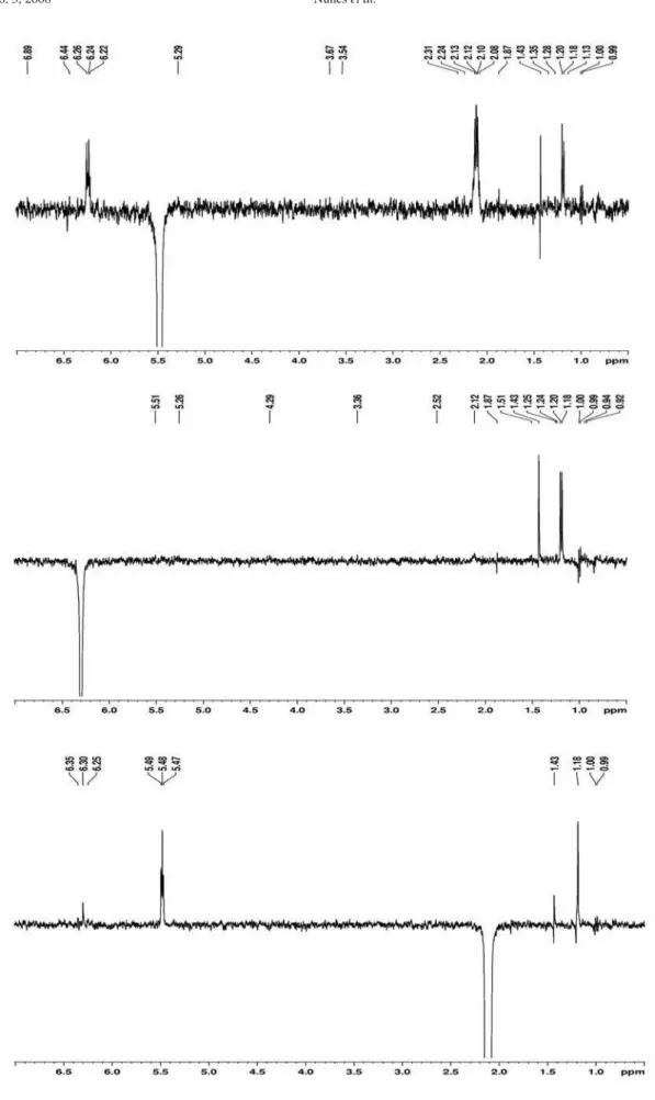
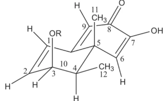
The relative stereochemistry of 3 was elucidated using nOe difference spectroscopy with the aid of geometry optimization using computational calculations. Thus, irradiation of H-3 at GH 5.48 enhanced the H-2 signal (GH 6.24), H-4 (GH 2.11), and 3H at C12 (GH 1.19); and when the signal at GH 6.30 (H-6) is irradiated, the resonances at GH 1.43 (3H at C11) and 1.19 (3H at C12) are increased, which indicates these groups of hydrogen are close in space. The optimized molecular geometry shows the methyl groups 3H at C11 and C12 almost equidistant to H-6 (Figure 2). Finally, irradiation of the H-4 signal (GH 2.11) produced an enhancement of the resonances at GH 5.48 (H-3) and 1.19 (3H at C12). All of these nOe effects observed are in agreement with the results of the computational calculations used to optimize the molecular geometry. As expected, the atoms that form the diosphenol substructure are all co-planar and the p-orbitals at double-bound $1,2 parallels those at$9,10. The present suggested relative stereochemistries at C-3, C-4 and C-5 are also in agreement with the molecular structure of other fungi eremophilane sesquiterpenes.18, 22, 23 The stereochemistry of C-4´and C-6´ at the octanoate ester was not determined in this work, since it is not directly deduced from nOe measurements.
These findings revealed 3 as a new eremophilane-type sesquiterpene with a branched unsaturated fatty acid attached to the C-3 position, named 2,4,6-trimethyloct-2-enoic acid, 1,2,6,8a-tetrahydro-7-hydroxy-1,8a-dimethyl-6-oxo-2-naphtalenyl ester. APCIMS spectrum of this compound showed the peak m/z 373 [M+1]+which is in accordance with the molecular formula C23H33O4.
Experimental
General procedure
CDCl3 as solvent and TMS as internal standard. IR spectra were run on a Perkin-Elmer 1000 FT-IR spectrometer using KBr pellets. Melting points were determined on a Mettler FP5 apparatus and are uncorrected. Gravity column chromatography was performed on Merck Kieselgel 60 (70-230 mesh). Low-resolution APCIMS data were acquired in positive ion mode, using a MICROMASS QUATTRO-LC instrument equipped with an ESI/ APCI “Z-spray” ion source. Molecular modeling of the sesquiterpene was conducted following the MM+ minimum energy optimization routines using the HyperChem24 for Windows (Release 3) program from Autodesk, Inc (Sausalito, CA).
Fungus material
L. theobromae (strain # 009) was isolated from infected guava in the Laboratory of Phytopathology from Embrapa Agroindústria Tropical, Ceará State, Brazil.
Fungus culture in rice and isolation of 1
Twenty seven Erlenmeyer flasks (250 mL), containing 100 g of rice (“Uncle Ben’s”) and 84 mL of distilled water per flask, were autoclaved twice at 121 oC for 60 min.
Small discs of the PDA medium from the Petri dish containing mycelium of L. theobromae was transferred under sterile conditions to 24 Erlenmeyer flasks containing sterilized rice and three flasks were kept as control. After 32 days of growth at 25 oC, 100 mL of CH
2Cl2:MeOH (3:7) was added to each flask and allowed to stand for 24 h. Blending of the material followed by filtration under reduced pressure provided 45.5 g of extract after solvent distillation. Vacuum chromatography of the extract on silica gel provided twelve fractions after elution with gradient mixture of hexane, CH2Cl2, EtOAc and MeOH. The fraction eluted with CH2Cl2/EtOAc 10% (512.0 mg) was chromatographed on silica gel by elution with Hexane/ EtOAc 10% and provided 66.8 mg of 1.
Fungus culture in Czapeck broth and isolation of 2 and 3
Small discs were cut from Petri dishes containing mycelium of L. theobromae in PDA medium and transferred under sterile conditions to 25 Erlenmeyer flasks (1000 mL), containing 300 mL of Czapeck medium per flask. Both broth and flask were previously autoclaved. Two flasks with no fungus were kept for control purposes. After 40 days of growth at 25 oC under static conditions, the liquid medium was separated from the mycelium by
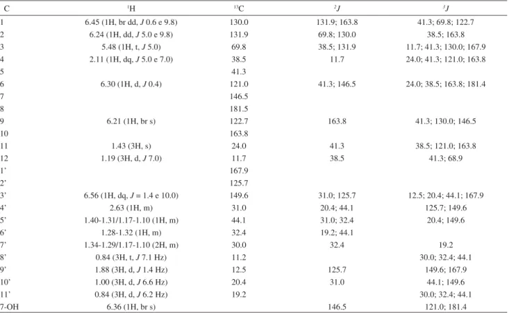
Table 1. 1H (400 MHz) and 13C (100 MHz) NMR data for 3 in CDCl 3
C 1H 13C 2J 3J
1 6.45 (1H, br dd, J 0.6 e 9.8) 130.0 131.9; 163.8 41.3; 69.8; 122.7
2 6.24 (1H, dd, J 5.0 e 9.8) 131.9 69.8; 130.0 38.5; 163.8
3 5.48 (1H, t, J 5.0) 69.8 38.5; 131.9 11.7; 41.3; 130.0; 167.9
4 2.11 (1H, dq, J 5.0 e 7.0) 38.5 11.7 24.0; 41.3; 121.0; 163.8
5 41.3
6 6.30 (1H, d, J 0.4) 121.0 41.3; 146.5 24.0; 38.5; 163.8; 181.4
7 146.5
8 181.5
9 6.21 (1H, br s) 122.7 163.8 41.3; 130.0; 146.5
10 163.8
11 1.43 (3H, s) 24.0 41.3 38.5; 121.0; 163.8
12 1.19 (3H, d, J 7.0) 11.7 38.5 41.3; 68.9
1’ 167.9
2’ 125.7
3’ 6.56 (1H, dq, J = 1.4 e 10.0) 149.6 31.0; 125.7 12.5; 20.4; 44.1; 167.9
4’ 2.63 (1H, m) 31.0 20.4; 44.1 125.7; 149.6
5’ 1.40-1.31/1.17-1.10 (1H, m) 44.1 31.0; 32.4 20.4; 149.6
6’ 1.28-1.32 (1H, m) 32.4 19.2; 44.1
7’ 1.34-1.29/1.17-1.10 (2H, m) 30.0 32.4 19.2
8’ 0.84 (3H, t, J 7.1 Hz) 11.2 30.0; 32.4; 44.1
9’ 1.88 (3H, d, J 1.4 Hz) 12.5 125.7 149.6; 167.9
10’ 1.00 (3H, d, J 6.6 Hz) 20.4 31.0 44.1; 149.6
11’ 0.84 (3H, d, J 6.2 Hz) 19.2 30.0; 32.4; 44.1
vacuum filtration. Liquid-liquid partition of the liquid medium with EtOAc and n-BuOH provided 1.0 (LMA) and 1.5 g (LMB) of extract, respectively. Extraction of mycelium with EtOH yielded 28.3 g of extract (ME). After TLC analysis, extracts LMA and ME were grouped and subjected to vacuum chromatography on silica gel by elution with gradient mixture of Hexane, CH2Cl2, EtOAc and MeOH. The fractions eluted with CH2Cl2/EtOAc 30%, CH2Cl2/EtOAc 50%, CH2Cl2/EtOAc 70% and EtOAc were grouped providing 910.0 mg of material which was chromatographed on silica gel by elution with a gradient mixture of Hexane, EtOAc and MeOH. Seventeen fractions (F1-F17) were obtained and F4 (90.4 mg), eluted with Hexane/EtOAc 10%, was purified on silica gel column after elution with gradient Hexane/Acetone mixture, providing 6.9 mg of 3. Fractions F8 and F9, obtained by elution with Hexane/EtOAc 30%, were grouped (33.6mg) and chromatographed on silica gel with gradient mixture of Hexane/Acetone as eluent. This procedure provided 4.0 mg of compound 2.
Physical and spectral data of 3
2,4,6-trimethyloct-2-enoic acid, 1,2,6,8a-tetrahydro-7-hydroxy-1,8a-dimethyl-6-oxo-2-naphtalenyl ester (3). Amorphous solid; mp 115.7-117.3 oC; [A]
D = +0.246 (c 0.05, CHCl3); IR Nmax/cm-1: 3299, 1708, 1643, 1211(KBr), APCIMS (Daughter ions, 10 eV): m/z 373 [M+1]+ (11%), 189 (100%), 171 (60%), 167 (58%); 1H and 13C NMR: see Table 1.
Acknowledgments
The authors gratefully thank FUNCAP/Pronex, CAPES/Procad and FAPESP for financial support, CAPES for sponsoring F. M. Nunes.
Supplementary Information
Suplementary data are available free of charge at http://jbcs.sbq.org.br, as PDF file.
References
1. Pinto, A. C.; Silva, D. H. S.; Bolzani, V. S. Lopes, N. P.; Epifanio, R. A.; Quim. Nova2002,25, 45.
2. Keller, N. P.; Turner, G.; Bennett, J. W.; Microbiology2005,3, 937.
3. Sutton, B.C.; The Coelomycetes, Commonwealth Mycological Institute Kew. 1980.
4. Freire, F.C.O.; Viana, F.M.P.; Cardoso, J.E.; Santos, A.A.;
Embrapa Agroindústria Tropical (Comunicado Técnico). 2004, 91.
5. Punithalingam, E. Bibliotheca Mycologica: Plant Diseases Attributed to Botryodiplodia Theobromae. J. Cramer, ed.: 1980.
6. Miersch, O.; Preiss, A.; Sembdner, G.; Schreiber, K.;
Phytochemistry1987,26,1037.
7. Miersch, O.; Schimidt, J.; Sembdner, G.; Schreiber, K.;
Phytochemistry1989,28,1303.
8. Nakamori, K.; Matsuura, H.; Yoshihara, T.; Ichihara, A.; Koda, Y.; Phytochemistry1994,35,835.
9. Miersch, O.; Schneider, G.; Sembdner, G.; Phytochemistry
1991,30,4049.
10. Aldridge, D. C.; Galt, S.; Giles, D.; Turner, W. B.; J. Chem. Soc. C1971, 1623.
11. Matsuura, H.; Nakamori, K.; Omer, E. A.; Hatakeyama, C.; Yoshihara, T.; Phytochemistry1998,49,579.
12. Yang, Q.; Asai, M.; Matsuura, H.; Yoshihara, T.; Phytochemistry
2000,54, 489.
13. Nago, H.; Matsumoto, H.; Biosci., Biotechnol., Biochem.1994,
58, 1262.
14. Matsuura, H.; Obara, N.; Chisaka, N.; Ichihara, A.; Yoshihara, T.; Biosci., Biotechnol., Biochem.1998,62, 2460.
15. Shirane, N.; Takenaka, H.; Ueda, K.; Hashimoto, Y.; Katoh, K.; Ishii, H.; Phytochemistry1996,41, 1301.
16. Janardhana, G. R.; Raveesha, K. A.; Shetty, S.; Food Chem. Toxicol.1999,37, 863.
17. Devys, M.; Bousquet, J.; Kollman, A.; Barbier, M.;
Phytochemistry1980,19, 2221.
18. Kim, S-k.; Hatori, M.; Ojika,M.; Sakagami, Y.; Marumo, S.;
Bioorg. Med. Chem.1998,6, 1975.
19. Singh, S. B.; Zink, D.; Polishook, J.; Valentino, D.; Shafiee, A.; Silverman, K.; Felock, P.; Teran, A.; Vilella, D.; Hazuda, D. J.; Linngham, R. B.; Tetrahedron Lett.1999,40, 8775.
20. McDonald, L. A.; Barbiere, L. R.; Berman, V. S.; Janso, J.; Lassota, P.; Carter, G. T.; J. Nat. Prod.2004,67, 1565. 21. Shiono, Y.; Murayama, T.; Z. Naturforsch., B: Chem. Sci.2005,
60, 885.
22. Sugawara, F.; Strobel, G.; Fisher, L. E.; Van Duyne, G. D.; Clardy, J.; Proc. Natl. Acad. Sci. U. S. A.1985,82, 8291. 23. Arciniegas, A.; Pérez-Castorena, A-L.; Reyes, S.; Contreras, J.
L.; de Vivar, A. R.; J. Nat. Prod.2003,66, 225. 24. Froimowitz, M.; BioTechniques2003,14, 1010.
Received: May 3, 2007 Web Release Date: February 29, 2008
Supplementary Information
0103 - 5053 $6.00+0.00
*e-mail: [email protected]
A New Eremophilane-type Sesquiterpene from the Phytopatogen Fungus
Lasiodiplodia theobromae
(Sphaeropsidaceae)
Fátima M. Nunes,
aMaria da Conceição F. de Oliveira,*
,aÂngela M. C. Arriaga,
aTelma L. G. Lemos,
aManoel Andrade-Neto,
aMarcos C. de Mattos,
aJair Mafezoli,
bFrancisco M. P. Viana,
cViviane M. Ferreira,
cEdson Rodrigues-Filho
dand Antônio G. Ferreira
daDepartamento de Química Orgânica e Inorgânica, Universidade Federal do Ceará, Campus do Pici,
CP 6044, 60455-970 Fortaleza-CE, Brazil
bCurso de Farmácia, Universidade de Fortaleza, Av. Washington Soares, 1321,
CP 1258, 60811-341 Fortaleza-CE, Brazil
cLaboratório de Fitopatologia, Embrapa – Agroindústria Tropical, Fortaleza-CE, Brazil
d Departamento de Química, Universidade Federal de São Carlos, Rodovia Washington Luiz, km 235,
CP 676, 13565-905 São Carlos-SP, Brazil
Figure S2.13C NMR spectrum of 1 (50 MHz, CDCl 3).
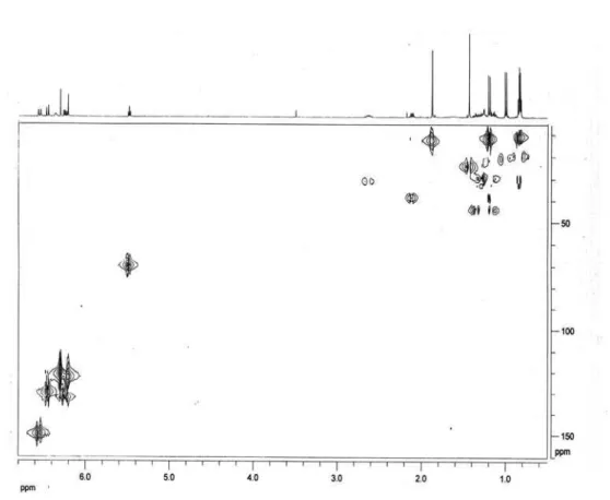
Figure S4.1H-13C HSQC 2D NMR correlation spectroscopy of 1 (400 MHz/100 MHz, CDCl 3).


![Figure S6. APCIMS [M+H]+ daugther ions spectrum of 1: (A) 20 eV (B) 10 eV.](https://thumb-eu.123doks.com/thumbv2/123dok_br/18992649.461186/9.892.90.785.124.626/figure-s-apcims-m-h-daugther-ions-spectrum.webp)