*e-mail:[email protected]
1. Introduction
Drug loaded delivery system is considered an emerging alternative for the regeneration of bone defects1. Such drug provide readily available bioactivity facilitating bone ingrowth, Steroidal saponins (SS) has been demonstrated to promote the proliferation of osteoblasts. Moreover, the biomedical potential of SS loaded biodegradable collagen microparticles as a drug delivery system has been demonstrated in our previous study2. For bone repair, an ideal delivery system should combine an osteoconductive scaffold as structural support for cells with a drug for cellular growth and differentiation3,4.
The challenge of such scaffolds to mimic physiological patterns lies in (i) an optional control of independent drug release kinetics of more than one direction. (ii) control of appropriate release rates upon tissue. (iii) the potentially
targeted drug delivery to speciic cells, and (iv) the preservation
of drug functionality and integrity during construction of the scaffold5,6.
So far little studies reported how the spatiotemporal control
of drug may affect tissue repair. The principal signiicance
has been described for spatially separated release and deposition of drug, showing an interesting correlation between drug pharmacokinetics and material structure7. Moreover, manufacturing processes of scaffolds with spatiotemporal control of drug in bone repair are challenging.
The extracellular matrices of hard tissue are composed of organic and inorganic phases. The inorganic phase consists primarily of hydroxyapatite (HA), whilst the organic phase consists mainly of type I collagen (COL1) and small amounts of ground substance, such as glycosaminoglycans, proteoglycans, glycoproteins and phosphatidylserine (PS),
all with unique distributions relecting their biological
roles. HA, COL and PS, when processed into biomaterial, results a bone scaffold with appropriate bio-degradation, excellent mechanical properties, and well bioactivity and biocompatibility, which has been demonstrated in our previous work8. Here we introduce a protocol to fabricate inhomogeneous HA/COL/PS (HCP) scaffolds with gradient distribution of SS loaded collagen microparticles. We discussed in detail the impact of scaffold structure on release kinetics of drug to estimate the spatiotemporal control of drug delivery. Cellular response was tested with MC3T3-E1 cells to assess the impact of drug release on the proliferation and migration of cells when seeded onto different surface of the scaffolds.
2. Material and Methods
2.1. Preparation of parafin spheres
Parafin spheres were used as porogen for the preparation
of HCP scaffolds. They were prepared as described before9.
Briely, gelatin (Sigma-Aldrich Co., Saint Louis, USA) was
dissolved at 80 °C in ultrapure water at a concentration
Microporous Nano-hydroxyapatite/collagen/phosphatidylserine Scaffolds Embedding
Collagen Microparticles for Controlled Drug Delivery in Bone Tissue Engineering
Chunrong Yanga,b*,Cong Fanga,b
aSchool of Materials Science and Engineering, Fujian University of Technology – FJUT,
Fuzhou 350118, China
bFujian Provincial Key Laboratory of Advanced Materials Processing and Application,
Fuzhou 350118, China
Received: July 22, 2015; Revised: September 7, 2015
Scaffolds featuring spatiotemporal control of drug release is highly desirable for bone tissue regeneration. The objective of this study was to construct a scaffold with gradient porosity and drug distribution and evaluate the effect of scaffold structure on drug release kinetics and cell bioactivity. Nano-hydroxyapatite/collagen/phosphatidylserine scaffolds embedded with steroidal saponin loaded collagen microparticles were prepared using a porogen leaching protocol. Morphological characterization showed that the scaffolds consisted of dense layer and loose layer, and pores were interconnective. The microparticles were entrapped at the center of the scaffolds follow a gradient distribution. Release kinetics correlated with the structure. The loose layer showed greater drug release amount as compared to the dense layer. Such differences in release kinetics have distinct effects on cell bioactivity. Cell proliferated much more in loose layer than that in the dense layer. Such spatial and temporal control over drug deposition and delivery within the scaffolds could provide opportunities for tissue regeneration associated with optimum drug doses at wound site, and lessen undesirable drug release and side-effects at uninjured site.
of 30 g/l and mixed with parafin (Yudao Co., Chengdu, China) upon melting at a inal concentration of 200 g/l. Phases was emulsiied for 20 min and then poured into ice water. The solidiied parafin spheres were collected and washed with ultrapure water. After drying, the parafin spheres with the diameter of 180-250 μm were collected
with standard sieves.
2.2. Preparation of HCP scaffolds
SS loaded collagen microparticles were prepared in previously research and the particle size of microparticles
ranged from several microns up to about 100 μm.
HCP scaffolds were prepared as follows. In brief,
parafin spheres of 180-250 μm diameter were illed into
20 ml cylindrical syringe, followed by sintered at 37 °C for
40 min in order to achieve suficiently interconnected pores
within the scaffold. After the heat treatment the syringe was annealed to room temperature.
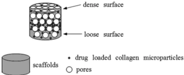
HCP scaffolds embedding collagen microparticles were prepared by mixing previously fabricated collagen
microparticles with the parafin spheres prior to heating
the spheres at 37 °C. After annealing to room temperature, nano-hydroxyapatite, phosphatidylserine, and collagen solution were added to the syringe and carefully aspirated to
ill the interspace between the spheres. The constructs were
in multiple layers and the pores were interconnective and gradient, which can be controlled by gradient distribution
of drug loaded collagen microparticles and parafin spheres
(Figure 1).
The preparations were then shock-frozen in liquid nitrogen,
and freeze dried at −30 °C for 45 h (ALPHA2-4, Christ,
Germany). Scaffolds were treated with 90% (v/v) methanol solution for 45 min and then treated with hexane for 20 h
to extract the parafin spheres. Hexane was replaced after
24 h and the resulting scaffolds were dried under vacuum for 10 h (YB-FD-1, Yibei Co., Shanghai, China) in order to completely dry the scaffolds and minimize residual hexane10.
2.3. Cross-section observation of HCP scaffolds
The cross-section of HCP scaffolds with embedded collagen microparticles was coated with gold and morphologically determined using scanning electron microscopy (SEM) (S-3400N, Hitachi Co., Tokyo, Japan).
2.4. In vitro drug release
The temporal and spatial release experiment of the HCP
scaffolds was performed in a continuous low system (Figure 2).
The in vitro simulated body luid employed for the release
medium was prepared by dissolving reagent-grade NaCl
(142 mmol•l–1), NaHCO
3 (4.2mmol•l
–1), KCl (3.0mmol•l–1), K2HPO4•3H2O(1mmol•l–1), CaCl
2 (2.6 mmol•l –1), Na
2SO4
(0.5 mmol•l–1), and MgCl
2•6H2O (1.6 mmol•l –1) in
deionized water. The solution was buffered at PH 7.4 with trishydroxymethylaminomethane [(CH2OH)3CNH2] and 1 N HCL at 37 °C.
To assess the effect of the structure of the scaffolds on the release kinetics of drug, the samples were laid down the
ilter at 37 °C with the loose surface as the input side and the
dense surface as the output side, or reversed (Figure 2). After
speciic time periods, the outlow solution was collected,
and the inlow solution was refreshed. The steroidal saponin
content of solution collected for each interval was determined by using the ultra performance liquid chromatography
(UPLC, Waters Co., Milford, USA), and steroidal saponins
were used as the standard.
2.5. In vitro cell bioactivity on HCP scaffolds
The osteoblast bioactivity of the HCP scaffolds in bone formation was investigated using MC3T3-E1 cells (Japan Riken Cell Collection). Cells were cultured in the DMEM-10% FBS medium (Gibco), and maintained in 5% CO2 at 37 °C. The cells (2 × 10³ cells/mL) were seeded on the dense or loose surface of 30 mg of specimens, placed on the bottom of 100 mm plastic tissue-culture dishes (Corning Co.,Midland,
USA) and remained undisturbed in a humidiied incubator
(37 °C and 5% CO2) .
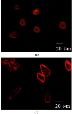
The morphology of cells grown on different surfaces of the scaffolds was observed using a confocal laser scanning microscope (CLSM, TCS SP5, Leica, Wetzlar, Germany).
After 5 h of incubation, the specimens with cells were ixed
by 4% paraformaldehyde in PBS at room temperature, followed by wash. They were then immersed in 1% rhodamine phalloidin for 10 min to stain cell. After rinsing with phosphate buffered saline (PBS), the stained specimens were placed on the glass-bottomed dish and examined, then
luorescence density of the cell was measured.
The growth kinetics of MC3T3-E1 cells on both surfaces of the scaffolds were assessed using a 3-(4,5-dimethyl-2-thiazolyl)-2,5-diphenyl-2H tetrazolium bromide (MTT)
(Sigma Co., Saint Louis, USA) assay. The cells were cultured
for 1, 3, or 5 days, respectivly. Subsequently, the medium was removed and 200 µl MTT solution was added to each well for incubation at 37 °C for 4 h. Then the MTT solution was removed and the insoluble blue formazan crystal was dissolved in dimethyl sulfoxide (DMSO) (Sigma Co., Saint
Louis, USA). Subsequently, optical density of the solution
was measured. The absorbance was directly proportional to cell proliferation.
Figure 1. Illustration showing gradient distribution of drug loaded collagen microparticles in HCP scaffolds.
2.6. Statistical analysis
The data were reported as the mean ± SD. Two-factor analyses of variance (ANOVAs) with Bonferroni–Dunn and Fisher’s post hoc tests for multiple comparisons as well as Student’s t test were performed using Sigma StatTM
Software for Windows Version 2.03, SPSS Inc. Signiicance
was accepted at a level of P<0.05.
3. Results and Discussion
3.1. Morphological characterization of HCP
scaffolds
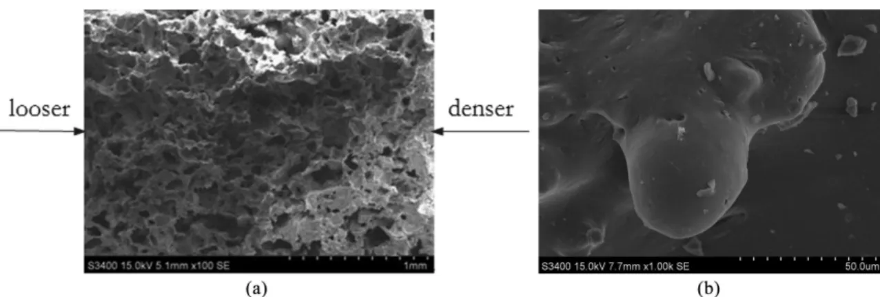
SEM observation of the HCP scaffolds showed a continuous and gradient structure of irregular interconnected pores, and consisted of dense layer and loose layer (Figure 3a).
Most of the pore sizes were around 100-300 μm. Parafin
leaching from SS loaded HCP matrices with hexane resulted in porous structure of high interconnectivity. Also matrix between the interconnecting pores was observed in this case. The embedded collagen microparticles to be well integrated into the HCP scaffolds, strictly locating in the intersections or walls of the matrices (Figure 3b). Also embedded collagen microparticles appeared to be coated with a thin layer of HCP matrices.
Such three-dimensional gradient structure may vary
release proile and enhance the eficiency of the drug in bone
regeneration by creating and maintaining appropriate space for progenitor cell migration, proliferation, and differentiation.
3.2. In vitro drug release studies
In vitro SS release kinetics of collagen microparticles embedded in HCP scaffolds, and the effect of the structure of the scaffolds on the release kinetics were evaluated for 30 days in Figure 4. Irrespective of the low direction, the
release proiles were triphasic consisting of (i) a typical burst release within the irst 12 h, followed by (ii) a period
of low release rates until day 7 (the dense surface to loose surface) and day 12 (the loose surface to dense surface), and
inally (iii) a marked increase in release from these days on.
The drug from the dense surface to loose surface of the scaffolds showed a burst release of 25.8±3.8% after 0.5 d.
The burst release from the loose surface to dense surface
was less prominent with 21.3±4.7%. These indings were further relected by the release rate throughout the observation
period. The average release rate of drug from the dense
surface to loose surface exceeded signiicantly (p<0.05)
the rate from the loose surface to dense surface. Almost no drug was released from scaffolds until day 26, turning into a continuous and slow release from then on. After 30 days, 73.7±4.0% of the initially loaded drug was released from the dense surface to loose surface of the scaffolds. On the other hand, much less, only 43.6±3.1% of the initially loaded drug was released from the loose surface to dense surface of the scaffolds.
The difference of release proiles of drug from scaffolds has been largely affected by the low direction. This outcome
was explained to result from the diffusional barrier through the scaffold matrix. From the loose surface to dense surface, drug diffusion was driven from low to high resistance, which induced slow release. In contrast, from the dense surface to loose surface diffusion was faster.
Figure 3. SEM images of the cross-section of the HCP scaffolds (a) and SS loaded collagen microparticles embedded in the scaffolds (b).
Such differences in release kinetics of drug from scaffolds with different exposed surfaces may have distinct effects on bone regeneration7. In this study, we could show that
the release proiles were spatiotemporal. This may be an
advantage as compared to homogeneous structure of scaffolds. This would direct the drug much into the loose layer of the scaffold from where they may be easily washed out and exposed to the defect tissue. Additionally, the smaller releas amount in another surface when exposed to well-surrounding site could avoid potential toxicity and reduce undesirable side-effects, such as hyperplasia, hypertrophy, metaplasia, ectopic bone formation, or unwanted vessel growth11. This leads to the conclusion that the constructed inhomogeneous scaffolds incorporating drug-loaded microparticles could allow spatially separated release of drug, a prerequisite for the regeneration of bone defects.
3.3. Cell bioactivity
A MC3T3-E1 cell proliferation assay was used to assess if the bioactivity of released drug was impacted by the inhomogeneous structure of scaffolds. According to the
luorescence intensity (Figure 5), it could be found that cell grown on the loose surface of the scaffolds spread fully, interconnected or formed dense cell clusters in the scaffold matrix after 5 h of culture. On the other hand, most cells grown on dense surface of the scaffolds were round or oval, and area of cell spreading was smaller.
This result was further conirmed by the cell-proliferation
(Figure 6). Compared to the loose surface, cell grown on the
dense surface of the scaffolds induced signiicantly lower
proliferation within the same culture time.
This outcome could be explained that released drug in combination with different surfaces of the scaffolds had substantial effects on the cell growth and proliferation. We previously demonstrated the SS on the bioactivity of MC3T3-E1 cells. The growth and proliferation of cells related to the release of SS from collagen microparticles. From above release experiments, we would have expected high proliferation rates on the loose layer of the scaffolds when considering the high release of drug. The HCP scaffolds may possibly act similar to the extracellular matrix, and provide a depot for drug that is released surface-bound to stimulate different cell growth rates. Therefore controlled release of drug could deliver optimum dose for the wound tissue to induce cell growth and promote tissue regeneration, while lessening the undesired release to uninjured areas to avoid side effects.
4. Conclusions
We developed an inhomogeneous and gradient HCP scaffold embedding collagen microparticles which we prior loaded with SS. Different surface of scaffold resulted in distinct drug release kinetics and cell population. Spatial control of drug deposition and temporal control of drug delivery within the scaffolds could provide opportunities for tissue regeneration associated with optimum drug doses at wound site, and reduce undesirable drug release and side-effects at uninjured site. Future, pre-clinical studies will be needed to investigate the performance of SS loaded scaffolds on bone in vivo.
Acknowledgements
This work was inancially supported by natural science
foundation project of Fujian (Grant no. 2014J05054) and
scientiic research starting foundation project of Fujian
university of technology (Grant no. GY-Z0854).
Figure 6. Proliferation of MC3T3-E1 cells on both surfaces of the HCP scaffolds. At the indicated time points, cell viability of the loose surface group was signiicantly higher than those of the dense surface group. Data are presented as means ± standard deviations for n=5.
References
1. Nagai N, Kumasaka N, Kawashima T, Kaji H, Nishizawa M and Abe T. Preparation and characterization of collagen microspheres for sustained release of VEGF. Journal of Materials Science. Materials in Medicine. 2010; 21(6):1891-1898. http://dx.doi. org/10.1007/s10856-010-4054-0. PMid:20232232.
2. Yang C and Wang J. Preparation and characterization of collagen microspheres for sustained release of steroidal saponins. Materials Research. 2014; 17(6):1644-1650. http://dx.doi. org/10.1590/1516-1439.307214.
3. Chen J, Wang Z, Wen Z, Yang S, Wang J and Zhang Q. Controllable self-assembly of mesoporous hydroxyapatite. Colloids and Surfaces. B, Biointerfaces. 2015; 127:47-53. http:// dx.doi.org/10.1016/j.colsurfb.2014.12.055. PMid:25638722.
4. Chen J, Yu Q, Zhang G, Yang S, Wu J and Zhang Q. Preparation and biocompatibility of nanohybrid scaffolds by in situ homogeneous formation of nanohydroxyapatite from biopolymer polyelectrolyte complex for bone repair applications. Colloids and Surfaces. B, Biointerfaces. 2012; 93:100-107. http://dx.doi. org/10.1016/j.colsurfb.2011.12.022. PMid:22297206.
5. Li F, Wu W, Xiang L, Weng G, Hong H, Jiang H, et al. Sustained release of VH and rhBMP-2 from nanoporous magnesium-zinc-silicon xerogels for osteomyelitis treatment and bone repair. International Journal of Nanomedicine. 2015; 22(10):4071-4080. http://dx.doi.org/10.2147/IJN.S82486. PMid:26124660.
6. Boukari Y, Scurr DJ, Qutachi O, Morris AP, Doughty SW, Rahman CV, et al. Physicomechanical properties of sintered scaffolds formed from porous and protein-loaded poly(DL-lactic-co-glycolic acid) microspheres for potential use in bone
tissue engineering. Journal of Biomaterials Science. Polymer Edition. 2015; 26(12):796-811. http://dx.doi.org/10.1080/092 05063.2015.1058696. PMid:26065672.
7. Li X, Wang J, Su G, Zhou Z, Shi J, Liu L, et al. Spatiotemporal control over growth factor delivery from collagen-based membrane. Journal of Biomedical Materials Research. Part A. 2012; 100(2):396-405. http://dx.doi.org/10.1002/jbm.a.33282. PMid:22083844.
8. Yang C, Wang Y and Chen X. Preparation and evaluation of biomimetric nano-hydroxyapatite-based composite scaffolds for bone-tissue engineering. Chinese Science Bulletin. 2012; 57(21):2787-2792. http://dx.doi.org/10.1007/s11434-012-5201-4.
9. Wenk E, Meinel AJ, Wildy S, Merkle HP and Meinel L. Microporous silk fibroin scaffolds embedding PLGA microparticles for controlled growth factor delivery in tissue engineering. Biomaterials. 2009; 30(13):2571-2581. http:// dx.doi.org/10.1016/j.biomaterials.2008.12.073. PMid:19157533. 10. Uebersax L, Merkle HP and Meinel L. Insulin-like growth
factor I releasing silk fibroin scaffolds induce chondrogenic differentiation of humanmesenchymal stem cells. Journal of Controlled Release. 2008; 127(1):12-21. http://dx.doi. org/10.1016/j.jconrel.2007.11.006. PMid:18280603.