JSCS–4562 Original scientific paper
Application of ultrasound and methanol for the rapid removal of
surfactant from MCM-41 molecular sieve
MOHAMMAD A. ZANJANCHI and SHAGHAYEGH JABARIYAN*
Department of Chemistry, Faculty of Science, University of Guilan, P. O. Box 1914, Rasht 41335, Iran
(Received 12 January 2013, revised 12 April 2013)
Abstract: Ultrasound waves were successfully applied for the removal of the template from mesoporous MCM-41 molecular sieve. The method uses 28 KHz ultrasound irradiation in a methanol solvent for disrupting the micellar aggregation of the surfactant molecules, cetyltrimethylammonium bromide, which fill the pores of as-synthesized MCM-41. After 15 min sonication at the moderate temperature of 40 °C, the majority of surfactant molecules had been removed from powder MCM-41. The template removal rate using ultrasound irradiation (15 min) is faster than the rate obtained via thermal calcination. In addition, a perfect hexagonal pore structure was obtained after template remo-val using ultrasound irradiation, according to characterization using X-ray diffraction (XRD) and nitrogen adsorption analyses, while high temperatures during calcination cause shrinkage that affected the surface properties of the materials. In the present procedure, the surfactant molecules are released into methanol and can be recovered for reuse. The effectiveness of the sonication-prepared MCM-41 as an adsorbent was confirmed by the adsorption of methyl-ene blue (MB).
Keywords: ultrasound; methanol; mesoporous; MCM-41; template removal; micelle.
INTRODUCTION
During the past decade, the study of the physical and chemical effects of ultrasound irradiation is a rapidly growing area of research.1–3 Ultrasound is the
name given to sound waves having frequencies higher than those to which the human ear can respond (16 kHz). The use of ultrasound may be divided broadly into two areas: i) high frequency ultrasound (2–10 MHz), and ii) low frequency
ultrasound (20–100 kHz). Over the last few years, the principal application of ultrasound was in synthesis (organic, organometallic and inorganic), polymer chemistry (degradation, initiation, and copolymerization) and some aspects of
catalysis. This field of scientific research that uses ultrasound is called sono-chemistry.4
Ultrasound waves consist of a cyclic succession of expansion and com-pression phases imparted by mechanical vibration. The expansion cycles exert a negative pressure and pull the molecules apart, while the compression cycles exert a positive pressure and push the liquid molecules together. When cyclic stress through repeated implosion exceeds the tensile strength of a liquid in the rarefaction regions, small vapor-filled voids, called cavitation bubbles, are formed. The compression of the bubbles during cavitation is more rapid than thermal transport, which generates a short-lived localized hot-spot. Experimental results have shown that hot-spots with high local temperatures of around 5000 K, pressures of roughly 1000 atm*, and combined with heating and extraordinarily rapid cooling rates of above 1010 K s–1 provide a unique means for driving
chemical reactions under extreme conditions.5–8
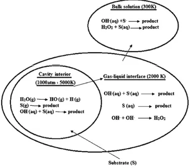
In sonochemical reactions, the ultrasonic energy influences the chemical reactions by providing huge heat (pyrolysis) or producing reactive free radicals. There are mainly three reaction sites (Fig. 1): a) cavity interior, b) gas–liquid interface and c) bulk liquid. Inside the cavitation bubble, water molecules are pyrolyzed forming •OH and •H in the gas phase. The substrate either reacts with
the hydroxyl radical or undergoes pyrolysis. In the interfacial region, a similar
* 1 atm = 101325 Pa
reaction occurs but in an aqueous phase. The additional reaction is the recom-bination of the OH radicals to form H2O2. In the bulk phase, the reactions are
between the substrate and the •OH or H
2O2. All these reactions are considered
homogeneous in sonochemistry.7
MCM-41, one of the members of the extended family of mesoporous sieves, possesses a hexagonal array of uniform mesopores. MCM-41 was discovered in 1992 by researchers at Mobil. In addition, it has been synthesized with uniform channels varying from approximately 15 Å to greater than 100 Å in size. Thus, it has potential applications in the field of adsorption and catalysis.9–11
Although the synthesis of MCM-41 is possible via a number of methods,
generally it is synthesized from the surfactant micellar template addition of an inorganic silica source, with cetyltrimethylammonium bromide (CTA+Br–) being
the most commonly used template.12,13
In order to make the porous network accessible in such systems, the template has to be removed from the mesoporous channels inside the particles. The most common method used in laboratories to remove the template is calcination at high temperatures.14–16 Although all organic templates are removed from the
structure using this method, high temperatures cause shrinkage, which affects the surface and the catalytic and adsorption properties of the materials. For this reason, methods and other conditions should be used for template removal.
Extraction with conventional solvent is another method for template remo-val.17,18 The efficiency of solvent extraction methods depends on the interaction
between organic molecules and inorganic framework. For mesoporous materials that are synthesized by the S+X–I+ and S0I0 way, the interaction between the organic phase and the inorganic network is weak. Therefore, the surfactant mole-cules in the pores of mesoporous materials can be removed by extraction with ethanol.19 Since solvent extraction methods due to the strong electrostatic
inter-actions between the surfactant and a network of micelle cannot be applied to ordered mesoporous materials prepared in the alkaline environment (S+I-way), ion exchange is widely used under this condition. In recent years, many different solutions, such as HCl,17,20 H2SO418
and
NH4NO321 in ethanol solvent wereused for the removal of surfactant. Although structural damage is minimized in the solvent extraction method, several extraction steps are required to remove the organic template completely and thus, large amounts of solvent and lots of extraction time are required.
Additionally, oxidation methods were used for template removal. In these methods, several oxidizing agents, such as KMnO4,22 UV–ozone or ozone,23–25
perchlorates,26 H2O2 and H2O2–UV/Fe27,28 have been used. Although template
extraction33,34 are other techniques to remove templates, but complex devices are
required.
Furthermore, in 2012, a new method for the removal of the surfactant from the pores of mesoporous materials employing ultrasonic irradiation in ethanolic solution was presented.35
However, a method for template removal should have the following pro-perties: efficient removal of the template, short operation time, reduced organic solvent consumption, regular (well-organized or systematic) structure retention and the possibility for surfactant recovery
In this work, methanol as solvent in the presence of ultrasonic waves was used to remove the template and the effect of ultrasonic wave and methanol solvent on the structure of MCM-41 were investigated. Additionally, the obtained results were compared with those of a calcined sample. The results indicated that the samples kept their porous structures and structural shrinkage was minimized. Moreover, the surface area and pore volume of the samples were higher than those for the calcined samples were. Thus, methanol as solvent in the presence of ultrasonic waves functioned effectively.
EXPERIMENTAL Preparation of hexagonal mesoporous silica
The surfactant-templated MCM-41 was synthesized by a familiar room temperature method.35,36
Template removal using ultrasound waves
The ultrasound-assisted template removal was performed in a 7500S ultrasonic device (Sairan Instrument Company, Iran) with an ultrasound power of 600 W, heating power of 800 W and a frequency of 28 kHz, equipped with a timer and a temperature controller. In general, 0.5 g of MCM-41 was dispersed in 75 mL of methanol in a beaker. Then, the suspension was immersed in water in the ultrasonic device and irradiated for 15 min at 40 °C. A mechanical stirrer at a speed of 300 rpm was used during the treatment. After ultrasonic irradiation, the sample was recovered by centrifugation, washed with cold methanol and dried at 60 °C for 6 h [MCM-41(US1)]. For certain samples, this procedure was repeated for two more successive steps [MCM-41(US2)].
Template removal using thermal calcination
For comparison, the template was also removed by thermal calcination. The fresh sample was heated under air at 550 °C and held for 5 h (MCM-41(C-550)). Then the sample was cooled to room temperature. The total time for the complete removal was 12.4 h.
Characterization of the as-synthesized, calcined and sonicated powders and the supernatant solutions
were performed under static air from room temperature to 600 °C at a heating rate of 10 °C min-1. In addition, the concentration of CTAB in the supernatant solution was determined by using a double-beam UV spectrophotometer at 375 nm and in the presence of 0.02 mL of 0.1 % picric acid in 0.002 M NaOH and 10 mL of chloroform per 1.0 mL of the supernatant.37 The specific surface areas of the sonicated and calcined MCM-41 samples were estimated based on the data provided by a Sibata surface area apparatus 1100. The sample was degassed at 250 °C for 2 h prior to nitrogen physisorption measurements. The FTIR spectra of the bare MCM- -41 and the treated MCM-41 samples were obtained using a Shimadzu FTIR-8900 spectro-photometer.
Adsorption test
Methylene blue (MB) solutions were prepared from a commercially available product (Merck, 1.59270) dissolved in distilled water. For the adsorption experiments, 100 mg of the as-synthesized MCM-41 or any of the sonicated MCM-41 samples were added into 100 mL of MB solutions at a concentration of 25 ppm and pH of 5.5. The solution was stirred for about 30 min for the sorption process to reach equilibrium.
The solution and the solid phase were separated by centrifugation. A Shimadzu UV-2100 spectrophotometer was employed for measuring the MB concentration.
RESULTS AND DISCUSSION
Structural characterization
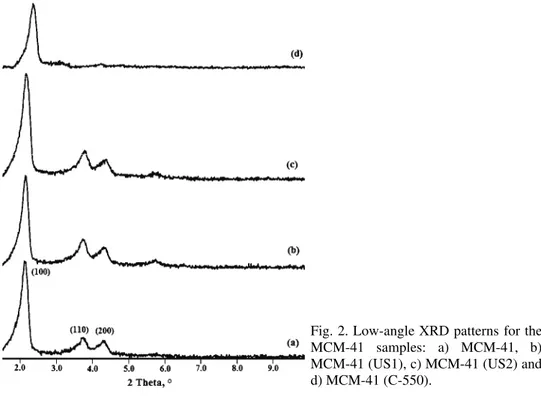
The XRD diffraction patterns of the as-prepared, sonicated and calcined MCM-41 materials are shown in Fig. 2. Three peaks were observed in all cases.
The first peak was detected at around 2θ = 2.150° and the two small intensity peaks were detected at 2θ 3.73 and 4.73°. These diffraction peaks were indexed as the 2D hexagonal arrangement with the reflection of (100), (110) and (200).38
The structural parameter (d-spacing) calculated from the diffraction angles is
given in Table I. The XRD patterns showed that ordered mesoporous structures were preserved in all the sonicated samples. However, the weaker reflections corresponding to the planes (110) and (200) could hardly be seen in the pattern of the calcined MCM-41(C-550) sample. The frequent disappearance of these weak reflections was assigned to disordering of the array of meso channels of MCM-41 samples subjected to a calcination step.39 The small shift of the main diffraction
peak toward a higher 2θ value for MCM-41 (C-550) was due to shrinkage of the sample that occurred because of the template removal by calcinations.40
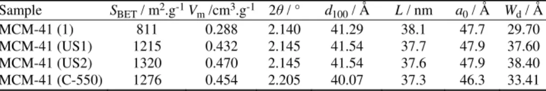
TABLE I. Comparison of the physicochemical properties of porous materials treated by ultrasound waves and thermal calcinations; SBET: apparent surface area calculated by the
BET method; Vm: pore volume; d100: LA-XRD spacing of the 100 reflection of a
hexa-gonal plane array of pores; L: crystallite size calculated by the Sherrer equation (L =
= kλ/βcos θ); a0: cell constant, a0 = 2d100/√3; Wd: pore size
Sample SBET / m2.g-1Vm /cm3.g-1 2θ / ° d100 / Å L / nm a0 / Å Wd / Å
MCM-41 (1) 811 0.288 2.140 41.29 38.1 47.7 29.70
MCM-41 (US1) 1215 0.432 2.145 41.54 37.7 47.9 37.60 MCM-41 (US2) 1320 0.470 2.145 41.54 37.6 47.9 38.40 MCM-41 (C-550) 1276 0.454 2.205 40.07 37.3 46.3 33.41
The degree of template removal was verified by BET analysis based on phy-sisorption of nitrogen and calculation of the specific surface area of the sonicated samples. Table I shows the results of the sonication in methanol solvent at 40 °C and for 15 min and thermal calcination for the removal of CTAB template from the pores of MCM-41. The table also contains the results of the treatment of MCM-41 by stirring in methanol at 40 °C in the absence of ultrasound irradiation (MCM-41 (1)). It is evident from Table I that stirring of MCM-41 in methanol solutions without sonication led to the partial removal of template from the pores of MCM-41, as indicated by the resulting pore volume and surface area. How-ever, using ultrasound irradiation for the treatment of MCM-41 in methanol at the same temperature (40 °C) for the same time (15 min), a greater increase in the surface area and pore volume resulted. In fact, the calculated specific surface area for MCM-41 (1) of 811 m2 g–1 increased to 1215 m2 g–1 for MCM-41 (US1) due
to the ultrasound irradiation.
Repeating the sonication under the same conditions affected the value of the surface area. Table I shows that the calculated surface area for MCM-41 (US1) was 1215 m2 g–1, which increased to 1320 m2 g–1 for MCM-41 (US2) that had
by thermogravimetric analysis (discussed later) and it was found that the amount of template remaining in the structure following 15 min sonication were very small. Therefore, at this stage it is obvious that although template removal from MCM-41 is feasible by the treatment with alcohols, such as methanol, applica-tion of ultrasound irradiaapplica-tion greatly increased the speed and the efficiency of the removal.36 The specific surface area for MCM-41 (C-550), prepared by thermal
treatment at 550 °C of the as-synthesized MCM- 41 was 1276 m2 g–1. The values
of the surface areas for the sonicated MCM-41 samples (around 1200 m2 g–1 or
higher) were comparable with that of the thermally treated sample (1276 m2 g–1).
This proximity is an indication that the employed sonication procedure was suc-cessful in the removal of the template from MCM-41.
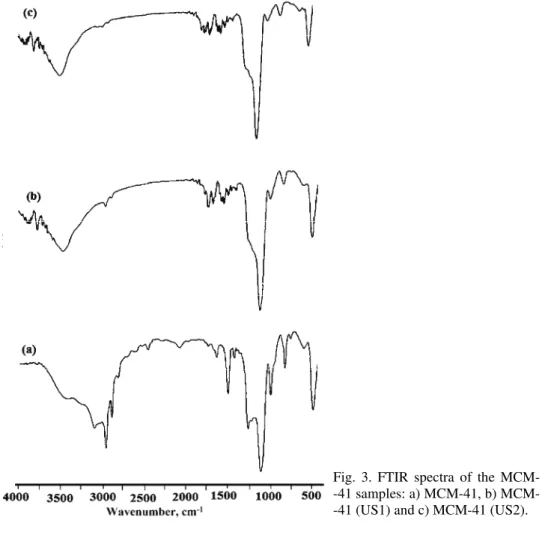
The FTIR absorbance spectra of the as-prepared and sonicated MCM-41 materials are shown in Fig. 3. A broad band around 3200–3500 cm−1 appears for
all samples, which is partially caused by the O–H stretching vibration mode of
adsorbed water molecules, the bending vibration mode of which is responsible for the band recorded at 1630 cm−1. The absorption bands at around 2858–2925
cm−1 and 1375–1475 cm−1 observed in the spectrum of the as-prepared MCM-41
could be assigned to C–H stretching and bending vibrations of the template CTAB.41 After treatment (sonication), the C–H vibration peaks are nearly
indis-cernible. This suggests the efficient removal of the surfactant template by the sonication methods. These bands were absent in the FTIR spectrum of MCM-41 (C-500) because of the complete removal of the template from MCM-41 by calcination.
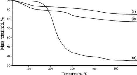
Thermogravimetric analysis was employed to determine quantitatively the degree of surfactant removal from the sonicated MCM-41 materials. The thermo-grams for the as-synthesized MCM-41 and for those sonicated at 40 °C in metha-nol for 15 min and for two consecutive 15-min periods are shown in Fig. 4. The thermograms were recorded from 20 to 600 °C. In the temperature range up to 150 °C, physisorbed water was released from the pores. From 150 °C to about 340 °C, the remaining template was successfully decomposed. At temperatures above 340 °C, the weight loss could be attributed to water released from silanol condensation and to the oxidation of a small amount of carbonaceous residues from incomplete template combustion. The amount of template could therefore be estimated from the weight loss between 150 and 340 °C.15 These amounts
were approximately 57, 7 and 1 % for MCM-41, MCM-41 (US1) and MCM-41 (US2), respectively. These data show that most of the template molecules had been released from MCM-41.
Mechanism of cationic surfactant extraction from the pores of MCM-41
The data in Table I revealed that ultrasound irradiation played essential roles in the removal of the template from the pores of MCM-41. It was already esta-blished that organic solvents such as methanol or ethanol cause disruption of the surfactant aggregates.42,43 Due to this disintegration, the surfactant monomers
are released into the organic solvent and can be eluted. It is known that the cri-tical micelle concentration (cmc) for CTAB in methanol is much higher than that in water. Therefore, the micelles of CTAB that were formed during the synthesis process of MCM-41 in aqueous media can be disrupted by treating MCM-41 in methanol. The ultrasound irradiation makes the disruption faster and more effi-cient via a synergistic effect.36
The role of ultrasound in the disintegration of the micelles could be attri-buted to the shear forces generated by bubble implosion during ultrasound irra-diation. There are several reports to describe this role of ultrasound and con-sequent forces in the disruption or segregation of the micelles.44–46 During
soni-cation, bubble implosion occurs, which results in liquid jets of high velocity forming shear forces. The shear forces break the integrity of the micelles and convert them to free surfactant molecules.
In addition, it should be taken into account that upon ultrasound irradiation, degradation of the surfactant molecules may also occur. From the previous studies, it could be learnt that only surfactant monomers are susceptible to deg-radation by a sonochemical process and their presence as micelles would shield them from the irradiation.47,48 In fact, the hydrophobic tail of the surfactant
cannot be directly exposed to a bubble ‘‘hot spot’’ when it is pointed into the core of the micelle. Therefore, provided that the ultrasound irradiation causes only micelle disruption, the intact template could be recovered at the end of the template removal procedure.
Investigation of the effects of sonication on the surfactant structure by UV and FTIR spectroscopy
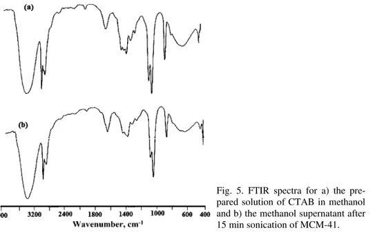
Due to decomposition of surfactant monomers during sonochemistry pro-cesses, the possibility of destruction of surfactant molecules by ultrasound irra-diation should be considered. Ultrasonic irrairra-diation was applied to disturb order in the micelles so surfactant molecules in the micelles de-aggregate. In order to obtain useful information about the structural integrity of the surfactant mole-cules that were removed from MCM-41 and transferred into the methanol sol-vent, further studies are required. The FTIR spectrum of the supernatant solution after treatment in the presence of ultrasound could be useful.
CTAB is presented in Fig. 5a. The two bands at 2891 and 2974 cm–1 correspond
to C–H vibrations of the surfactant molecules in methanol. On comparing the FTIR spectra, it is readily observable that the C–H stretching vibration bands do not significantly change. This shows that the CTAB molecules had preserved their structure after sonication in methanol. However, these results are based on FTIR spectroscopy (qualitative analysis); hence, a more comprehensive study and quantitative analysis are required.
To obtain the necessary information about the structure and composition of the surfactant and determine the amount of surfactant present in the supernatant solutions after exposure of samples to ultrasonic waves, a special spectrophoto-metric method was used. In this method, the solutions remaining from MCM-41 samples that had been irradiated by ultrasound waves for 2, 4, 6, 8, 10 and 12 min at 40 °C in methanol were studied spectrophotometrically.
After ultrasound irradiation, the micelles in the methanol environment dis-rupted slowly and consequently by addingpicricacid, ion pairs were formed with the monomers of surfactant and the ion pairs transferred from the aqueous phase to the organic phase. The absorbance of the organic phase was read at λmax =
= 375 nm.
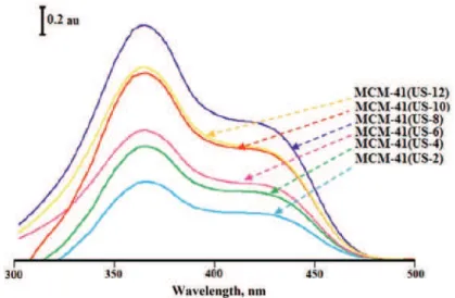
According to UV–Vis spectra (Fig. 6), it wasfound that the micelles in the MCM-41 structure break from each other and monomers are released into the methanol. Although the spectrum of samples MCM-41 (US2), MCM-41 (US4), MCM-41 (US6) and MCM-41 (US8) showed increases in the concentration of surfactant CTA+ with increasing sonication time, the spectra of samples MCM-
-41 (US10) and MCM-41 (US12) showed gradually decreasing concentration of the surfactant. Reduction in the surfactant concentration means that degradation of surfactant molecules occurred at longer sonication times.
Fig. 6. UV–Vis spectra for the methanol supernatants after various times.
Thus, it can be stated that ultrasonic vibrations increase the molecular move-ment of the surfactant molecules away from each other following micelle dis-ruption in a methanolic solution. Consequently, surfactant micelles are released from MCM-41 mesoporous pores into organic solvents. Since the hydrophobic tail of the organic template is pointed into the core of the micelle, they will not be damaged by the ultrasonic vibrations, which can only cause accelerated disrupt-tion of micelles in methanol solvent. When the micelles are converted to mono-mers, they may be attacked by hydroxyl radicals through their hydrophilic heads in bulk solution; as a result, they lose their own nature as a CTAB surfactant monomer. The alkyl chain of the surfactant may be separated from the head of the alkyl ammonium; hence, the C–H stretching vibration bands in the FTIR spectra do not change but the CTAB molecules are not identified by spectropho-tometric methods.
Performance of the sonicated MCM-41 materials in MB adsorption
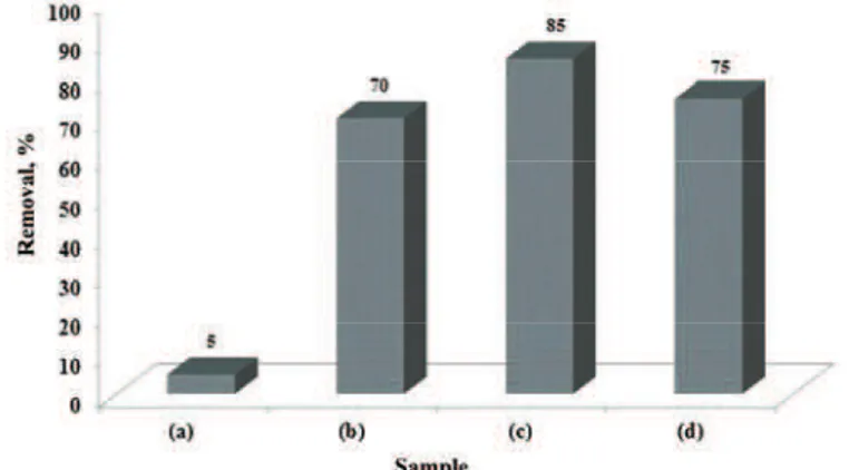
that of MCM-41 (C-550) and MCM-41 (US2). While, the adsorption of MB onto the as-synthesized MCM-41 was much lower than onto sonicated MCM-41 samples (Fig. 7). These results suggest that the absence of CTA+ inside of
soni-cated MCM-41 affects the sorption of MB by these adsorbents. The pHpzc value
of 4.1 for MCM-41 (C-550) clearly shows that at the employed experimental conditions (pH ≈5.5), the surface charge was negative and the electrostatic interaction between MB and MCM-41 (C-550) or the sonicated MCM-41 sample was very likely.
Fig. 7. MB removal by the adsorbents: a) MCM-41, b) MCM-41 (US1), c) MCM-41 (US2) and d) MCM-41 (C-550).
CONCLUSIONS
И З В О Д
ПРИМЕНАУЛТРAЗВУКАИМЕТАНОЛАЗАБРЗОУКЛАЊАЊE СУРФАКТАНТА
САПОВРШИНЕ MCM-41
MOHAMMAD. A. ZANJANCHI и SHAGHAYEGH. JABARIYAN
Department of Chemistry, Faculty of Science, University of Guilan, P. O. Box 1914, Rasht 41335, Iran
Ултразвучниталасисууспешнопримењенизауклањањетемплатасамезопорозног молекулскогсита MCM-41. Примењенојеултразвучноозрачивањеод 28 kHz уметанолу каорастварачудабисеспречиламицеларнаагрегацијамолекуласурфактанта, цетил
-триметиламонијум-бромида, којијеиспуњаваопоре MCM-41 приликомсинтезе. Сони
-кацијомутрајањуод 15 min наумеренојтемепературиод 40 °C, већинамолекуласурф
-актанта је уклоњена са праха MCM-41. Брзинауклањањa темплата приултразвучном озрачивању (15 min) јевећаодбрзинеуклањања којасепостижекалцинацијомипри томједобијенаперфектноуређенахексагоналнаструктурапораштојепотврђенопри
-меном дифракције X-зрачења (XRD) и адсорпцијом азота, док загревање до високих температурарезултује ускупљањупораштоимаутицај наповршинскa својствамате
-ријала. Утокуовепроцедуре, молекулисурфактантасеослобађајууметанолимогусе поновокористити. Ефикасностнаовајначинприпремљеног MCM-41 каоадосорбентаје потврђенаадсорпцијомметиленскогплавог (MB).
(Примљено 12. јануара, ревидирано 12. априла 2013)
REFERENCES
1. G. J. A. Chiffoleau, T. A. Steinberg, M. Veidt, G. F. Stickley, Ultrasonics39 (2001) 173 2. B. G. Pollet, Int. J. Hydrogen Energy35 (2010) 11986
3. C. Yu, Q. Shu, C. Zhang, Z. Xie, Q. Fan, J. Porous Mater. 19 (2012) 3
4. T. J. Mason, J. P. Lorimer, Sonochemistry, Ellis Harwood, New York, 1988, p. 1 5. P. Chowdhury, T. Viraraghavan, Sci. Total Environ. 407 (2010) 2474
6. T. J Mason, Foreword, to Theoretical and Experimental Sonochemistry Involving Inor-ganic Systems, P. Pankaj, M. Ashokkumar, Eds., Springer, New York, 2010
7. Y. Adewuyi, Ind. Eng.Chem. Res. 40 (2001) 4681 8. K. S. Suslick, Sci. Am. 260 (1989) 80
9. Q. Huo, D. I. Margolese, U. Ciesla, P. Feng, T. E. Gier, P. Sieger, R. Leon, P. M. Petroff, F. Schüth, D. I. Stucky, Nature368 (1994) 317
10. G. Øye, W. R. Glomm, T. Vralstad, S. Volden, H. Magnusson, M. Stöcker, J. Sjöblom, Adv. Colloid Interface Sci. 123–126 (2006) 17
11. J. Y. Ying, C. P. Mehnert, M. S. Wong, Angew. Chem. Int. Ed. 38 (1999) 56 12. M. Kruk, M. Jaroniec, A. Sayari, J. Phys. Chem., B101 (1997) 583
13. M. L. Occelli, S. Biz, J. Mol. Catal., A151 (2000) 225
14. M. T. J. Keene, R. D. M. Gougeon, R. Denoyel, R. K. Harris, J. Rouquerol, P. L. Llewellyn, J. Mater. Chem. 9 (1999) 2843
15. F. Kleitz, W. Schmidt, F. Schüth, Microporous Mesoporous Mater. 65 (2003) 1 16. J. Goworek, A. Kierys, R. Kusak, Microporous Mesoporous Mater. 98 (2007) 242 17. S. Hitz, R. Prins, J. Catal. 168 (1997) 194
18. W. A. Gomes, L. A. M. Cardoso, A. R. E. Gonzaga, L. G. Aguiar, H. M. C. Andrade, Mater. Chem. Phys. 93 (2005) 133
19. P. T. Tanev, T. Pinnavaia, J. Chem. Mater. 8 (1996) 2068
21. N. Lang, A. Tuel, Chem. Mater. 16 (2004) 1961
22. A. H. Lu, W. C. Li, W. Schmidt, F. Schüth, J. Mater. Chem, 16 (2006) 3396. 23. M. T. J. Keene, R. Denoyel, P. L. Llewellyn, Chem. Commun. (1998) 2303
24. E. Meretei, J. Halász, D. Méhn, Z. Kónya, T. I. Korányi, J. B. Nagy, I. Kiricsi, J. Mol. Struct. 651–653 (2003) 323
25. K. C. Hsu, K. J. Chao, S. F. Chen, H. K. Li, P. Y. Wu, Thin Solid Films517 (2008) 686 26. H. Cai, D. Zhao, Microporous Mesoporous Mater. 118 (2009) 513
27. J. Kecht, T. Bein, Microporous Mesoporous Mater. 116 (2008) 123
28. L. Xiao, J. Li, H. Jin, R. Xu, Microporous Mesoporous Mater. 96 (2006) 413
29. B. Tian, X. Liu, C. Yu, F. Gao, Q. Luo, S. Xie, B. Tu, D. Zhao, Chem. Commun. (2002) 1186
30. T. L. Lai, Y. Y. Shu, Y. C. Lin, W. N. Chen, C. B. Wang, Mater. Lett. 63 (2009) 1693 31. P. Pootawang, N. Saito, O. Takai, Thin Solid films519 (2011) 7030
32. Y. Liu, Y. Pan, Z. J. Wang, P. Kuai, C. Liu, J. Catal. Commun. 11 (2010) 551
33. Z. Huang, L. Huang, S. C. Shen, C. C. Poh, K. Hidajat, S. Kawi, S. C. Ng, Microporous Mesoporous Mater. 80 (2005) 157
34. Z. Huang, L. Xu, J. H. Li, S. Kawi, A. H. Goh, Sep. Purif. Technol. 77 (2011) 112 35. S. Jabariyan, M. A. Zanjanchi, Ultrason. Sonochem. 19 (2012) 1087
36. M. A. Zanjanchi, S. Asgari, Solid State Ionics171 (2004) 277
37. A. Gurses, M. Yalcin, M. Sozbilir, C. Dogar, Fuel Process. Technol. 81 (2003) 57 38. J. S. Beck, J. C. Vartuli, W. J. Ruth, M. E. Leonowiez, C. T. Kresge, K. D. Schmitt, C. T.
Chu, D. H. Olson, E. W. Sheppard, S. B. Higgins, J. B. Higgins, J. L. Schlenker, J. Am. Chem. Soc. 114 (1992) 10834
39. L. Huang, H. Xiao, Y. Ni, Colloids Surf., A 247 (2004) 247
40. M. Jaroniec, M. Kruk, H. J. Shin, R. Ryoo, Y. Sakamoto, O. Terasaki, Microporous Mesoporous Mater. 48 (2001) 127
41. B. Zohra, K. Aicha, S. Fatima, B. Nourredine, D. Zoubir, Chem. Eng. J. 136 (2008) 295 42. M. Cantero, S. Rubio, D. Pérez-Bendito, J. Chromatogr., A1120 (2006) 260
43. L. Sun, C. Zhang, L. Chen, J. Liu, H. Jin, H. Xu, L. Ding, Anal. Chim. Acta638 (2009) 162
44. N. Miyoshi, T. Takeshita, V. Misik, P. Riesz, Ultrason. Sonochem. 8 (2001) 367
45. A. Vercet, R. Oria, P. Marquina, S. Crelier, P. Lopez-Buesa, J. Agric. Food Chem. 50 (2002) 6165
46. A. Madadlou, M. E. Mousavi, Z. Emam-Djomeh, M. Ehsani, D. Sheehan, Ultrason. Sonochem. 16 (2009) 644