J of Evidence Based Med & Hlthcare, pISSN- 2349-2562, eISSN- 2349-2570/ Vol. 2/Issue 43/Oct. 26, 2015 Page 7616
OMENTOPLASTY IN CASES OF BUE
RGER’S DISEASE WITH POST
LUMBAR SYMPATHECTOMY
Rajendra Prasad Das1, Niranjan Sahoo2, Sushanta Kumar Das3, Paresh Kumar Gouda4
HOW TO CITE THIS ARTICLE:
Rajendra Prasad Das, Niranjan Sahoo, Sushanta Kumar Das, Paresh Kumar Gouda. “Omentoplasty in Cases of Buerger’s Disease with Post Lumbar Sympathectomy”. Journal of Evidence based Medicine and Healthcare; Volume 2, Issue 43, October 26, 2015; Page: 7616-7620, DOI: 10.18410/jebmh/2015/1030
ABSTRACT: Twelve cases of Buerger’s disease with post lumbar sympathectomy were subjected to omentoplasty for the purpose of neovascularisation of distal part to alleviate rest pain and healing of ulcer after resurfacing with SSG. All the cases didn’t improve after lumbar sympathectomy with rest pain and ulceration of distal part were subjected to omentoplasty to improve the rest pain and healing of ulcer. The mean period of surveillance was 2 years 4 months. All the 12 cases were male with habitual tobacco smokers for 15 to 20 years who developed impending gangrene of toes with ulceration to established gangrene of toes with ulceration. Out of 12 cases 7 had gangrene of toes with non-healing ulcer and rest pain and 5 had impending gangrene of the toes with ulceration. The results of post omentoplasty showed significant gradual improvement in rest pain and healing status of ulcer, thereby negating the role of amputation with salvage of limb. The principle behind omentoplasty is initiation of the process of neovascularisation from the vascularised omental pedicle. The surgical skill plateau of learning curve can be reached within a shorter duration, thereby making it a choice of surgery in cases with poor result following lumbar sympathectomy in Buerger’s disease as a process of salvaging the limb.
KEYWORDS: Buerger’s disease, Post lumbar sympathectomy, Omentoplasty, Limb neovascularization.
INTRODUCTION: Buerger’s disease (thromboangitis obliterans) is a limb threatening condition occuring in productive age group of patients and its management is a challenging problem.1,2 It
affects medium sized vessels of lower limb.3 The therapeutic approach has been consisting of
medical or surgical treatment. Surgical treatment options have consisted of lumbar sympathectomy, direct arterial surgery, adrenalectomy.4,5 With exception of smoking none of
these measures is curative. However in patients with critical limb ischaemia surgery is required to salvage the limb.6 In lumbar sympathectomy relapses are frequent due to normalisation of
vasomotor tone within two weeks to six months of operation. Arterial reconstruction is usually impossible due to distal nature of the disease and carries a high failure rate.7,8 Omentoplasty
(greater omentum made into a pedicle flap with intact vascularity as right gastroepiploic or left gastroepiploic artery as required to reach upto the distal foot) is a variable alternative for limb salvage and also significantly improves signs and symptoms.9,10
J of Evidence Based Med & Hlthcare, pISSN- 2349-2562, eISSN- 2349-2570/ Vol. 2/Issue 43/Oct. 26, 2015 Page 7617 improving circulation of surrounding tissues has been well established. Hence omentum has been used to revascularise the ischaemic limb.
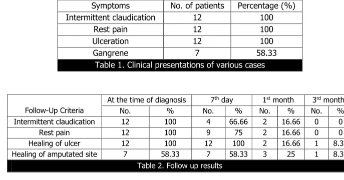
MATERIALS AND METHODS: The study was conducted in the department of general surgery and plastic surgery M.K.C.G. Medical College, Berhampur from March 2013 to July 2015. The study was conducted in a total of 12 patients who have been diagnosed as Buerger’s disease and undergone lumbar sympathectomy previously. The patients were male with either BEEDI smokers (a beedi is a rolled up tobacco without filter) or cigarette smokers. The patients on an average smoked 18 to 20 beedis or cigarettes per day for more than 20 years. The chosen cases did not have posterior tibial, anterior tibial and dorsalis pedis artery pulsations. In the entire cases non-healing ulcer was present over the toes with rest pain. All the cases complained intermittent claudication, rest pain and ulcers of limb. Of which 7 cases have gangrene of toes with non-healing ulcer over the stump (Table no. 1).
All the patients underwent doppler scan of lower limbs prior to surgery. It was done mainly to demonstrate the block and the flow in the distal vessels.
SURGICAL TECHNIQUE: The abdomen was opened by a midline incision. The greater omentum was freed from the transverse colon along the vascular plane and then from the greater curvature of the stomach taking care to preserve the gastroepiploic arcades and the blood vessels between the arcades. In doing so the pedicle is based on either the right or the left gastroepiploic aryery based on dominance and site of lesion (fig. 1). Then the omentum was lengthened equal to the length of lower limb by spreading the omentum by dissection according to the arterial arcade of Omega (fig. 2 and 3). The omentum was fixed to the parietal wall with non-absorbable sutures to prevent the rotation of the gut around it. An incision was made 2 cm. above the inguinal ligament and omentum was then taken out from this incision. Abdomen was closed. A series of transverse incisions of about 1 inch was made on medial aspect of the thigh, leg and ankle at the distances of about 10 cm. from one another (fig. 4). Tunnel was made in the subfascial plane through the incisions and the lengthened omentum was spread in the tunnel from the upper most incision down to the lower most and at times spread over the wound and subsequent resurfacing with SSG (fig. 5). The wounds were closed.
Amputation was done in case of gangrene of toes and then resurfaced with split thickness skin grafting.
The patients were examined for signs and symptoms preoperatively. After the surgery the improvement of signs and symptoms compared at a follow up of 7 days, 15 days, 1 month and 3 months post operatively with longest surveillance period of more than 2 years to a minimum of 3 months in the recent case. All the patients improved with alleviation of rest pain and healing of ulcers.
J of Evidence Based Med & Hlthcare, pISSN- 2349-2562, eISSN- 2349-2570/ Vol. 2/Issue 43/Oct. 26, 2015 Page 7618 wound was inspected for healing and graft take as in the case. In 8 cases there was no intermittent claudication anymore and rest pain was relieved in 3 cases. On 1st month post
omentoplasty, 10 cases were free from all the symptoms with ulcer healing. Out of 7 cases who presented with gangrene, in 4 cases there was complete healing. After 3rd month of follow up all
the cases were free from any complaints. In all cases except one, there was complete healing of ulcer and grafted site. At 6th month of evaluation all 12 cases were free from all the complaints
with healing of the ulcerated or the grafted site (table no. 2).
DISCUSSION: The most common age group of the patients was 41 to 50 years. Rest pain with ulcer over the toes was the most common symptoms observed in the patients with other symptoms present in varying proportions. All the candidates were post lumbar sympathectomy cases those did not get relieved from symptoms for a long period after lumbar sympathectomy. Later on they developed non-healing ulcer and gangrene of toes. But after omentoplasty patients were followed up on 7days, 15 days, 1 month and 3 months and subsequently 3 month post operatively. It has been found to be effective in relieving the various symptoms of Buerger’s disease to a great extent particularly after 3 months of follow up.
This may be due to the neoangiogenesis induced by the presence of vascularised pedicled omentum in the lower limb. The reason is that the omentum causes new capillary growth in both superficial and deep ischaemic tissues.
CONCLUSION: It is thus the pedicled omentoplasty which is a safe, less expensive and simple surgical treatment of choice for Buerger’s disease with distressing symptoms and the end stage ischaemia of the lower limbs. Though the surgical skill to harvest vascularised pedicled omental flap requires a tremendous knowledge but can be reached to a surgical skill plateau within a reasonable shorter time.
Symptoms No. of patients Percentage (%) Intermittent claudication 12 100
Rest pain 12 100
Ulceration 12 100
Gangrene 7 58.33
Table 1. Clinical presentations of various cases
Follow-Up Criteria
At the time of diagnosis 7th day 1st month 3rd month No. % No. % No. % No. % Intermittent claudication 12 100 4 66.66 2 16.66 0 0
Rest pain 12 100 9 75 2 16.66 0 0 Healing of ulcer 12 100 12 100 2 16.66 1 8.33 Healing of amputated site 7 58.33 7 58.33 3 25 1 8.33
J of Evidence Based Med & Hlthcare, pISSN- 2349-2562, eISSN- 2349-2570/ Vol. 2/Issue 43/Oct. 26, 2015 Page 7620 REFERENCES:
1. Buerger L. Thromboangitis Obliterans; a study of the vascular leasions leading to spontaneous gangrene. Am J Med Sci 1908; 136; 567-580.
2. Olin JW. Thromboangitis Obliterans( Buerger’s disease). New Eng J Med 2000; 343: 864-869.
3. Talwar S. Choudhury SK, Bhan A. Buerger’s disease. Indian Journal of Cariology 1998; 1:31-34.
4. Talwar S, Prasad P. Single-stage lumbar sympathectomy and omentopexy: a new surgical approach towards patients with Buerger’s disease. Trop Dr. 2001; 31: 75-5.
5. Nakajima N. The change in concept and surgical treatment on Buerger’s disease-personal experience and review. Int J Cardiol 1998; 66(Suppl.1); S273-S280.
6. Sasajima T, Yoshihiko K, Izumi Y, Inaba M, Goh K. Plantar or Dorsalis pedis artery bypass in Buerger’s disease. Ann Vasc Surg. 1994; 8(3): 248-57.
7. Nishikimi N. Fate of limbs with failed vascular reconstruction in Buerger’s disease patients. Int J Cardiol. 2000; 75: S183-5.
8. Goldsmith HS, Griffith AL, Catsimpoolas N. Increased vascular perfusion after administration of omental lipid fraction. Surg Gynecol Obstet 1986; 162: 579-583.
9. Pai A, Bhat S, Shankar Ram HS, Pai SR, Pai SG. Omental Transplantation for Peripheral Vascular Disease- Our Experience. International Journal of Collaborative Research on Internal Medicine & Public Health. 2011; 3(10): 797-809.
10.Prajapati R, Sethiya A, Gupta A. Single stage lumbar sympathectomy with omentopexy through midline approach: surgical treatment of choice for Buerger’s disease. International Journal of Medical and Applied Sciences. 2014; 3(1): 28-35.
4. Resident, Department of General Surgery, M. K. C. G. Medical College, Berhampur, Odisha.
NAME ADDRESS EMAIL ID OF THE CORRESPONDING AUTHOR:
Dr. Rajendra Prasad Das, Associate Professor,
Department of Plastic Surgery, M. K. C. G. Medical College, Berhampur-760004, Odisha. E-mail: [email protected]
Date of Submission: 27/09/2015. Date of Peer Review: 28/09/2015. Date of Acceptance: 05/10/2015. Date of Publishing: 21/10/2015.
AUTHORS:
1. Rajendra Prasad Das 2. Niranjan Sahoo 3. Sushanta Kumar Das 4. Paresh Kumar Gouda
PARTICULARS OF CONTRIBUTORS:
1. Associate Professor, Department of Plastic Surgery, M. K. C. G. Medical College, Berhampur, Odisha. 2. Assistant Professor, Department of
General Surgery, M. K. C. G. Medical College, Berhampur, Odisha.