https://doi.org/10.1177/2040622318821622 https://doi.org/10.1177/2040622318821622 Ther Adv Chronic Dis 2019, Vol. 10: 1–15 DOI: 10.1177/ 2040622318821622 © The Author(s), 2019. Article reuse guidelines: sagepub.com/journals-permissions
Therapeutic Advances in Chronic Disease
journals.sagepub.com/home/taj 1 Introduction
Impaired swallowing, also called dysphagia, is one of the most critical problems in patients with neuro-muscular diseases (NMDs) and can be related to increased morbidity and mortality.1,2 In adult patients with NMDs, dysphagia is present in 34.9–
80%,1,3–10 depending on several factors including the
genetic mutation, symptoms, age at onset, rate of progression, and prognosis.2,11,12 Early signs related to dysphagia, such as ‘wet voice’, silent aspiration, or loss of weight, are often discreet and unclear13,14 and overall prevalence and incidence are challenging due
also to the lack of standardized assessment proce-dures.1–5,9 Specific disorders such as bulbar and pro-gressive respiratory muscle weakness, often associated with NMDs, disrupt the ability to swal-low safely and efficiently and may lead to severe complications, such as malnutrition, dehydration, aspiration pneumonia, and other pulmonary
seque-lae.2,5,6,10,15,16 Therefore, assessment of swallowing
problems appears to be a high priority for NMD patient’s caregivers. Dysphagia detection should contribute to earlier management, and possible pre-vention, of comorbidities, including the impact on
Screening and evaluation tools of dysphagia
in adults with neuromuscular diseases:
a systematic review
Nicolas Audag , Christophe Goubau, Michel Toussaint and Gregory Reychler Abstract
Background: The purpose of this systematic review was to summarize the different dysphagia screening and evaluation tools, and to identify their measurement properties in adults with neuromuscular diseases (NMDs).
Methods: A systematic review was performed based on the Preferred Reporting Items for Systematic Reviews and Meta-Analyses (PRISMA) guidelines. The search strategy was conducted across three databases (PubMed, CINAHL and ScienceDirect). Measurement properties of each tools and the Quality Index, developed by Downs and Black, were considered for the different investigated studies.
Results: The search strategy produced 2221 articles. After removal of duplicates and full-text analysis, 19 studies were included. Most of the publications focused on amyotrophic lateral sclerosis (ALS; n = 10) and Duchenne muscular dystrophy (DMD; n = 4). A total of 12 tools, listed as instrumental and noninstrumental examinations, were retrieved. A total of five of them used videofluoroscopic swallow study (VFSS). Measurement properties of the tools are not completely described in detail in many studies. The neuromuscular disease swallowing status scale, a noninstrumental tool, is the only one that assessed all measurement properties in ALS patients. The median score reported for the Quality Index was 16. Conclusions: This systematic review identified 12 different tools for the screening and evaluation of dysphagia in adults with NMD. Majority of the studies presented VFSS as a valid and reliable examination to assess dysphagia in ALS and DMD. Other tools were mainly evaluated in ALS patients, but further studies are needed to complete their measurement properties. In other NMDs, no firm conclusion can be made because of insufficient data and heterogeneity of NMDs.
Keywords: dysphagia, neuromuscular diseases, screening, evaluation, tools, amyotrophic lateral sclerosis, impaired swallowing
Received: 8 March 2018; revised manuscript accepted: 22 October 2018.
Correspondence to: Nicolas Audag Service de Médecine Physique, Cliniques Universitaires Saint-Luc, Avenue Hippocrate, 10, 1200 Brussels, Belgium nicolas.audag@uclouvain. be Christophe Goubau Unité de Pneumologie Pédiatrique, Cliniques Universitaires Saint-Luc, Brussels, Belgium Michel Toussaint Inkendaal Rehabilitation Hospital, Vlezenbeek, Belgium Gregory Reychler Institut de Recherche Expérimentale et Clinique (IREC), Pôle de Pneumologie, ORL & Dermatologie, Université Catholique de Louvain, Brussels, Belgium Service de Médecine Physique et Réadaptation, Cliniques Universitaires Saint-Luc, Brussels, Belgium Service de Pneumologie, Cliniques Universitaires Saint-Luc, Brussels, Belgium Systematic Review
quality of life.2,17–19 Previous guidelines and studies considered fiberoptic endoscopic evaluation of swal-lowing (FEES) or videofluoroscopic swalswal-lowing study (VFSS) as the standard criteria for the evalua-tion of swallowing problems in adult patients with non-NMD disorders.20–23 However, such evidence does not exist in adult patients with NMDs and different assessment strategies are utilized depend-ing on the center, the country and the usual practices.2,20,24,25 International guidelines concern-ing patients with NMDs only mentioned that patients with a clinical history of swallowing difficul-ties or recurrent chest infections should have a spe-cialist assessment by a speech and language therapist (SLT) if the swallow is thought to be unsafe.24,25 An ideal swallowing assessment tool should offer quan-titative measures and be indicated for patients with NMDs with symptoms or underlying conditions potentially associated with dysphagia to eliminate or minimize the complications of dysphagia.2,26,27 For this evaluation, it also appears important to quantify the severity and progression of dysphagia. For asymptomatic patients at risk of dysphagia, screen-ing tools need to detect early symptoms or swallow-ing abnormalities and identify those that need more definitive swallowing assessment.28,29 Adequate rehabilitation and quantification of treatment effi-ciency for clinical purposes or research is possible with both. They indicate a precise cutoff score and, ideally, be cost-effective, easy to interpret and not too time-consuming.3,19,27,28,30–32 The purpose of this systematic review was to summarize the differ-ent dysphagia screening and evaluation tools, and to identify their measurement properties in adults with neuromuscular diseases.
Materials and methods
The Preferred Reporting Items for Systematic Reviews and Meta-Analyses (PRISMA) guidelines were followed during the stages of design, analysis, and reporting of this systematic review.33,34 The
protocol has been registered in PROSPERO (Registration No. CRD42016033690). The research strategy followed the same pattern and the same criteria as our previous systematic review in children with NMDs.35 The full search strategy is highlighted in Supplement 1. Online databases were screened from inception to June 2018. The PICOS (participant, intervention/exposure, comparator, outcome and study design) approach was applied for data extraction (Table 1). After removing dupli-cates, abstracts were selected based on relevance by two independent investigators (N.A. and G.R.). Full-text articles were assessed when inclusion was uncertain from the title and abstract. Where there was disagreement, a consensus meeting was organ-ized to determine eligibility. Articles were excluded if they included insufficient information on the instrument used. Study details and data were extracted by N.A. and G.R. Data extracted included the name of the tests, sample characteristics (includ-ing sample size, age group and disease severity), test protocols, outcomes, and correlations. Measurement properties of investigated tools, defined following the COSMIN statement, were reported when avail-able and were described in two categories: ‘instru-mental’ and ‘noninstru‘instru-mental’ examinations.36 As described by Mann, we classified the different pub-lications as cohort, cross-sectional, or case-control studies.37 The Quality Index, developed by Downs and Black for assessing methodological quality and bias, was applied by the two same investigators.38,39 This tool covers 27 questions relating to the study description and external and internal validity, with a total maximum score of 28.40 Each study was assigned a grade of ‘excellent’ (24–28 points), ‘good’ (19–23 points), ‘fair’ (14–18 points) or ‘poor’ (<14 points).40
Results
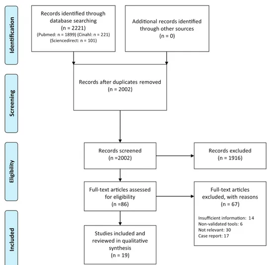
A total of 2221 references were retrieved in the different databases (Figure 1). After removal of
Table 1. The PICOS (participant, intervention/exposure, comparator, outcome and study design).
Criterion Description
Population Patients with neuromuscular diseases.
Adult patients.
Intervention Validation of one or more tools for screening or evaluation of dysphagia.
Comparison Comparison between a minimum of two tools.
Outcomes Psychometrics properties, characteristics of each tool.
duplicates, and full-text analysis, 19 studies met the inclusion criteria of this systematic review. All the studies were published from 1997 to 2017. Out of the 19 included studies, 6 were Japanese. The other were conducted in European countries (10), USA (2) and Turkey (1). All the studies used obser-vational research methods. A total of four involved adult and child patients in their studied samples.41–44 Studies characteristics are summarized in Table 2. The Downs and Black index ranged from 12 to 19 with a median total score of 16. Most studies were classified as ‘fair’.7,10,14,41–43,45–54 Two were assigned as ‘poor’55,56 and only one study presented a ‘good’ quality index44 (Table 3).
Table 4 summarizes the different tools used to study dysphagia for each separate NMD. Out of the 19 studies, 10 focused on amyotrophic lateral sclerosis (ALS), and 4 on Duchenne muscular dystrophy (DMD). Other pathologies studied
included myotonic dystrophy type 1 (DM1), inclusion body myositis (IBM), myasthenia gravis (MG), spinal muscular atrophy (SMA), polymy-ositis/dermatomyositis (PM/DM), Friedreich’s ataxia (FA) and spinal and bulbar muscular atro-phy (SBMA). With regards to the tools, most pub-lications (n = 5) used VFSS.10,42,48,55,56 Surface electromyography14,43,54 (sEMG) and FEES10,45 were found in three and two studies, respectively. Other tools listed with measurement properties and characteristics are listed in Supplement 2. Instrumental examinations
Validity
The VFSS, also reported as the modified barium swallowing examination, is the most commonly assessed tool in NMDs for dysphagia
assess-ment10,42,48,55,56 or as reference to compare the
other techniques.7,41,46,49–54 In ALS, two generic
scales were used for VFSS analysis, the dysphagia outcome severity scale (DOSS)57 and the penetra-tion aspirapenetra-tion score (PAS).48,59 Murono and col-leagues suggested that evaluation of swallowing kinematics is one of the major advantages of VFSS.55 In patients with ALS, they observed that the oral phase seemed the most affected and phar-yngeal contraction was correlated with PAS and may play a role in penetration or aspiration even in patients without bulbar symptoms (p < 0.01). However, they showed that aspiration or penetra-tion is not common in those patients in the first stages of the disease.55 In Briani and colleagues, VFSS findings were compared with FEES and manometry findings.10 VFSS had a significantly
greater sensitivity (92%), to highlight swallowing impairment, especially when silent in ALS and SMA patients.10 In contrast, all of the nondysphagic patients also showed radiological swallowing abnor-malities indicating a specificity of 0% in this study.10 In DMD patients, Hanayama and colleagues sug-gested that VFSS abnormalities were related to advanced age, except for impaired oral holding.42 VFSS is a better indicator for the oral phase of swallowing (p < 0.05) and the pooling of contrast fluid in the valleculae (p < 0.05) than the ‘dyspha-gia questionnaire’, a standard set of questions related to the frequency of 10 symptoms of upper gastrointestinal dysfunction.42,60 In MG, the abnor-mal laryngeal elevation observed during VFSS was
Table 2. Characteristics of included studies (n = 19).
Study Study
design Number of cases (n = 1.101) Number of controls Underlying diseases
Cosentino and colleagues57 CSS 26 30 ALS
Hiraoka and colleagues56 CSS 25 n/a ALS
Plowman and colleagues58 CSS 70 n/a ALS
Plowman and colleagues53 CSS 70 n/a ALS
Olthoff and colleagues54 CCS 20 n/a IBM
Wada and colleagues44 CSS 218 n/a ALS, DMD
Murono and colleagues59 CSS 19 n/a ALS
Pilz and colleagues48 CS 45 10 DM1
Aydogdu and colleagues14 CS 364 297 ALS, DM1, MG, PM/DM
Mano and colleagues55 CSS 47 38 SBMA
Archer and colleagues47 CSS 15 12 DMD
Archer and colleagues46 CSS 15 12 DMD
Paris and colleagues49 CCS 20 n/a ALS
Cox and colleagues7 CSS 43 n/a IBM
Hanayama and colleagues45 CSS 31 n/a DMD
Higo and colleagues60 CCS 11 n/a MG
Kidney and colleagues51 CSS 25 n/a ALS
Briani and colleagues10 CSS 23 n/a ALS, SMA
Mari and colleagues50 CS 14 n/a ALS, DM1, FA
ALS, amyotrophic lateral sclerosis; CCS, case-control study; CS, cohort study; CSS, cross-sectional study; DM1, myotonic dystrophy type 1; DMD, Duchenne muscular dystrophy; FA, Friedreich’s ataxia; IBM, inclusion body myositis; MG, myasthenia gravis; n/a, not available; PM/DM, polymyositis/dermatomyositis; SBMA, Spinal and bulbar muscular atrophy; SMA, spinal muscular atrophy.
significantly correlated with aspiration (p = 0.001) and may lead to predicting aspiration and pneumo-nia.56 However, across the different studies evaluat-ing VFSS, a great variety of methodological settevaluat-ings was observed in terms of thickness, viscosity and volume of contrast fluids used (Table 5).
Manometry was used in patients with ALS and SMA and showed that an abnormal upper esopha-geal sphincter (UES) opening and hypotonia of the proximal pharynx were the most sensitive physio-logical signs of dysphagia (80%).10 When compared with controls, the patients of this study presented a greater number of incomplete UES openings and a
significantly extended ‘intra-bolus’ pharyngeal pres-sure.10 As demonstrated in VFSS, dysphagia appeared to be linked to the presence of a defective oropharyngeal phase of swallowing. It should be noted that the specificity of pharyngo-esophageal manometry is weak in this study (20%).10 According to Briani and colleagues, FEES examination was not sensitive in highlighting swallowing alterations both in dysphagic and in nondysphagic patients (53%) but had a good specificity to rule out other organic causes of dysphagia (85%).10
Teams from the United Kingdom (UK), Turkey and Italy used sEMG to assess swallowing and
Table 3. Quality Index developed by Downs and Black. Scores for each included study.
Study Reporting External
validity Bias Confounding Power Total Grades+
(11)* (3)* (7)* (6)* (1)* (28)*
Cosentino and colleagues57 8 1 3 2 0 14 Fair
Hiraoka and colleagues56 6 1 3 2 0 14 Fair
Plowman and colleagues58 8 1 5 2 0 16 Fair
Plowman and colleagues53 8 1 5 2 0 16 Fair
Olthoff and colleagues54 8 0 5 2 0 15 Fair
Wada and colleagues44 8 2 5 3 0 18 Fair
Murono and colleagues59 6 0 5 2 0 13 Poor
Pilz and colleagues48 9 1 5 1 0 16 Fair
Aydogdu and colleagues14 8 1 4 3 0 16 Fair
Mano and colleagues55 9 1 3 3 1 17 Fair
Archer and colleagues47 9 1 6 3 0 19 Good
Archer and colleagues46 8 1 5 2 0 16 Fair
Paris and colleagues49 10 0 5 1 0 16 Fair
Cox and colleagues7 6 3 4 2 0 15 Fair
Hanayama and colleagues45 7 3 5 1 0 16 Fair
Higo and colleagues60 6 1 4 1 0 12 Poor
Kidney and colleagues51 7 1 5 2 0 15 Fair
Briani and colleagues10 10 0 5 2 0 17 Fair
Mari and colleagues50 6 3 6 2 0 17 Fair
*Maximum score that can be given for each item with the Quality Index developed by Downs and Black.
+Grading maximal score with Quality Index developed by Downs and Black: Excellent (24–28 points); Good (19–23 points); Fair (14–18 points); Poor
dysphagia in patients with DMD, ALS, DM1, PM/DM and MG.14,43,54 Aydogdu and col-leagues used the dysphagia limit (DL) to com-pare the peak duration and relative timing of muscle activity during swallowing of four mus-cle groups (orbicularis oris, masseter, submen-tal, and infrahyoid muscles) in ALS, DM1, PM/DM and MG patients.14 DL is a quantita-tive and noninvasive method for the assessment of swallowing impairment. It represents the vol-ume at which two or more swallows become necessary to swallow the whole bolus. Values for DL were compared with the clinical degrees of dysphagia, graded from 1 (no clinical signs and symptoms of dysphagia) to 4 (obvious clin-ical signs and symptoms of dysphagia, includ-ing aspiration).61 Patients with clinical dysphagia had abnormal DL for <20 ml of water.14 DL was significantly correlated with the improvement or worsening of dysphagia for ALS, PM/DM and MG (p < 0.001) but not for DM1 patients (p > 0.05). Also, DL was useful
to assess the effect of treatment as well as changes of dysphagia over time.14 Archer and colleagues tested peak activity of four muscle groups during swallowing between DMD patients with dysphagia, those with preserved muscle function, and healthy controls.43 Although there were no differences in the tim-ing or duration of muscle activity between the three groups, these comparative data provided insight into the relative activity of the orbicula-ris oorbicula-ris, masseter, infrahyoid, and submental muscles during swallowing. Thus, a distinction between a preserved and disordered swallowing function in patients with DMD could be made.43 Compared with controls, patients with DMD had to use a greater maximal muscle activity to swallow [masseter (p < 0.01), orbic-ularis oris (p < 0.05), submental (p < 0.05)], indicating muscle weakness. However, no dif-ferences were found between DMD with dys-phagia and those patients with an intact swallowing function.43
Table 4. Tools used to study dysphagia in each NMD.
Underlying
diseases Tools Study
ALS VFSS, sEMG (DL), FEES, V-VST, Man., VCA, 3SwT, NdSSS, MTP, EAT-10
Murono and colleagues55; Aydogdu and colleagues14; Mari and
colleagues47; Paris and colleagues46; Plowman and colleagues49,50,62;
Kidney and colleagues48; Wada and colleagues41; Briani and
colleagues10; Cosentino and colleagues54; Hiraoka and colleagues53;
Plowman and colleagues49,50,62
DMD VFSS, SSQ, sEMG,
NdSSS Archer and colleagues
43; Archer and colleagues44; Hanayama and
colleagues42; Wada and colleagues41
DM1 FEES, sEMG (DL),
3SwT Pilz and colleagues
45; Aydogdu and colleagues14; Mari and
colleagues47
MG sEMG (DL), VFSS Higo and colleagues56; Aydogdu and colleagues14
IBM Standard Questionnaire, RT-MRI
Cox and colleagues7; Olthoff and colleagues51
SMA VFSS, Man., FEES Briani and colleagues10
PM/DM sEMG (DL) Aydogdu and colleagues14
FA 3SwT Mari and colleagues47
SBMA MTP Mano and colleagues52
3SwT, 3-ounce water swallow test; ALS, amyotrophic lateral sclerosis; DL, dysphagia limit; DM1, myotonic dystrophy type 1; DMD, Duchene muscular dystrophy; EAT-10, eating assessment tool; FA, Friedreich’s ataxia; FEES, fiberoptic endoscopic evaluation of swallowing; IBM, inclusion body myositis; Man., pharyngo-esophageal manometry; MG, myasthenia gravis; MTP, maximum tongue pressure; NdSSS, neuromuscular disease swallowing status scale; NMD, neuromuscular disease; PM/DM, polymyositis/dermatomyositis; RT-MRI, real-time magnetic resonance imaging; SBMA, spinal and bulbar muscular atrophy; sEMG, surface electromyography; SMA, spinal muscular atrophy; SSQ, Sydney Swallow Questionnaire; VCA, voluntary cough airflow; VFSS, videofluoroscopic swallow study; V-VST, volume-viscosity swallow test.
Tabl e 5. Pr ot oc ols used in VF SS s tudies. Articl es Consis tencies/f ood Fr equency/bolus Crit eria used f or diagnosing dysphagia
Cox and colleagues
7
n/a
0, 3, 6 or 9 ml of opaque fluid
Signs subdivided in two categories: IP or AR.
Hanayama and colleagues
45 Liquid: 70% water + 30% Omnipaque Jelly swallow: strawberry jelly + 30% of volume Omnipaque Solid: Barium
pancakes (60% flour/40% barium powder)
Liquid: 1 × 30 ml Jelly swallow: 3 × 5 ml Solid:
1 piece of cake cut in 3 pieces and put into the patient’s mouth Impaired oral hold, oral residuals, pooling in the valleculae, pooling in the valleculae after repeated swallow, pooling in the pyriform sinus, pooling in the pyriform sinus after repeated swallow, supraglottic penetration, pharyngo-oral regurgitation.
Higo and colleagues
60
‘Appropriate and safe bolus textures’ (thin liquid or semisolid)
1, 3, 5 ml or self-regulated
Bolus transport from the mouth to the pharynx, bolus holding in the oral cavity, velopharyngeal seal, tongue base movement, pharyngeal constriction, laryngeal elevation, upper esophageal sphincter opening, and bolus stasis at the pyriform sinus
+
lung aspiration.
Murono and colleagues
59
140% barium mixture
+
other contrast
and consistencies (n/a)
3 ml
All 15 physiologic components except for a component of esophageal clearance, proposed by Martin-Harris and Jones, were thoroughly evaluated from the perspective of all six oral components and all eight pharyngeal components.
Kidney and colleagues
51
Varied food from liquid to solid
n/a
PAS, DOSS.
Hiraoka and colleagues
56
Yogurt (282 mPas) containing contrasting agent
3 g
Qualitative evaluation: (1). Tongue function, (2). Residue accumulation; Quantitative evaluation (1). Bolus formation and oral transit time (second): (2). Pharyngeal transit time (second).
Plowman and colleagues
58
Liquid:
Varibar thin barium sulphate
suspension, (EZ-EM, Inc., Westbury, NY, USA). Pudding:
EZ-pudding, (EZ-EM, Inc.).
Thin liquid:
n/a
A standardized bolus presentation protocol: (1). two 1-cc boluses of liquid contrast; (2). one 3-cc of thin liquid contrast; (3). one 3-cc of pudding; (4). one 20-cc bolus of liquid contrast; (5). 90 cc sequential swallows of thin liquid contrast; (6). in the anterior-posterior view, the patient was administered a 20-cc bolus of liquid contrast.
PAS
Plowman and colleagues
53
Idem as Plowman and colleagues
58
Idem as Plowman and colleagues
58
Idem as Plowman and colleagues.
58
Olthoff and colleagues
54
Liquid contrast agent (either Imeron 350 or Gastrolux)
1
×
20 ml
Relevant parameters of swallowing included bolus control and transport, velo-pharyngeal closure, laryngeal penetration, aspiration, and bolus retention in the pharyngeal tract.
Mano and colleagues
55
40% weight per volume barium sulphate
3 ml
PAS
+
residue quantification.
Briani and colleagues
10
Fluid/Semisolid
Chosen by the patient
Oral stasis; loss of barium in the mouth vestibula during swallowing; repetitive tongue movements; incomplete or inadequate velopharyngeal closure; presence of aspiration; functionality of upper esophageal sphincter and lower esophageal sphincter; pharyngo-esophageal motility.
AR, aspir ation-r elat ed dysphagia; c c, cubic c entimet er; DOS S, dysphagia out come se verity sc al
e; IP, dysphagia due t
o impair
ed pr
opulsion; mP
as, millipasc
al sec
onds; n/a, not av
ailabl e; P AS, penetr ation and aspir ation sc al e.
In 2017, Cosentino and colleagues investigated electrophysiologically submental/suprahyoid activity (SHEMG) and laryngeal-pharyngeal movements (LPMs) during swallowing in patients with ALS (p = 0.004).54 They detected reduced SHEMG and LPM in patients with dysphagic ALS. A strong to moderate correlation was observed between these two indices and the PAS and DOSS score, respectively. Moreover, a decrease in swallowing reproducibility could be a preclinical sign of dysphagia and, beyond a cer-tain threshold, a pathological hallmark of oro-pharyngeal dysphagia. Interestingly, SHEMG was the only electrophysiological parameter cor-related with the disease duration and could be an expression of the progressive degeneration of the motor neurons occurring in the course of the dis-ease (r = 0.494, p = 0.010).54
Overall, two studies assessed the validity of maxi-mum tongue pressure (MTP) in ALS and SBMA patients.52,53 Both used a digital tongue pressure manometer equipped with a balloon probe (JMS Co. Ltd., Hiroshima, Japan) and asked the partici-pants to compress the balloon upward onto their palates for 7 seconds, three times at 1-minute intervals, using the maximum voluntary effort of the tongue. The maximum value of these three measurements was considered as the MTP for each patient.52,53 Hiraoka and colleagues evaluated the relationship between MTP and the characteris-tics of swallowing disorders in patients with ALS.53 They showed that MTP was significantly lower in the patients with ALS with reduced tongue func-tion (p = 0.002) or with pharyngeal residue (p = 0.006) than in the patients with normal character-istics. Bolus formation and oral and pharyngeal transit time observed in VFSS were significantly prolonged among those with reduced MTP (p < 0.01).53 In SBMA, Mano and colleagues described a decrease in patients within 3 years from the onset of the disease compared with healthy controls (p < 0.001). In comparison with VFSS, the values of tongue pressure in the patients with laryngeal pen-etration were significantly smaller than those of the patients without (p = 0.018).52
Real-time magnetic resonance imaging (RT-MRI) allowed precise time measurements and identifi-cation of the respective tissue morphology.51 In 20 patients, penetration was seen in 10% and 30% of them by RT-MRI and FEES or VFSS, respec-tively. Bolus retention in the pharyngeal tract is believed to be the most sensitive indicator of func-tional deficits in swallowing and was reliably
identified by all three modalities (FEES, VF, RT-MRI). The Bland–Altman plots did not reveal any systematic deviation among those methods.51 Finally, Plowman and colleagues identified the cough volume acceleration (CVA), peak expira-tory flow rate (PEFR) and peak expiraexpira-tory flow rise time (PEFRT) as significant predictors of penetration/aspiration status in 70 ALS patients.62 Following this study, ALS patients with a CVA less than 45.3 l/s2 were 5.6 times more likely to penetrate/aspirate. A PEFR lower than 4.0 l and a PEFRT greater than 80 ms increased 3.6 and 3.2 times the risk of penetration/aspiration, respec-tively.62 Those cutoffs for CVA, PEFR and PEFRT had sensitivities of 91.3%, 82.6%, and 73.9% respectively and, specificities of 82.2%, 73.9%, and 78.3% for identifying ALS penetra-tor/aspirators.62
Reliability
Reliability was evaluated in five studies focused on five different NMD populations. Kidney and col-leagues investigated inter- and intra-rater reliabil-ity of VFSS in relation to the DOSS and PAS, and analysis of videofluoroscopy was undertaken in ALS patients. These results indicated acceptable test–retest reliability when using the DOSS and the Aspiration-Penetration Rating Scale (APRS) with inter-rater reliability of 92% and 95% of agreement, respectively and intra-rater reliability scored at 98% and 100% of agreement, respec-tively.48 Pilz and colleagues described inter- and intra-rater reliability as sufficient for all FEES var-iables (piecemeal deglutition, delayed initiation pharyngeal reflex, post-swallow vallecular and pyriform pooling, laryngeal penetration or tra-cheal aspiration), with a weighted kappa > 0.61 in DM1 patients.45 For all FEES variables, bolus consistency significantly influenced the likelihood of observing mild or severe swallowing impair-ment: patients were more likely to have impaired swallowing with thin liquids than with thickened fluids (odds ratio > 1).45 Archer and colleagues have suggested that physiological changes in the swallowing muscles activity in DMD can be dis-tinguished between preserved and disordered swallowing function with sEMG.43 They repeated the sEMG procedure in order to examine the reproducibility of the results. The mean differ-ences between the repeated sEMG assessments were amplitude: 29.12 ± 21.69% MVC, dura-tion: 0.69 ± 0.62 s, and relative timing: 0.40 ± 0.24 s. Mano and colleagues performed the
test–retest of MTP in 24 patients with SBMA on two different occasions at an interval of 21.3 ± 3.9 days. They found no statistically significant differ-ence. The intraclass correlation coefficient was 0.986 (p < 0.001), indicating an excellent test– retest reliability of the tongue pressure. The RT-MRI was compared with FEES and VFSS in a cohort of patients with IBM.51 The inter-rater agreement was assessed for bolus transport and retention. The Krippendorff α was 0.39 (RT-MRI), 0.55 (VF), and 0.67 (FEES) for bolus transport, and 0.51 (RT-MRI), 0.52 (VFSS), and 0.52 (FEES) for retention.51
Responsiveness
Responsiveness has not been evaluated in studies concerning instrumental examinations.
Noninstrumental examination Validity
Validity was assessed in five studies about nonin-strumental examinations.
Plowman and colleagues compared the eating assessment tool 10 (EAT-10), a 10-question self-administered, symptom-specific dysphagia out-come tool to score patients with ALS.50 The total EAT-10 score was calculated with scores ranging from 0 (no impairment) to 40 (severe impair-ment) and demonstrated good discriminant abil-ity to accurately identify ALS penetrator/ aspirators (PAS ⩾ 3) with a cutoff score of 3 [area under the curve (AUC): 0.77, sensitivity: 88%, specificity: 57%].63 It demonstrated excellent accuracy at identifying aspirators (PAS ⩾ 6) uti-lizing a cutoff score of 8 (AUC: 0.88, sensitivity: 86%, specificity: 72%). On average, EAT-10 scores were five times higher in ALS aspirators than in those patients who demonstrated safe swallowing.50 The neuromuscular disease swal-lowing status scale (NdSSS), an 8-stage scale for dysphagia in patients with progressive NMD, was correlated with PAS significantly in patients with ALS (r = −0.51) but in DMD patients (r = 0.22).41 Another examination, the volume-viscos-ity swallow test (V-VST) showed a high sensitiv-ity and specificsensitiv-ity (93% and 80%, respectively) for screening and diagnosing oropharyngeal dys-phagia in patients with ALS.46 The V-VST involved administering increasing fluid volumes of different textures/thicknesses and the assess-ment of efficiency and safety of swallowing.
In DMD patients, Archer and colleagues showed that the Sydney swallow questionnaire (SSQ),19 a self-administered questionnaire based on 17 ques-tions using visual analogue scales, detected dys-phagia with good sensitivity and specificity (78 and 83%, respectively) at a cutoff score of 234 (total maximum score = 1700).44 In this study, the dysphagic status of DMD participants were defined with a clinical history of dysphagia and the functional oral intake score. The 3-ounce water swallow test (3SwT), validated previously in other patient groups (e.g. stroke, head and neck surgery, Parkinson’s disease, dementia),58,64,65 was com-pared with VFSS and a 25-item form (one of them is ‘history of cough’), identifying dysphagia. The 3Swt showed a higher specificity than clinical signs (86 versus 30%) but a lower sensitivity (52%) compared with VFSS in patients with NMD.47 The association of ‘history of cough’ (coughing episode reported by the patient) with the 3SwT gave an increase in both positive and negative pre-dictive values (84 and 76%, respectively).47 Finally, Cox and colleagues assessed impaired food propulsion or aspiration-related problems in patients with IBM by comparison of a standard questionnaire, previously validated in patients with Parkinson’s disease,66 with VFSS results.7 The authors showed that the questionnaire had a better sensitivity and positive predictive value for impaired food propulsion than for aspiration-related problems (70 versus 63% and 92 versus 65%, respectively) and that two questions reliably predicted the presence of impaired food propul-sion on VFSS, namely ‘Does food get stuck in your throat?’ and ‘Do you have to swallow repeat-edly in order to get rid of food?’. However, dys-phagia was more frequently designated by VFSS when used as a reference than the standard ques-tionnaire results (79 versus 65%).7
Reliability
Inter- and intra-rater reliabilities were only assessed for the NdSSS, in 50 patients with DMD and 84 patients with ALS. For inter- and intra-rater reliabilities, the weighted kappas were 0.95 and 1.00, respectively, for DMD; and 0.98 and 0.98, respectively, for ALS.41
Responsiveness
Responsiveness was determined with the standard-ized response mean for the NdSSS and was large in ALS (1.21) and moderate in DMD (0.65).67
Discussion
The purpose of this systematic review was to summarize the different dysphagia screening and evaluation tools and to identify their measure-ment properties in adults with NMDs. A total of 19 articles were identified based on strong crite-ria. Evaluation tools were mainly found for patients with ALS with eight different tools.10,14,41,46–50,53–55 Validity has been assessed in the majority of the studies in instrumental and noninstrumental examinations even if specificity and sensibility were frequently lacking. Reliability has been verified for VFSS, FEES, sEMG, MTP, RT-MRI and NdSSS but was incomplete mainly in noninstrumental examinations.41,43,45,48,51,52 Responsiveness has been reported only for NdSSS in patients with ALS and DMD.42
VFSS was used as primary outcome in 5 studies
10,42,48,55,56 and as comparator in 10 out of the
retrieved studies.7,41,46,49–54 Among them, we observed a great heterogeneity in the VFSS proce-dures and analysis used as highlighted in Table 5. This disparity was already found in prior reviews of swallowing assessment in other dis-eases.20,31,32,68,69 In addition, previous studies reported poor inter-rater reliability for VFSS, depending on the procedure and bolus consist-ency used to assess swallowing in adult patients.70,71 Equally, five reviews in non-NMD adult patients emphasized that consensus on the terminology and study protocols (e.g. fluid thickness used dur-ing studies) is needed for more useful comparison with VFSS,31,32,69,72 like for sEMG or FEES.14,43 The importance of bolus properties was high-lighted by Ciccero and colleagues73 They insisted on the considerable changes in physiology observed depending of the bolus consistency (volume, vis-cosity, solid or fluid), size, method of ingestion (e.g. cup, straw) and chemosensory input (taste, smell, sensory receptors) in the normal swallow.73 Those properties need to be considered by the cli-nician during dysphagia assessments when consid-ering what is normal from what is pathological.73,74 Classically, patients with solid food dysphagia are more likely to have disorders of esophageal phase, whereas those with difficulty with liquids are more likely to have oropharyngeal dysphagia.75 However, in NMD patients, this dichotomy may be artificial because it is well known that those with oropharyn-geal dysphagia can have dysphagia for liquids and solids in the different phases of swallowing, specific patterns of dysphagia depending on the underly-ing disease.2,3,75 For example, DMD patients may
have difficulties with chewing and oropharyngeal transport of solid foods, as well as pharyngeal resi-due without aspiration is more common and is likely due to muscle weakness.2 In ALS, difficulties may likely be inability to hold bolus, reduced mas-tication, residue in the oral cavity and delayed swallow reflex.76 Moreover, all patients with NMDs may also have difficulty with management of excessive thick mucus, which may contribute to breathing discomfort. The use of bolus with different properties seems also extremely helpful in the assessment of patients with NMDs in order to assess the further therapeutic and dietetic man-agement. In the different studies included, those data were lacking, neither for cutoff level nor standardization are present specifically for the dif-ferent NMDs.
Several details need to be highlighted about instru-mental examinations. First, the fatigability, an important factor in patients with NMDs with mus-cle weakness, was generally not taken into account during the assessments.4,77 We can hypothesize that tools only give a snapshot and do not consider fac-tors such as fatigability or ventilatory support needed.4,44 Secondly, some assessments are inva-sive and can cause discomfort to the patient (e.g. manometry and FEES), and potentially involves exposure to radiation (e.g. in VFSS and RT-MRI). Finally, examinations that include large volumes of liquids may put the patient at risk of aspiration and choking.27,44 In addition, VFSS, MRI or FEES are often not possible in daily routine or in out-of-hos-pital settings such as physiotherapist and SLT prac-tices due to the required expertise, the patient compliance, and the evolution of the swallowing disorder which can be sometimes rapidly progres-sive.29 In regular follow-up assessments, even if the specific equipment needed may be considerate expensive,2 the cost burden is often distributed among departments (radiology, gastroenterology, or otolaryngology) and implementation can be only limited in underdeveloped countries.31
Noninstrumental examinations, done subjectively or by self-administration, can be interesting alter-natives for screening, follow up or complementa-tion of instrumental evaluacomplementa-tions in the diagnosis. These may be useful, in particular, in the assess-ment of the fluctuating nature of the motor and emotional symptoms in NMDs and their impact on quality of life.78
From a clinical point of view, following the defi-nition outlined previously,3,19,27,28,30–32 we can
consider that noninstrumental examinations (SSQ, V-VST, EAT-10 and 3SwT), voluntary cough airflow (VCA) and sEMG represent ideal screening or follow-up tools with high sensitivity and specificity and are quick, cost-effective, and easy to interpret.28 Moreover, they give accurate cutoff scores and are more effective to detect early swallowing problems or risk factors. Instrumental examinations (VFSS and FEES) showed good validity and should be used for diagnosis or when aspiration status is unclear from noninvasive approaches. Following previous studies, VFSS also presented an important role in the follow up of aspiration occurrence and the evaluation of the improvements related to the treatment, and maybe the reduction of symptoms. On the other side, FEES can provide real-time visual feedback during swallowing therapy contrary to VFSS. FEES may be helpful to test many therapeutic interventions and many strategies without a time limit when the patient’s intake and quality of life depend on a diet upgrade. FEES allows the assessment of a meal in a functional, real-life situation. Fatigue increases can also be checked as the meal progresses, but also if positioning, rate of intake, and method of feeding impact the safety. Moreover, the study of Andersen and col-leagues has shown the interest of FEES in the assessment of the laryngeal responses during treat-ment with mechanical insufflation-exsufflation.79 Further studies are needed to standardize assess-ment procedures (tools, settings and chronology) according to the evolution and the specificities of each adult patient with an NMD. Moreover, to evaluate deglutition in patients with NMDs, min-imal clinically important differences, which reflect changes related to clinical interventions, could improve those different examinations.
It would also be relevant to adapt tools to the tim-ing of occurrence and evolution of the disease. Indeed, dysphagia may vary with the natural course of the disease and either be present from the early stages of the disease progression or appear with time in end-stage patients. For example, Andrenelli and colleagues showed that patients with ALS and dysphagia had a global dysfunction of the oral and pharyngeal swallowing phases with more difficul-ties for swallowing thin liquids when conversely, DM1 patients with dysphagia were younger, tended to obesity and had greater muscular impair-ment than those without dysphagia and will have difficulties in swallowing a solid bolus.16 In another way, dysphagia in DMD worsens with age, with
increasing mastication effort.80 Recognition of spe-cific clinical profiles for the different disease and stage will support and guide the detection of swal-lowing disorders in patients with NMDs. Furthermore, with new treatments (like noninva-sive ventilation), life expectancy of patients with NMDs continues to increase. It will be more and more important in the future to deal with associ-ated complications such as swallowing impair-ments and treatment such as noninvasive ventilation or airway clearance technique.1,12,81,82 The main limitation of this systematic review was the quality level of the included studies. Indeed, most of them were rated as ‘fair’ and we have noted the absence of randomized controlled tri-als. Also, some methodological issues need to be discussed. As observed by Olthoff and colleagues, the thickness and volume of the bolus used is sometimes different in the evaluation of the tools in the same study and firm conclusion must be made carefully.51 Also, the positioning of the patients during the different assessments was poorly documented and is a source of bias. Most of the assessment is in a posture which does not reflect actual conditions of swallowing in daily life. The impact of positioning and head control has been demonstrated to be an important param-eter.83 Only Hanayama and colleagues and Hiraoka and colleagues evaluated the patients while seated in their own wheelchairs.42,53 The characteristics and measurement properties of the tools are not always described in detail in many studies (Supplement 2), and it is often difficult to distinguish among screening, evaluation and diagnosis.69,84 Frequently, the objectives of the different tools were not clearly defined by the dif-ferent authors. Screening is defined as the pre-sumptive identification of unrecognized disease in an apparently asymptomatic population by means of tests, examinations or other procedures that can be applied rapidly and easily to the target population.31,85,86 This screening can be done by questionnaires, observations, physical evidence, among others.87 Validated self-reported question-naires have been increasingly used in clinical research as well as clinical practice to capture the individual’s perspective regarding their disease symptoms.88 When identified by the screening tool, the patient should be referred for diagnosis of swallowing disorders, conducted from clinical evaluation and supplemented, when necessary. Diagnosis aims to determine presence of dyspha-gia, its severity and changes that it may cause, and the rehabilitation plan.
Conclusion
This systematic review identified 12 different tools for the screening and evaluation of dysphagia in adults with NMDs. Among them, NdSSS, a non-instrumental tool, is the only one who assessed all measurement properties in patients with ALS. The majority of the studies presented VFSS as a valid and reliable examination to assess dysphagia in patients with ALS and DMD. Other tools were mainly evaluated in patients with ALS, but further studies are needed to complete their measurement properties. In other NMDs, no firm conclusion can be made because of insufficient data and het-erogeneity of NMDs. Recognition of specific clini-cal profiles for the different disease and stage will support and guide the detection of swallowing dis-orders. Each tool presents specific measurement properties to be more designed for diagnosis or screening and could be used appropriately accord-ing to the evolution and the specificities of each adult patient with an NMD.
Funding
The author(s) disclosed receipt of the following financial support for the research, authorship, and/or publication of this article: N.A. received funding from Fund Eliane Lagast and Association Belge Contre les Maladies neuro-Musculaires-Aide à la Recherche asbl for his PhD scholarship. C.G. is supported by the Fondation Saint-Luc. G.R. received funding from the Institut de Recherche Expérimentale et Clinique, Université Catholique de Louvain.
Conflict of interest statement
The author(s) declared no potential conflicts of interest with respect to the research, authorship, and/or publication of this article.
Supplemental material
Supplemental material for this article is available online.
ORCID iD
Nicolas Audag https://orcid.org/0000-0002 -8352-4138
References
1. Hill M, Hughes T and Milford C. Treatment for swallowing difficulties (dysphagia) in chronic muscle disease. Cochrane Database Syst Rev 2004; 2: Cd004303.
2. Britton D, Karam C and Schindler JS. Swallowing and Secretion Management in
Neuromuscular Disease. Clin Chest Med 2018; 39: 449–457.
3. Willig TN, Paulus J, Lacau Saint Guily J,
et al. Swallowing problems in neuromuscular
disorders. Arch Phys Med Rehabil 1994; 75: 1175–1181.
4. Toussaint M, Davidson Z, Bouvoie V, et al. Dysphagia in Duchenne muscular dystrophy: practical recommendations to guide management.
Disabil Rehabil 2016; 5: 1–11.
5. Pane M, Vasta I, Messina S, et al. Feeding problems and weight gain in Duchenne muscular dystrophy. Eur J Paediatr Neurol 2006; 10: 231–236.
6. Knuijt S, Kalf JG, de Swart BJ, et al. Dysarthria and dysphagia are highly prevalent among various types of neuromuscular diseases. Disabil
Rehabil2014; 36: 1285–1289.
7. Cox FM, Verschuuren JJ, Verbist BM, et al. Detecting dysphagia in inclusion body myositis.
J Neurol 2009; 256: 2009–2013.
8. Aloysius A, Born P, Kinali M, et al. Swallowing difficulties in Duchenne muscular dystrophy: indications for feeding assessment and outcome of videofluroscopic swallow studies. Eur J
Paediatr Neurol 2008; 12: 239–245.
9. Jones K, Pitceathly RD, Rose MR, et al. Interventions for dysphagia in long-term, progressive muscle disease. Cochrane Database
Syst Rev 2016; 2: Cd004303.
10. Briani C, Marcon M, Ermani M, et al. Radiological evidence of subclinical dysphagia in motor neuron disease. J Neurol 1998; 245: 211–216.
11. Gozal D. Pulmonary manifestations of neuromuscular disease with special reference to Duchenne muscular dystrophy and spinal muscular atrophy. Pediatr Pulmonol 2000; 29: 141–150.
12. Simonds AK. Recent advances in respiratory care for neuromuscular disease. Chest 2006; 130: 1879–1886.
13. Winterholler C. Diagnosis and treatment of dysphagia in patients with neuromuscular disease.
Pneumologie 2008; 62(Suppl. 1): S35–S38.
14. Aydogdu I, Kiylioglu N, Tarlaci S, et al. Diagnostic value of “dysphagia limit” for neurogenic dysphagia: 17 years of experience in 1278 adults. Clin Neurophysiol 2015; 126: 634–643.
15. Altman KW, Richards A, Goldberg L, et al. Dysphagia in stroke, neurodegenerative disease, and advanced dementia. Otolaryngol Clin North
16. Andrenelli E, Galli FL, Gesuita R, et al.
Swallowing impairments in Amyotrophic Lateral Sclerosis and Myotonic Dystrophy type 1: looking for the portrait of dysphagic patient in neuromuscular diseases. NeuroRehabilitation 2018; 42: 93–102.
17. Dusick A. Investigation and management of dysphagia. Semin Pediatr Neurol 2003; 10: 255–264.
18. Queija Ddos S, Portas JG, Dedivitis RA,
et al. Swallowing and quality of life after total
laryngectomy and pharyngolaryngectomy. Braz J
Otorhinolaryngol 2009; 75: 556–564.
19. Wallace KL, Middleton S and Cook IJ. Development and validation of a self-report symptom inventory to assess the severity of oral-pharyngeal dysphagia. Gastroenterology 2000; 118: 678–687.
20. Espitalier F, Fanous A, Aviv J, et al.
International consensus (ICON) on assessment of oropharyngeal dysphagia. Eur Ann
Otorhinolaryngol Head Neck Dis 2018; 135(1
Suppl.): S17–S21.
21. Coscarelli S, Verrecchia L and Coscarelli A. Endoscopic evaluation of neurological dysphagic patients. Acta Otorhinolaryngol Ital 2007; 27: 281–285.
22. Martin-Harris B and Jones B. The
videofluorographic swallowing study. Phys Med
Rehabil Clin N Am 2008; 19: 769–785, viii.
23. Rugiu MG. Role of videofluoroscopy in evaluation of neurologic dysphagia.
Acta Otorhinolaryngologica Italica 2007; 27:
306–316.
24. Hull J, Aniapravan R, Chan E, et al. British Thoracic Society guideline for respiratory management of children with neuromuscular weakness. Thorax 2012; 67: 1–40.
25. National Clinical Guideline Centre. National Institute for Health and Care Excellence: Clinical Guidelines. In: Motor neurone disease: assessment
and management. London: National Institute for
Health and Care Excellence (UK) Copyright (c) National Clinical Guideline Centre, 2016. 26. Nilsson H, Ekberg O, Olsson R, et al.
Quantitative assessment of swallowing in healthy adults. Dysphagia 1996; 11: 110–116.
27. Logemann JA, Veis S and Colangelo L. A screening procedure for oropharyngeal dysphagia.
Dysphagia 1999; 14: 44–51.
28. Logemann JA. Screening, diagnosis, and management of neurogenic dysphagia. Semin
Neurol 1996; 16: 319–327.
29. Britton D, Benditt JO, Merati AL, et al. Associations between laryngeal and cough dysfunction in motor neuron disease with bulbar involvement. Dysphagia 2014; 29: 637–646. 30. Higgins JPT and Green S. Cochrane handbook
for systematic reviews of interventions, Version 5.1.0 [updated March 2011]. The Cochrane
Collaboration, 2011.
31. Etges CL, Scheeren B, Gomes E, et al. Screening tools for dysphagia: a systematic review. Codas 2014; 26: 343–349.
32. Kertscher B, Speyer R, Palmieri M, et al. Bedside screening to detect oropharyngeal dysphagia in patients with neurological disorders: an updated systematic review.
Dysphagia 2014; 29: 204–212.
33. Moher D, Liberati A, Tetzlaff J, et al. Preferred reporting items for systematic reviews and meta-analyses: the PRISMA statement. BMJ 2009; 339: b2535.
34. Liberati A, Altman DG, Tetzlaff J, et al. The PRISMA statement for reporting systematic reviews and meta-analyses of studies that evaluate healthcare interventions: explanation and elaboration. BMJ 2009; 339: b2700. 35. Audag N, Goubau C, Toussaint M, et al.
Screening and evaluation tools of dysphagia in children with neuromuscular diseases: a systematic review. Dev Med Child Neurol 2017; 59: 591–596.
36. Terwee CB, Mokkink LB, Knol DL, et al. Rating the methodological quality in systematic reviews of studies on measurement properties: a scoring system for the COSMIN checklist. Qual Life Res 2012; 21: 651–657.
37. Mann CJ. Observational research methods. Research design II: cohort, cross sectional, and case-control studies. Emerg Med J 2003; 20: 54–60.
38. Downs SH and Black N. The feasibility of creating a checklist for the assessment of the methodological quality both of randomised and non-randomised studies of health care interventions. J Epidemiol Community Health 1998; 52: 377–384.
39. Deeks JJ, Dinnes J, D’Amico R, et al. Evaluating non-randomised intervention studies. Health
Technol Assess 2003; 7: iii–x, 1–173.
40. O’Connor SR, Tully MA, Ryan B, et al. Failure of a numerical quality assessment scale to identify potential risk of bias in a systematic review: a comparison study. BMC Res Notes 2015; 8: 224.
41. Wada A, Kawakami M, Liu M, et al. Development of a new scale for dysphagia in patients with progressive neuromuscular diseases: the neuromuscular disease swallowing status scale (NdSSS). J Neurol 2015; 262: 2225–2231. 42. Hanayama K, Liu M, Higuchi Y, et al.
Dysphagia in patients with Duchenne muscular dystrophy evaluated with a questionnaire and videofluorography. Disabil Rehabil 2008; 30: 517–522.
43. Archer SK, Garrod R, Hart N, et al. Dysphagia in Duchenne muscular dystrophy assessed objectively by surface electromyography.
Dysphagia 2013; 28: 188–198.
44. Archer SK, Garrod R, Hart N, et al. Dysphagia in Duchenne muscular dystrophy assessed by validated questionnaire. Int J Lang Commun
Disord 2013; 48: 240–246.
45. Pilz W, Baijens LW, Passos VL, et al. Swallowing assessment in myotonic dystrophy type 1 using fiberoptic endoscopic evaluation of swallowing (FEES). Neuromuscul Disord 2014; 24: 1054–1062. 46. Paris G, Martinaud O, Hannequin D, et al.
Clinical screening of oropharyngeal dysphagia in patients with ALS. Ann Phys Rehabil Med 2012; 55: 601–608.
47. Mari F, Matei M, Ceravolo MG, et al. Predictive value of clinical indices in detecting aspiration in patients with neurological disorders. J Neurol
Neurosurg Psychiatry 1997; 63: 456–460.
48. Kidney D, Alexander M, Corr B, et al.
Oropharyngeal dysphagia in amyotrophic lateral sclerosis: neurological and dysphagia specific rating scales. Amyotroph Lateral Scler Other Motor
Neuron Disord 2004; 5: 150–153.
49. Plowman EK, Watts SA, Robison R, et al. Voluntary cough airflow differentiates safe versus unsafe swallowing in amyotrophic lateral sclerosis. Dysphagia 2016; 31: 383–390. 50. Plowman EK, Tabor LC, Robison R, et al.
Discriminant ability of the eating assessment tool-10 to detect aspiration in individuals with amyotrophic lateral sclerosis. Neurogastroenterol
Motil 2016; 28: 85–90.
51. Olthoff A, Carstens PO, Zhang S, et al.
Evaluation of dysphagia by novel real-time MRI.
Neurology 2016; 87: 2132–2138.
52. Mano T, Katsuno M, Banno H, et al. Tongue pressure as a novel biomarker of spinal and bulbar muscular atrophy. Neurology 2014; 82: 255–262.
53. Hiraoka A, Yoshikawa M, Nakamori M,
et al. Maximum tongue pressure is associated
with swallowing dysfunction in ALS patients.
Dysphagia 2017; 32: 542–547.
54. Cosentino G, Alfonsi E, Mainardi L, et al. The importance of the reproducibility of oropharyngeal swallowing in amyotrophic lateral sclerosis. An electrophysiological study. Clin
Neurophysiol 2017; 128: 792–798.
55. Murono S, Hamaguchi T, Yoshida H, et al. Evaluation of dysphagia at the initial diagnosis of amyotrophic lateral sclerosis. Auris Nasus Larynx 2015; 42: 213–217.
56. Higo R, Nito T and Tayama N.
Videofluoroscopic assessment of swallowing function in patients with myasthenia gravis. J
Neurol Sci 2005; 231: 45–48.
57. O’Neil KH, Purdy M, Falk J, et al. The dysphagia outcome and severity scale. Dysphagia 1999; 14: 139–145.
58. Garon BR, Engle M and Ormiston C. Reliability of the 3-Oz water swallow test utilizing cough reflex as sole indicator of aspiration. Neurorehabil
Neural Repair 1995; 9: 139–143.
59. Rosenbek JC, Robbins JA, Roecker EB, et al. A penetration-aspiration scale. Dysphagia 1996; 11: 93–98.
60. Jaffe KM, McDonald CM, Ingman E, et al. Symptoms of upper gastrointestinal dysfunction in Duchenne muscular dystrophy: case-control study. Arch Phys Med Rehabil 1990; 71: 742–744. 61. Ertekin C, Aydogdu I, Yuceyar N, et al.
Pathophysiological mechanisms of oropharyngeal dysphagia in amyotrophic lateral sclerosis. Brain 2000; 123: 125–140.
62. Plowman E, Watts S, Robison R, et al. Voluntary cough airflow differentiates safe versus unsafe swallowing in amyotrophic lateral sclerosis.
Dysphagia 2016; 31: 383–390.
63. Belafsky PC, Mouadeb DA, Rees CJ, et al. Validity and reliability of the Eating Assessment Tool (EAT-10). Ann Otol Rhinol Laryngol 2008; 117: 919–924.
64. DePippo KL, Holas MA and Reding MJ. Validation of the 3-oz water swallow test for aspiration following stroke. Arch Neurol 1992; 49: 1259–1261.
65. Suiter DM and Leder SB. Clinical utility of the 3-ounce water swallow test. Dysphagia 2008; 23: 244–250.
66. Wintzen AR, Badrising UA, Roos RA, et al. Dysphagia in ambulant patients with Parkinson’s disease: common, not dangerous. Can J Neurol
67. Middel B and van Sonderen E. Statistical significant change versus relevant or important change in (quasi) experimental design: some conceptual and methodological problems in estimating magnitude of intervention-related change in health services research. Int J Integr
Care 2002; 2: e15.
68. Bours GJ, Speyer R, Lemmens J, et al. Bedside screening tests vs. videofluoroscopy or fibreoptic endoscopic evaluation of swallowing to detect dysphagia in patients with neurological disorders: systematic review. J Adv Nurs 2009; 65: 477–493. 69. Calvo I, Conway A, Henriques F, et al. Diagnostic
accuracy of the clinical feeding evaluation in detecting aspiration in children: a systematic review. Dev Med Child Neurol 2016; 58: 541–553. 70. Ekberg O. Dysphagia diagnosis and treatment.
Berlin, Heidelberg: Springer, 2012. 71. Stoeckli SJ, Huisman TA, Seifert B, et al.
Interrater reliability of videofluoroscopic swallow evaluation. Dysphagia 2003; 18: 53–57.
72. Perry L and Love CP. Screening for dysphagia and aspiration in acute stroke: a systematic review. Dysphagia 2001; 16: 7–18.
73. Cichero JAY and Murdoch BE. Dysphagia:
foundation, theory and practice. Chichester,
England; New York: Wiley, 2006. 74. Logemann JA. The dysphagia diagnostic
procedure as a treatment efficacy trial. Clin
Commun Disord 1993; 3: 1–10.
75. Groher ME and Crary MA. Dysphagia: clinical
management in adults and children. Maryland
Heights, Mo.: Mosby Elsevier, 2010. 76. Jani MP and Gore GB. Swallowing
characteristics in Amyotrophic Lateral Sclerosis.
Neurorehabilitation 2016; 39: 273–276.
77. Solomon NP. What is orofacial fatigue and how does it affect function for swallowing and speech?
Sem Speech Lang 2006; 27: 268–282.
78. Manor Y, Giladi N, Cohen A, et al. Validation of a swallowing disturbance questionnaire for
detecting dysphagia in patients with Parkinson’s disease. Mov Disord 2007; 22: 1917–1921. 79. Andersen TM, Sandnes A, Fondenes O, et al.
Laryngeal responses to mechanically assisted cough in progressing amyotrophic lateral sclerosis. Respir Care 2018; 63: 538–549. 80. van den Engel-Hoek L, de Groot IJ, Sie LT,
et al. Dystrophic changes in masticatory muscles
related chewing problems and malocclusions in Duchenne muscular dystrophy. Neuromuscul
Disord 2016; 26: 354–360.
81. Kohler M, Clarenbach CF, Bahler C, et al. Disability and survival in Duchenne muscular dystrophy. J Neurol Neurosurg Psychiatry 2009; 80: 320–325.
82. Eagle M, Baudouin SV, Chandler C, et al. Survival in Duchenne muscular dystrophy: improvements in life expectancy since 1967 and the impact of home nocturnal ventilation.
Neuromuscul Disord 2002; 12: 926–929.
83. Calvo I, Sunday KL, Macrae P, et al. Effects of chin-up posture on the sequence of swallowing events. Head Neck 2017; 39: 947–959.
84. McCullough GH, Rosenbek JC, Wertz RT,
et al. Utility of clinical swallowing examination
measures for detecting aspiration post-stroke. J
Speech Lang Hear Res 2005; 48: 1280–1293.
85. Speyer R, Cordier R, Kertscher B, et al. Psychometric properties of questionnaires on functional health status in oropharyngeal dysphagia: a systematic literature review. Biomed
Res Int 2014; 2014: 458678.
86. World Health Organization. WHO guidelines approved by the guidelines review committee, https://www.ncbi.nlm.nih.gov/books/NBK132015/ (2008, accessed 7 September 2018).
87. Speyer R. Oropharyngeal dysphagia: screening and assessment. Otolaryngol Clin North Am 2013; 46: 989–1008.
88. Filipek PA, Accardo PJ, Baranek GT, et al. The screening and diagnosis of autistic spectrum disorders. J Autism Dev Disord 1999; 29: 439–484.
Visit SAGE journals online journals.sagepub.com/ home/taj