PROCEEDINGS OF THE 5TH INTERNATIONAL CONFERENCE ON BIODENTAL ENGINEERING,
PORTO, PORTUGAL, 22–23 JUNE 2018
Biodental Engineering V
Editors
J. Belinha
Instituto Politécnico do Porto, Porto, Portugal
R.M. Natal Jorge, J.C. Reis Campos, Mário A.P. Vaz &
João Manuel R.S. Tavares
CRC Press/Balkema is an imprint of the Taylor & Francis Group, an informa business
© 2019 Taylor & Francis Group, London, UK
Typeset by V Publishing Solutions Pvt Ltd., Chennai, India
All rights reserved. No part of this publication or the information contained herein may be reproduced, stored in a retrieval system, or transmitted in any form or by any means, electronic, mechanical, by photocopying, recording or otherwise, without written prior permission from the publisher.
Although all care is taken to ensure integrity and the quality of this publication and the information herein, no responsibility is assumed by the publishers nor the author for any damage to the property or persons as a result of operation or use of this publication and/or the information contained herein.
Library of Congress Cataloging-in-Publication Data
Names: International Conference on Biodental Engineering (5th: 2018: Porto, Portugal), author. | Belinha, Jorge, editor. | Jorge, Renato M. Natal editor. |
Campos, J.C. Reis, editor. | Vaz, Mario A.P., editor. | Tavares, Joao Manuel R.S., editor. Title: Biodental engineering V: proceedings of the 5th International
Conference on Biodental Engineering, Porto, Portugal, 22–23 June 2018 / editors, J. Belinha, R.M. Natal Jorge, J.C. Reis Campos, Mario A.P. Vaz & Joao Manuel R.S. Tavares.
Description: London, UK; Boca Raton, FL: Taylor & Francis Group, [2019] | Includes bibliographical references and index.
Identifiers: LCCN 2019000506 (print) | LCCN 2019001080 (ebook) | ISBN 9780429265297 (ebook) | ISBN 9780367210878 (hardcover: alk. paper) Subjects: | MESH: Dental Materials | Biocompatible Materials | Dental Implants | Biomedical Technology | Medical Informatics | Tissue Engineering | Congress
Classification: LCC RK652.5 (ebook) | LCC RK652.5 (print) | NLM WU 190 | DDC 617.6/95--dc23 LC record available at https://lccn.loc.gov/2019000506
Published by: CRC Press/Balkema
Schipholweg 107C, 2316 XC Leiden, The Netherlands
e-mail: [email protected]
www.crcpress.com – www.taylorandfrancis.com ISBN: 978-0-367-21087-8 (Hbk)
v
Biodental Engineering V – Belinha et al. (Eds) © 2019 Taylor & Francis Group, London, ISBN 978-0-367-21087-8
Table of contents
Preface ix
Acknowledgement xi
Scientific committee xiii
Numerical analysis of titanium hybrid-plates in atrophic maxilla 1
M. Prados-Privado, H. Diederich, S.A. Gehrke & J.C. Prados-Frutos
Effects of introducing gap constraints in the masticatory system: A finite element study 5 S.E. Martinez Choy, J. Lenz, K. Schweizerhof & H.J. Schindler
Influence of temperature on the dimensional stability of an addition silicone 11 C.F. Almeida, F. Dantas, A. Portela, M. Vasconcelos & J.C. Reis Campos
Computational analysis for stress intensity factor (KI) measuring in metal-ceramic interface 15 E.M.M. Fonseca, J.F. Piloto, M.G. Fernandes & R.M. Natal Jorge
Thermal stimulation of dentinal tubules 19
P.A.G. Piloto & J.F. Piloto
Analysis tool of skills’ acquisition in fine motor skill 25
R.D. Lopes, S. Castro, M.J. Ponces, N. Ramos & M. Vaz
A new approach in 3D finite element analysis in restorative dentistry 27
C. Özcan, C. Muraille, P. Lestriez & Y. Josset
Biological behavior of titanium, zirconia or PEEK dental implant-abutments 31 M.B. Sordi, S.N.D. Sarwer-Foner, F.H. Schünemann, K. Apaza-Bedoya,
G.M.P. Juanito, B. Henriques, R.S. Magini & C.A.M. Benfatti
Dental implants fatigue life: A probabilistic fatigue study 43
M. Prados-Privado, S.A. Gehrke, R. Rojo & J.C. Prados-Frutos
Micromorphology, microstructure and micro-Raman spectroscopy of a case
of amelogenesis imperfecta 47
S. Arroyo Bote, A. Villa-Vigil, M.C. Manzanares Céspedes & E. Brau-Aguadé
Influence of resin composite cement on the final color of fixed rehabilitation 53 J.F. Piloto, C.A.M. Volpato, P. Rocha, P.J. Almeida, C. Silva & P. Vaz
Clinical determination of chewing side 57
I. Fediv, A. Carvalho, A. Correia & P. Fonseca
Finishing and polishing of acrylic resins used in provisional restorations 61 S. Matos, F. Araujo, A. Correia & S. Oliveira
3D analysis of the clinical results of VISTA technique combined with connective
tissue graft 65
D.S. Martins, L. Azevedo, N. Santos, T. Marques, C. Alves & A. Correia
3D analysis of rest seats in clinical environment 75
M. Pimenta, F. Araujo, T. Marques, P. Fonseca & A. Correia
Hyaluronic acid vs chlorhexidine in mandibular molar extractions 79
vi
Research of oral health information by patients of a University dental clinic 83 H. Costa, B. Oliveira, A. Oliveira & A. Correia
Effect of bleaching on microleakage of class V composite resin restorations – in vitro study 89 T. Pereira, A. Azevedo, M. Vasconcelos, P. Mesquita, M.T. Carvalho & C.F. Almeida
Influence of the Er,Cr:YSGG laser and radial firing tips on the push-out bond strength
of glass fiber posts 95
A.I. Araújo, M. Martins, J.C. Reis Campos, A. Barros, A. Azevedo & T. Oliveira
Trigeminal nerve – interdisciplinarity between the areas of dentistry and audiology 101 F. Gentil, J.C. Reis Campos, M. Parente, C.F. Santos, B. Areias & R.M. Natal Jorge
Facial nerve—a clinical and anatomical review 105
F. Gentil, J.C. Reis Campos, M. Parente, C.F. Santos, B. Areias & R.M. Natal Jorge
Prenatal ultrasound features in a case of Arnold Chiari malformation 109 B. Fernandes, I. Côrte-Real, P. Vaz, R. Nogueira, F. Valente & A.C. Braga
Relevance of facial features in ultrasound diagnosis of Holoprosencephaly 111 B. Fernandes, I. Côrte-Real, P. Mesquita, M.H. Figueiral, P. Vaz & F. Valente
Orthodontic stainless steel wire and nickel release 113
S. Castro, M.J. Ponces, J.D. Lopes, M. Vasconcelos, J.C. Reis Campos & C. Pollmann
Influence of thermocycling and colorants in the color of a bis-acryl composite
resin—in vitro study 115
M.G.F. de Macedo, C.A.M. Volpato, B.A.P.C. Henriques, P.C. Vaz, F.S. Silva & C.F.C.L. Silva
Flexible prosthesis in polyamide: Literature revision 119
R.F.A. da Costa, M.H. Figueiral, M. Sampaio-Fernandes, S. Oliveira & J.C. Reis Campos
Application of chitosan in dentistry—a review 123
J.M.S. Gomes, J. Belinha & R.M. Natal Jorge
Computational simulation of the vestibular system using a meshless particle method 129 C.F. Santos, M. Parente, J. Belinha, R.M. Natal Jorge & F. Gentil
Using meshless methods to simulate the free vibrations of the cupula under
pathological conditions 135
C.F. Santos, M. Parente, J. Belinha, R.M. Natal Jorge & F. Gentil
Development of an image processing based algorithm to define trabecular bone
mechanical properties using the fabric tensor concept 141
M. Marques, J. Belinha, R.M. Natal Jorge & A.F. Oliveira
A homogenization multiscale procedure for trabecular bone tissue using meshless methods 147 M. Marques, J. Belinha, R.M. Natal Jorge & A.F. Oliveira
Bone remodeling mathematical models using advanced discretization techniques: A review 155 M.M.A. Peyroteo, J. Belinha, L.M.J.S. Dinis & R.M. Natal Jorge
Predicting the trabecular architecture in the vicinity of natural teeth: A comparison
between finite elements and meshless methods 161
M.M.A. Peyroteo, J. Belinha, L.M.J.S. Dinis & R.M. Natal Jorge
Comparing the stress distribution between atrophic maxillary rehabilitation techniques
using FEM 167
K.F. Vargas, G.A.R. Caldas, J. Belinha, R.M. Natal Jorge, P.A.G. Hernandez, A. Ozkomur, R. Smidt, M.M. Naconecy & L.E. Schneider
The numerical analysis of 4-On-Pillars technique using meshless methods 171 K.F. Vargas, G.A.R. Caldas, J. Belinha, R.M. Natal Jorge, P.A.G. Hernandez,
vii
Numerical analysis of support structures on an adhesive dental bridge 177 G.A.R. Caldas, J. Belinha & R.M. Natal Jorge
Predicting in-silico structural response of dental restorations using meshless methods 183 G.A.R. Caldas, J. Belinha & R.M. Natal Jorge
Using meshless methods to analyse bone remodelling after the insertion
of a femoral implant 189
A.T.A. Castro, M.M.A. Peyroteo, J. Belinha & R.M. Natal Jorge
Using meshless methods to predict in-silico the stress distribution around
bone sarcoma 195
A.T.A. Castro, J. Belinha, E.M.M. Fonseca, R.M. Natal Jorge, V.C.C. Oliveira & A.F. Oliveira
Using meshless methods to predict the biomechanical behaviour of red blood cells 201 S.D. Ferreira, J. Belinha & R.M. Natal Jorge
The computational mechanical simulation of healthy and pathological red blood cells
with meshless methods 207
S.D. Ferreira, J. Belinha & R.M. Natal Jorge
Predicting the stress distribution in the mandible bone due to the insertion of implants:
A meshless method study 213
H.I.G. Gomes, J. Belinha & R.M. Natal Jorge
Studying the mandible bone tissue remodelling in the vicinity of implants using
a meshless method computational framework 219
H.I.G. Gomes, J. Belinha & R.M. Natal Jorge
Computational structural analysis of dental implants using radial point interpolation
meshless methods 225
C.C.C. Coelho, J. Belinha & R.M. Natal Jorge
The biomechanical simulation of a zygomatic bar implant using meshless methods 231 C.C.C. Coelho, J. Belinha & R.M. Natal Jorge
Wound healing angiogenesis: An overview on mathematical models 237
A.C. Guerra, J. Belinha & R.M. Natal Jorge
The influence of a blood clot in hemodynamics: A meshless method study 245 M.I.A. Barbosa, J. Belinha & R.M. Natal Jorge
ix
Biodental Engineering V – Belinha et al. (Eds) © 2019 Taylor & Francis Group, London, ISBN 978-0-367-21087-8
Preface
Dentistry is a branch of medicine with peculiarities and diverse areas of action, being commonly considered as an interdisciplinary area. The development, validation and clinical use of better and more advanced techniques and technologies has led to greater demand and more interest.
Biodental Engineering V contains the full papers presented at the 5th International Conference on Biodental Engineering (BIODENTAL 2018, Porto, Portugal, 22–23 June 2018). The conference had two workshops, one of them dealing with computational imaging combined with finite element method, the other dealing with bone tissue remodelling models. Additionally, the conference had three special sessions and sixty contributed presentations.
The topics discussed in Biodental Engineering V include: Aesthetics
Bioengineering Biomaterials
Biomechanical disorders Biomedical devices
Computational bio-imaging and visualization Computational methods
Dental medicine Experimental mechanics Signal processing and analysis Implantology
Minimally invasive devices and techniques Orthodontics
Prosthesis and orthosis Simulation
Software development Telemedicine
Tissue engineering Virtual reality
The purpose of the Series of BIODENTAL Conferences on Biodental Engineering, initiated in 2009, is to perpetuate knowledge on bioengineering applied to dentistry, by promoting a comprehensive forum for discussion on recent advances in related fields in order to identify potential collaboration between researchers and end-users from different sciences.
The conference co-chairs would like to take this opportunity to express their gratitude to the conference sponsors, all members of the conference scientific committee, invited lecturers, session-chairs and to all authors for submitting and sharing their knowledge.
J. Belinha R.M. Natal Jorge J.C. Reis Campos Mário A.P. Vaz João Manuel R.S. Tavares
xi
Biodental Engineering V – Belinha et al. (Eds) © 2019 Taylor & Francis Group, London, ISBN 978-0-367-21087-8
Acknowledgements
The editors and the Conference co-chairs acknowledge the support towards the organization of the 5th International Conference on Biodental Engineering BIODENTAL 2018 and the publishing of this Book of Proceedings to the following organizations:
− Universidade do Porto (UP)
− Faculdade de Engenharia da Universidade do Porto (FEUP)
− Faculdade de Medicina Dentária da Universidade do Porto (FMDUP) − Instituto Politécnico do Porto (IPP)
− Instituto Superior de Engenharia do Porto (ISEP)
− Instituto de Ciência e Inovação em Engenharia Mecânica e Engenharia Industrial (INEGI) − Laboratório de Biomecânica do Porto (LABIOMEP)
− Fundação para a Ciência e a Tecnologia (FCT)
− Project NORTE-01-0145-FEDER-000022—SciTech—Science and Technology for Competitive and Sustainable Industries, cofinanced by Programa Operacional Regional do Norte (NORTE2020), through Fundo Europeu de Desenvolvimento Regional (FEDER)
− Associação Portuguesa de Mecânica Teórica Aplicada e Computacional (APMTAC) − Câmara Municipal do Porto
− Espaço Atmosfera M—Associação Mutualista Montepio − Centros auditivos Widex
xiii
Biodental Engineering V – Belinha et al. (Eds) © 2019 Taylor & Francis Group, London, ISBN 978-0-367-21087-8
Scientific committee
All works submitted to BIODENTAL 2018 were evaluated by an International Scientific Committee composed by 58 expert researchers from recognized institutions:
André Correia, Instituto de Ciências da Saúde, Viseu, UC Portuguesa, Portugal António Completo, Universidade de Aveiro, Portugal
Carla Roque, IDMEC, Portugal
Cláudia Barros Machado, CESPU, Portugal
Cornelia Kober, Hamburg University of Applied Sciences, Germany Daniela Iacoviello, Sapienza University of Rome, Italy
Elza Maria Morais Fonseca, Instituto Politécnico do Porto, Portugal Estevam Las Casas, Universidade Federal de Minas Gerais, Brazil Fernanda Gentil, IDMEC—FEUP, Portugal
Gerhard A. Holzapfel, Graz University of Technology, Austria Helena Figueiral, FMDUP, Portugal
Ioannis Misirlis, University of Patras, Greece
João Batista Novaes Júnior, Universidade Federal de Minas Gerais, Brazil João Eduardo P.C. Ribeiro, Instituto Politécnico de Bragança, Portugal João Manuel Tavares, FEUP, Portugal
João Paulo Flores Fernandes, University of Minho, Portugal Joaquim Gabriel, Universidade do Porto, Portugal
Jorge Belinha, Instituto Politécnico do Porto, Portugal John Middleton, Cardiff University, UK
Kazem Alemzadeh, University of Bristol, UK
Leopoldo Forner Navarro, Universitat de València, Spain Luis Geraldo Vaz, UNESP, Brazil
Marco Parente, FEUP, Portugal
Maria Cristina Manzanares Céspedes, Universitat de Barcelona, Spain Mário Vaz, FEUP, Portugal
Mildred Ballin Hecke, Universidade Federal do Paraná, Brazil Pablo Jesús Rodríguez Cervantes, Universitat Jaume I, Spain Paula Vaz, FMDUP, Portugal
Paulo Alexandre Gonçalves Piloto, Instituto Politécnico de Bragança, Portugal Paulo Melo, FMDUP, Portugal
Paulo Rui Fernandes, Instituto Superior Técnico, Portugal Pedro Martins, INEGI, Portugal
Pedro Miguel Gomes Nicolau, University of Coimbra, Portugal Reis Campos, FMDUP, Portugal
Renato Natal Jorge, FEUP, Portugal Sampaio Fernandes, FMDUP, Portugal Stephen Richmond, Cardiff University, UK
Yongjie (Jessica) Zhang, Carnegie Mellon University, USA António Ramos, Universidade de Aveiro, Portugal
Henrique Almeida, Instituto Politécnico de Leiria, Portugal Teresa Pereira Leite, USF Alcaides, Portugal
Vicente Campos, CHLC Lisboa, Portugal
Margarida Sampaio Fernandes, FMDUP, Portugal Fernando Guerra, Universidade de Coimbra, Portugal
xiv
Patrícia Fonseca, Instituto de Ciências da Saúde, Viseu, UC Portuguesa, Portugal Amaya Pérez del Palomar, University of Zaragoza, Spain
Urbano Santana-Mora, University of Santiago de Compostela, Spain Urbano Santana-Penin, University of Santiago de Compostela, Spain Mª Jesús Mora, University of Santiago de Compostela, Spain Filipe Silva, University of Minho, Portugal
Sílvia Barbeiro, University of Coimbra, Portugal Susana Oliveira, FMDUP, Portugal
José Mario Rocha, FMDUP, Portugal Eduardo Campos, BySteel, UK Teresa Oliveira, FMDUP, Portugal César Leal Silva, FMDUP, Portugal Henrique Campos, HXC-France
1
Biodental Engineering V – Belinha et al. (Eds) © 2019 Taylor & Francis Group, London, ISBN 978-0-367-21087-8
Numerical analysis of titanium hybrid-plates in atrophic maxilla
M. Prados-Privado
Carlos III University, Madrid, Spain ASISA Dental SA, Madrid, Spain
H. Diederich
Private Practice, Luxembourg, Luxembourg
S.A. Gehrke
BioTecnos, Montevideo, Uruguay
J.C. Prados-Frutos
Rey Juan Carlos University, Madrid, Spain
ABSTRACT: The use of osseointegrated implants in severely atrophied maxilla has important limita-tions. Two different titanium hybrid-plates are evaluated with finite elements as another viable alternative for atrophic maxilla rehabilitation. The three-dimensional model analyzed is based on a real clinical case. An axial force of 100 N was applied in each plate. The model was subjected to a rigid fixation restriction in the upper and lateral maxilla to prevent displacement in the x, y and z axes. A non-penetration condition between plates and maxilla was added to prevent interferences during the execution process. Von Mises stresses on plates and principal stress on maxilla were obtained with a maximum value of 180 MPa in plates and 80 MPa in maxilla. According to these results, it is possible to conclude that this technique can be considered a viable alternative for atrophic maxilla rehabilitation although it is necessary more studies to corroborate the clinical results.
Scortecci in the early 1980’s when he proposed the Diskimplant® (Scortecci & Bourbon 1990).
The aim of this study is to evaluate the biome-chanical behavior of CF@O plates on a completely edentulous and atrophic maxilla.
2 MATERIAL AND METHODS
The three-dimensional model analyzed in this study correspond to the real clinical case shown in
Figure 1.
Figure 1 represents the final solution to a real case which was employed in a 58-year-old female 1 INTRODUCTION
The reconstruction of an atrophic maxilla has been always a challenge because of anatomical and clinical factors (Ali et al. 2014, van der Mark et al. 2011). The most common techniques in atrophic maxilla rehabilitation are bone grafting (Chia-pasco et al. 2014), pterygoid (Cucchi et al. 2017) or zygomatic implants (Aparicio et al. 2014), bone re-generation (Gultekin et al. 2017, Kaneko et al. 2016) and, finally, short implants (Alqutaibi et al. 2016).
Hard tissue augmentation provides an adequate bone volume for ideal implant placement and to support soft tissue for optimal esthetics and func-tion. Zygomatic implants present a viable alterna-tive because of their design and length. Pterygoid implants have the advantage of allowing anchor-age in the pterygomaxillary region, eliminat-ing the need for sinus lifts or bone grafts. And, finally, short implants are widely used because of their efficiency on implant treatment in atrophic jaw and maxilla (Anitua et al. 2008, Anitua et al. 2014).
The protocol employed in this study is called Cor-tically Fixed @ Once (CF@O). It is an alternative to conventional implant placement for atrophied max-illa and mandible. This technique has its origins in basal implantology, which was developed by Dr.
Figure 1. Model employed to reproduce the 3D finite element model.
2
who wanted fixed teeth in the maxilla in a compro-mised bone.
2.1 Plates
The plates used in the CF@O protocol are very thin, lightweight and highly flexible and therefore may be adapted to any bone anatomy.
In this study two plates have been employed, which are detailed in Figure 2, where HENGG means Highly Efficient No Graft Gear:
The HENGG-1 plate is appropriated for atro-phied maxilla. The HENGG-2 plate is recom-mended for premaxilla, and the retromolar region.
2.2 Finite element model
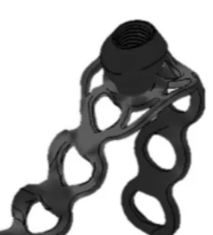
Geometry of the maxilla was obtained using CT and transformed to STL format. Maxilla file was imported to SolidWorks 2016 (Dassault Systèmes, SolidWorks Corp., Concord, MA, USA), where the assembly with the four plates was done. All three-dimensional plates were adjusted to the anatomic characteristics of the maxilla. The final reconstruction of the three-dimensional model is detailed in Figure 3.
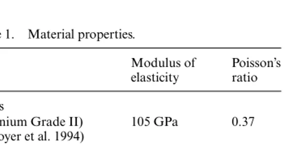
All materials were considered isotropic, lin-ear, elastic and homogeneous with the properties detailed in Table 1.
2.3 Mesh, boundary conditions and loading
configuration
The 3D model was meshed in SolidWorks 2016 (Dassault Systèmes, SolidWorks Corp., Concord, MA, USA) with a fine mesh and all regions of stress concentration that were of interest were manually refined. The convergence criterion was a change of less than 5% in von Mises stress in the model (Peixoto et al. 2017).
A rigid fixation restriction in the upper and lat-eral maxilla to prevent displacement in the x, y and
z axes was applied. A non-penetration condition
was also added to prevent interferences between plates and maxilla during the execution. Finally, an axial load of 100 N (Shimura et al. 2016) was directly applied to the area where the prosthesis is fixed to the plate.
3 RESULTS
Figure 4 shows the von Mises stress on plates. The difference between the maximum von Mises stress in HENGG-1 right and left plates is 3%, while the difference between HENGG-2 right and left is 2%.
In view of Figures 5 and 6, which are the plates placed on the molar region, it can be seen that the plate located in the left of the maxilla has bigger stresses on the area situated in the palate than the plate located in the right of the maxilla.
Figures 7 and 8, which represent the plates placed on the premaxilla, detail that both plates have the highest stresses on the area situated in the palate.
All plates showed a similar distribution patterns of maximum principal stress over the atrophic maxilla. Figure 2. Plates employed in this study: (a) HENGG-1
and (b) HENGG-2.
Figure 3. Three-dimensional assembly.
Table 1. Material properties.
Modulus of elasticity
Poisson’s ratio Plates
(Titanium Grade II) (Boyer et al. 1994) 105 GPa 0.37 Cortical bone (Bhering et al. 2016) 13.7 GPa 0.3 Trabecular bone (Bhering et al. 2016) 1.37 GPa 0.3
Figure 4. Stress distribution on plates [MPa].
3
The difference of principal stress value between the four regions in contact with plates is a 5%, with a mean maximum value of 80 MPa in plates’ region.
4 DISCUSSION
The biomechanical behavior of CF@O plates on a completely edentulous and atrophic maxilla has been evaluated employing finite element methods.
The accuracy of results in numerical studies depends on the precision of the model analyzed, the material properties and the constraining con-ditions (Van Staden et al. 2006). CT was used to obtain the geometry of the atrophic maxilla while plates were provided by the manufacturer.
This study has some limitations and assump-tions. All materials were considered homogeneous,
isotropic, and linearly elastic. Although these assumptions do not occur in clinical practice, they are common in finite element studies due to the challenges in establishing the properties of living tissues. These assumptions are consistent with other numerical studies (Prados-Privado et al. 2017, Almeida et al. 2015). Another limitation was the use of static loads although cyclic loads occur during chewing movements. This limitation is also validated in the several finite element analyses to study biomechanical behavior (Gümrükçü et al. 2017). However, this study analyzed biomechani-cal behavior of a new alternative to conventional treatments for atrophic maxilla.
There are several studies available in the litera-ture that employed different techniques to treat edentulous and atrophic maxilla, such as basal disk implants (Odin et al. 2012) or bone augmen-tation (Baldan et al. 2017).
Küçükkurt et al. compare the biomechanical behavior of different sinus floor elevation for den-tal implants placement (Küçükkurt et al. 2017). Under the condition of vertical loadings, von Mises stresses in mesial implants were lower than our results in plates in the case of lateral sinus lift-ing. However, plates analyzed in this study obtain lower von Mises stresses than prosthetic distal can-tilever application and short implant placement. Regarding to distal implants, our plates obtained lower stresses than prosthetic distal cantilever.
Ultimate stress limits in cortical bone have been described as 170 MPa in compression and 100 MPa in tension (Pérez et al. 2012). Based on these limits, the values observed in this model were lower than those considered physiologic to bone tissue.
A good biomechanical behavior of plates is understood when a homogeneous stress is transferred to the bone. In this case, the maxi-mum difference between all four plates’ region is 5%, meaning that the principal stress trans-ferred from plates to maxilla can be considered homogeneous.
Küçükkurt et al. obtained similar maximum principal stress in maxilla than our results in the case of short implant placement and higher princi-pal stress than our results in prosthetic distal canti-lever application (Küçükkurt et al. 2017).
Further studies simulating these titanium hybrid-plates alternatives for atrophic maxilla and jaw that include dynamic forces that occur during chew-ing and consider the anisotropic and regenerative properties of bone are needed. Furthermore, some in vivo clinical trials are necessary to validate the model and to confirm the efficiency of this protocol. A numerical study of the combination of prosthesis-plates-implant under different functional conditions (bruxism and other parafunctions) just like the antagonist arcade. Finally, a simulation of blood flow and bone regeneration around the plates is also necessary.
Figure 6. Stress distribution on plate HENGG-1 right.
Figure 7. Stress distribution on plate HENGG-2 left.
4
5 CONCLUSION
Based on the results provided by this numerical analysis, it is possible to conclude that in terms of clinical application, these titanium hybrid-plates have a better behavior than conventional treat-ments as prosthetic distal cantilever application and short implant placement. Titanium hybrid-plates distributed load to the maxilla with similar values as the case of short implants but with higher values than prosthetic distal cantilever application. In any case, resistance limits of bone and titanium were not exceeded. Finally, this technique can be considered a viable alternative for atrophic maxilla rehabilitation although it is necessary more studies to corroborate the clinical results.
REFERENCES
Ali, S., Karthigeyan, S., Deivanai, M., Kumar, A. 2014. Implant Rehabilitation For Atrophic Maxilla: A Review. The Journal of Indian Prosthodontic Society 13(3): 196–207.
Almeida, E.O., Rocha, E.P., Júnior, A.C.F., Anchieta, R.B. et al. 2015. Tilted and Short Implants Supporting Fixed Prosthesis in an Atrophic Maxilla: A 3D-FEA Bio-mechanical Evaluation. Clinical Implant Dentistry and
Related Research 17(Suppl 1): e332–e342.
Alqutaibi, A.Y., Altaib, F. 2016. Short Dental Implant Is Considered as a Reliable Treatment Option for Patients with Atrophic Posterior Maxilla. Journal of
Evidence-Based Dental Practice 16(3): 173–175.
Anitua, E., Orive, G., Aguirre, J.J., Andía, I. 2008. Five-Year Clinical Evaluation of Short Dental Implants Placed in Posterior Areas: A Retrospective Study.
Jour-nal of Periodontology 79(1): 42–48.
Anitua, E., Piñas, L., Begoña, L., Orive, G. 2014. Long-Term Retrospective Evaluation of Short Implants in the Posterior Areas: Clinical Results after 10–12 Years.
Journal of Clinical Periodontology 41(4): 404–411.
Aparicio, C., Manresa, C., Francisco, K., Claros, P., Alán-dez, J., González-Martín, O. et al. 2014. Zygomatic Implants: Indications, Techniques and Outcomes, and the Zygomatic Success Code. Periodontology 2000 66(1): 41–58.
Baldan, R.C.F., Coracin, F.L., Lins, L., Mello, W.R., San-tos, P.S. 2017. Atrophic Maxilla Reconstruction With Fresh Frozen Allograft Bone, Titanium Mesh, and Platelet-Rich Fibrin: Case Report. Transplantation
Pro-ceedings 49(4): 893–897.
Bhering, C.L.B., Mesquita, M.F., Kemmoku, D.T., et al. 2016. Comparison between Four and All-on-Six Treatment Concepts and Framework Material on Stress Distribution in Atrophic Maxilla: A Prototyping Guided 3D-FEA Study. Materials Science &
Engineer-ing. C, Materials for Biological Applications 69: 715–725.
Boyer, R., Welsch, G., Collings, E.W. 1994. Materials
Properties Handbook: Titanium Alloys. Ohio: ASM
International.
Chiapasco, M., Zaniboni, M. 2014. Methods to Treat the Edentulous Posterior Maxilla: Implants With Sinus
Grafting. Journal of Oral and Maxillofacial Surgery 67(4): 867–871.
Cucchi, A., Vignudelli, E., Franco, S., Corinaldesi, G. 2017. Minimally Invasive Approach Based on Pterygoid and Short Implants for Rehabilitation of an Extremely Atrophic Maxilla. Implant Dentistry 26(4): 1–6. Gultekin, B.A., Cansiz, E., Borahan, M.O. 2017. Clinical
and 3-Dimensional Radiographic Evaluation of Autog-enous Iliac Block Bone Grafting and Guided Bone Regeneration in Patients With Atrophic Maxilla.
Jour-nal of Oral and Maxillofacial Surgery 75(4): 709–722.
Gümrükçü, Z., Korkmaz, Y.T., Korkmaz, F.M. 2017. Bio-mechanical Evaluation of Implant-Supported Prosthe-sis with Various Tilting Implant Angles and Bone Types in Atrophic Maxilla: A Finite Element Study.
Comput-ers in Biology and Medicine 86: 47–54.
Kaneko, T., Nakamura, S., Hino, S., Norio, H., Shi-moyama, T. 2016. Continuous Intra-Sinus Bone Regen-eration after Nongrafted Sinus Lift with a PLLA Mesh Plate Device and Dental Implant Placement in an Atrophic Posterior Maxilla: A Case Report.
Interna-tional Journal of Implant Dentistry 2(1): 16.
Küçükkurt, S., Alpaslan, G., Kurt, A. 2017. Biomechani-cal Comparison of Sinus Floor Elevation and Alterna-tive Treatment Methods for Dental Implant Placement.
Computer Methods in Biomechanics and Biomedical Engineering 20(3): 284–293.
Odin, G., Misch, C.E., Binderman, I., Scortecci, G. 2012. Fixed Rehabilitation of Severely Atrophic Jaws Using Immediately Loaded Basal Disk Implants After In Situ Bone Activation. Journal of Oral Implantology 38: 611–616.
Peixoto, H.E., Camati, P.R., Faot, F., Sotto-Maior, B. et al. 2017. Rehabilitation of the Atrophic Mandible with Short Implants in Different Positions: A Finite Ele-ments Study. Materials Science & Engineering. C,
Mate-rials for Biological Applications 80: 122–128.
Pérez, M.A., Prados-Frutos, J.C., Bea, J.A., Doblaré, M. 2012. Stress Transfer Properties of Different Commer-cial Dental Implants: A Finite Element Study. Computer
Methods in Biomechanics and Biomedical Engineering
15: 263–273.
Prados-Privado, M., Bea, J.A., Rojo, R., Gehrke, S.A., Calvo-Guirado, J.L., Prados-Frutos, J.C. 2017. A New Model to Study Fatigue in Dental Implants Based on Probabilistic Finite Elements and Cumulative Damage Model. Applied Bionics and Biomechanics. 2017: 1–8. Shimura, Y., Sato, Y., Kitagawa, N., Omor, M. 2016.
Biomechanical Effects of Offset Placement of Dental Implants in the Edentulous Posterior Mandible.
Inter-national Journal of Implant Dentistry 2(1):17.
Scortecci, G. & Bourbon, B. 1990. [Dentures on the Dis-kimplant]. Revue Francaise Des Prothesistes Dentaires 13: 31–48.
van der Mark, E.L., Bierenbroodspot, F., Baas, EM., de Lange, J. 2011. Reconstruction of an Atrophic Maxilla: Comparison of Two Methods. British Journal of Oral
and Maxillofacial Surgery 49(3): 198–202.
Van Staden, R.C., Guan, H., Loo, Y.C. 2006. Applica-tion of the Finite Element Method in Dental Implant Research. Computer Methods in Biomechanics and
Numerical analysis of titanium hybrid-plates in atrophic maxilla
Ali, S. , Karthigeyan, S. , Deivanai, M. , Kumar, A. 2014. Implant Rehabilitation For Atrophic Maxilla: A Review. The Journal of Indian Prosthodontic Society 13(3): 196207.
Almeida, E.O. , Rocha, E.P. , Jnior, A.C.F. , Anchieta, R.B. et al. 2015. Tilted and Short Implants Supporting Fixed Prosthesis in an Atrophic Maxilla: A 3D-FEA Bio-mechanical Evaluation. Clinical Implant Dentistry and Related Research 17(Suppl 1): e332e342.
Alqutaibi, A.Y. , Altaib, F. 2016. Short Dental Implant Is Considered as a Reliable Treatment Option for Patients with Atrophic Posterior Maxilla. Journal of Evidence-Based Dental Practice 16(3): 173175.
Anitua, E. , Orive, G. , Aguirre, J.J. , Anda, I. 2008. Five-Year Clinical Evaluation of Short Dental Implants Placed in Posterior Areas: A Retrospective Study. Journal of Periodontology 79(1): 4248.
Anitua, E. , Pias, L. , Bego a, L. , Orive, G. 2014. Long-Term Retrospective Evaluation of Short Implants in the Posterior Areas: Clinical Results after 1012 Years. Journal of Clinical Periodontology 41(4): 404411.
Aparicio, C. , Manresa, C. , Francisco, K. , Claros, P. , Al ndez, J. , Gonzlez-Martn, O. et al. 2014. Zygomatic Implants: Indications, Techniques and Outcomes, and the Zygomatic Success Code. Periodontology 2000 66(1): 4158.
Baldan, R.C.F. , Coracin, F.L. , Lins, L. , Mello, W.R. , Santos, P.S. 2017. Atrophic Maxilla Reconstruction With Fresh Frozen Allograft Bone, Titanium Mesh, and Platelet-Rich Fibrin: Case Report. Transplantation Proceedings 49(4): 893897. Bhering, C.L.B. , Mesquita, M.F. , Kemmoku, D.T. , et al. 2016. Comparison between All-on-Four and All-on-Six
Treatment Concepts and Framework Material on Stress Distribution in Atrophic Maxilla: A Prototyping Guided 3D-FEA Study. Materials Science & Engineering. C, Materials for Biological Applications 69: 715725.
Boyer, R. , Welsch, G. , Collings, E.W. 1994. Materials Properties Handbook: Titanium Alloys. Ohio: ASM International. Chiapasco, M. , Zaniboni, M. 2014. Methods to Treat the Edentulous Posterior Maxilla: Implants With Sinus Grafting. Journal of Oral and Maxillofacial Surgery 67(4): 867871.
Cucchi, A. , Vignudelli, E. , Franco, S. , Corinaldesi, G. 2017. Minimally Invasive Approach Based on Pterygoid and Short Implants for Rehabilitation of an Extremely Atrophic Maxilla. Implant Dentistry 26(4): 16.
Gultekin, B.A. , Cansiz, E. , Borahan, M.O. 2017. Clinical and 3-Dimensional Radiographic Evaluation of Autogenous Iliac Block Bone Grafting and Guided Bone Regeneration in Patients With Atrophic Maxilla. Journal of Oral and Maxillofacial Surgery 75(4): 709722.
Gmrk, Z. , Korkmaz, Y.T. , Korkmaz, F.M. 2017. Bio-mechanical Evaluation of Implant-Supported Prosthesis with Various Tilting Implant Angles and Bone Types in Atrophic Maxilla: A Finite Element Study. Computers in Biology and Medicine 86: 4754.
Kaneko, T. , Nakamura, S. , Hino, S. , Norio, H. , Shimoyama, T. 2016. Continuous Intra-Sinus Bone Regeneration after Nongrafted Sinus Lift with a PLLA Mesh Plate Device and Dental Implant Placement in an Atrophic Posterior Maxilla: A Case Report. International Journal of Implant Dentistry 2(1): 16.
Kkkurt, S. , Alpaslan, G. , Kurt, A. 2017. Biomechanical Comparison of Sinus Floor Elevation and Alternative Treatment Methods for Dental Implant Placement. Computer Methods in Biomechanics and Biomedical Engineering 20(3): 284293. Odin, G. , Misch, C.E. , Binderman, I. , Scortecci, G. 2012. Fixed Rehabilitation of Severely Atrophic Jaws Using Immediately Loaded Basal Disk Implants After In Situ Bone Activation. Journal of Oral Implantology 38: 611616. Peixoto, H.E. , Camati, P.R. , Faot, F. , Sotto-Maior, B. et al. 2017. Rehabilitation of the Atrophic Mandible with Short Implants in Different Positions: A Finite Elements Study. Materials Science & Engineering. C, Materials for Biological Applications 80: 122128.
Prez, M.A. , Prados-Frutos, J.C. , Bea, J.A. , Doblar, M. 2012. Stress Transfer Properties of Different Commercial Dental Implants: A Finite Element Study. Computer Methods in Biomechanics and Biomedical Engineering 15: 263273.
Prados-Privado, M. , Bea, J.A. , Rojo, R. , Gehrke, S.A. , Calvo-Guirado, J.L. , Prados-Frutos, J.C. 2017. A New Model to Study Fatigue in Dental Implants Based on Probabilistic Finite Elements and Cumulative Damage Model. Applied Bionics and Biomechanics. 2017: 18.
Shimura, Y. , Sato, Y. , Kitagawa, N. , Omor, M. 2016. Biomechanical Effects of Offset Placement of Dental Implants in the Edentulous Posterior Mandible. International Journal of Implant Dentistry 2(1):17.
Scortecci, G. & Bourbon, B. 1990. [Dentures on the Diskimplant]. Revue Francaise Des Prothesistes Dentaires 13: 3148. van der Mark, E.L. , Bierenbroodspot, F. , Baas, E.M. , de Lange, J. 2011. Reconstruction of an Atrophic Maxilla: Comparison of Two Methods. British Journal of Oral and Maxillofacial Surgery 49(3): 198202.
Van Staden, R.C. , Guan, H. , Loo, Y.C. 2006. Application of the Finite Element Method in Dental Implant Research. Computer Methods in Biomechanics and Biomedical Engineering 9(4): 257270.
Effects of introducing gap constraints in the masticatory system: A finite element study
Ebrahim, S. , Montoya, L. , Busse, J.W. , Carrasco-Labra, A. , Guyatt, G.H. 2012. The effectiveness of splint therapy in patients with temporomandibular disorders: A systematic review and meta-analysis. The Journal of the American Dental Association 143: 847857.Hansson, T. , berg, T. 1977. Arthrosis and deviation in form in the temporomandibular joint: A macroscopic study on a human autopsy material. Acta Odontologica Scandinavica 35:167874.
Hukins, W. , Aspden, R. , Yarker, Y. 1984. Fiber reinforcement and mechanical stability in articular cartilage. Engineering in Medicine 13: 153156.
Koolstra, J.H. & van Eijden, T.M.G.J. , Weijs, W.A. , Naeije, M. 1988. A three-dimensional mathematical model of the human masticatory system predicting maximum possible bite forces. Journal of Biomechanics 21:563576.
Koolstra, J.H. & van Eijden, T.M.G.J. 2005. Combined finite-element and rigid-body analysis of human jaw joint dynamics. Journal of Biomechanics 38:24312439.
Koolstra, J.H. & van Eijden T.M.G.J. 2007. Consequences of Viscoelastic Behavior in the Human Temporomandibular Joint Disc. J Dent Res 2007 86:11981202.
Lettry, S. , Seedhom, B.B. , Berry, E. , Cupponea, M. 2003. Quality assessment of the cortical bone of the human mandible. Bone 32:3544.
Llodra-Calvo, J.C. , Bravo-Prez, M. , Corts-Martinicorena, F.J. 2000. Encuesta de Salud Oral en Espaa. Revista Consejo Odontlogos Estomatlagos 7:1963.
Macfarlane, T.V. , Blinkhorn, A.S. , Davies, R.M. , Kincey, J. Worthington, H.V. 2002. Oro-facial pain in the community: prevalence and associated impact. Community Dentistry and Oral Epidemiology 30: 5260.
Martinez Choy, S.E. , Lenz, J. , Schweizerhof, K. , Schmitter, M. , Schindler, H.J. 2017. Realistic kinetic loading of the jaw system during single chewing cycles: a finite element study. Journal of Oral Rehabilitation 44:375384.
Mow, V.C. , Huiskes, R. 2005. Basic Orthopaedic Biomechanics and Mechanobiology. Philadelphia: Lippincott Williams & Wilkins.
Osborn, J.W. 1989. The temporomandibular ligament and the articular eminence as constraints during jaw opening. Journal of Oral Rehabilitation 16: 323333.
Perez del Palomar, A. , Doblare, M. 2006a. The effect of collagen reinforcement in the behaviour of the temporomandibular joint disc. Journal of Biomechanics 39:10751085.
Perez del Palomar, A. , Doblare, M. 2006b. Finite element analysis of the temporomandibular joint during lateral excursions of the mandible. Journal of Biomechanics 39:21532163.
Perez del Palomar, A. , Doblare, M. 2007. An accurate simulation model of anteriorly displaced TMJ discs with and without reduction. Medical Engineering & Physics 29:216226.
Radin, E.L. , Paul I.L. 1971. Response of Joints to Impact Loading. Arthritis and Rheumatism 14(3): 356362.
van Ruijven, L.J. , Weijs, W.A. 1990. A new model for calculating muscle forces from electromyograms. European Journal of Applied Physiology 61: 479485.
Schindler, H.J. , Lenz, J. Trp, J.C. , Schweizerhof, K. , Rues, S. 2009. Small unilateral jaw gap variations: equilibrium changes, co-contractions and joint forces. Journal of Oral Rehabilitation 36:710718.
Tanaka, E. , Hanaoka, K. , Tanaka, M. , van Eijden, T. , Iwabe, T. , Ishino, Y. , Sasaki, A. , Tanne K. 2003. Viscoelastic properties of bovine retrodiscal tissue under tensile stress-relaxation. Eur J Oral Sci 111:518522.
Influence of temperature on the dimensional stability of an addition silicone
Amin, W.M. , Al-Ali, M.H. , Al Tarawneh, S.K. , Taha, S.T. , Saleh, M.W. & Ereifij, N. 2009. The effects of disinfectants on dimensional accuracy and surface quality of impression materials and gypsum casts. J Clin Med Res, 1, 8189.
Berg, J.C. , Johnson, G.H. , Lepe, X. & Adan-Plaza, S. 2003. Temperature effects on the rheological properties of current polyether and polysiloxane impression materials during setting. J Prosthet Dent, 90, 150161.
Chen, S.Y. , Liang, W.M. & Chen, F.N. 2004. Factors affecting the accuracy of elastometric impression materials. J Dent, 32, 603609.
Corso, M. , Abanomy, A. , Di Canzio, J. , Zurakowski, D. & Morgano, S.M. 1998. The effect of temperature changes on the dimensional stability of polyvinyl siloxane and polyether impression materials. J Prosthet Dent, 79, 626631.
Endo, T. & Finger, W.J. 2006. Dimensional accuracy of a new polyether impression material. Quintessence Int, 37, 4751. Fano, V. , Gennari, P.U. & Ortalli, I. 1992. Dimensional stability of silicone-based impression materials. Dent Mater, 8, 105109.
Faria, A.C. , Rodrigues, R.C. , Macedo, A.P. , Mattos Mda, G. & Ribeiro, R.F. 2008. Accuracy of stone casts obtained by different impression materials. Braz Oral Res, 22, 293298.
Forrester-Baker, L. , Seymour, K.G. , Samarawickrama, D. , Zou, L. , Cherukara, G. & Patel, M. 2005. A comparison of dimensional accuracy between three different addition cured silicone impression materials. Eur J Prosthodont Restor Dent, 13, 6974.
Giordano, R. , 2nd 2000. Impression materials: basic properties. Gen Dent, 48, 510512, 514, 516.
Hassan, A.K. 2006. Dimensional accuracy of 3 silicone dental impression materials. East Mediterr Health J, 12, 632636. Kim, K.M. , Lee, J.S. , Kim, K.N. & Shin, S.W. 2001. Dimensional changes of dental impression materials by thermal changes. J Biomed Mater Res, 58, 217220.
Pamenius, M. & Ohlson, N.G. 1992. Determination of thermal properties of impression materials. Dent Mater, 8, 140144. Perry, R. 2013. Dental impression materials. J Vet Dent, 30, 116124.
Rubel, B.S. 2007. Impression materials: a comparative review of impression materials most commonly used in restorative dentistry. Dent Clin North Am, 51, 629642, vi.
Computational analysis for stress intensity factor (KI) measuring in metal-ceramic interface
Ariel J. Raigrodski DMD, MS , Gerard J. Chiche DDS . 2001. The safety and efficacy of anterior ceramic fixed partial dentures: A review of the literature. The Journal of Prosthetic Dentistry 86(5): 520525.ASTM E399-90 , 2003: Standard Test Method for Plane-Strain Fracture Toughness of Metallic Materials, ASTM Standards, Vol 03.01.
Esben Byskov . 1970. The Calculation of Stress Intensity Factors Using the Finite Element Method with Cracked Elements. Int. J. Fracture Mechanics 6(2): 159167.
Ismail, A.E. 2014. Stress Intensity Factors of Eccentric Cracks in Bi-Material Plates. Applied Mechanics and Materials 663: 98102.
Kelly, J.R. , J.A. Tesk and J.A. 1995. Failure of All-ceramic Fixed Partial Dentures in vitro and in vivo: Analysis and Modeling. Journal of Dental Research 74(6): 12531258.
Livia Goulart Tovar , 2013. Anlise Computacional de Propagao de Trinca em Interface de Corpo de Prova Metalocermico. Universidade Federal do Rio de Janeiro, DEM/POLI/UFRJ.
Nik Ismail . 2014. Stress Intensity Factors of Edge Cracks in Dissimilar Joint Plates. Thesis, Univ. of Malaysia. Paris P. , Sih G. . 1965. Stress Analysis of Cracks. 67th Annual Meeting of the ASTM.
Qing Li , Ionut Ichim , Jeff Loughran , Wei Li , Michael Swain , Jules Ki . 2006. Numerical Simulation of Crack Formation in All Ceramic Dental Bridge. Key Engineering Materials 312: 293298.
Rola M. Shadid , Nasrin R. Sadaqah , Layla Abu-Naba , Wael M. Al-Omari . 2013. Porcelain fracture of metal-ceramic tooth-supported and implant-supported restorations: A review. Open Journal of Stomatology 3: 411418.
Rooke, D.P. , D.J. Cartwright . 1976. Compendium of Stress Intensity Factors, London HMSO.
Stefan Hajdu . 2014. The Investigation of the Stress State near the Crack Tip of Central Cracks Through Numerical Analysis. Procedia Engineering 69: 477485.
Xian-Kui Zhu , James A. Joyce . 2012. Review of fracture toughness (G, K, J, CTOD, CTOA) testing and standardization. Eng. Fracture Mechanics 85:146.
Thermal stimulation of dentinal tubules
Brown, W.S. , Dewey, W.A. , & Jacobs, H.R. 1970. Thermal Properties of Teeth. Journal of Dental Research, 49(4), 752755.
Coutinho, E.T. , Dalmeida, J.R.M. , Paciornik, S. 2007. Evaluation of microstructural parameters of human dentin by digital image analysis. Materials Research, v. 10, n. 2: 153159.
El Karim, I.A. , Linden, G.J. , Curtis, T.M. , About, I. , McGahon, M.K. , Irwin, C.R. , & Lundy, F.T. 2011. Human odontoblasts express functional thermo-sensitive TRP channels: Implications for dentin sensitivity. Pain, 152(10), 22112223.
Le Fur-Bonnabesse, A. , A. Bodere , C. Helou , V. Chevalier and J.P. Goulet . 2017. Dental pain induced by an ambient thermal differential: pathophysiological hypothesis. J Pain Res 10: 28452851.
Lin, M. , Liu, S. , Niu, L. , Xu, F. , & Lu, T.J. 2011. Analysis of thermal-induced dentinal fluid flow and its implications in dental thermal pain. Archives of Oral Biology, 56(9), 846854.
Lin, M. , Xu, F. , Lu, T.J. , & Bai, B.F. 2010. A review of heat transfer in human toothExperimental characterization and mathematical modeling. Dental Materials, 26(6), 501513.
Matthews, B. 1977. Responses of Intradental Nerves to Electrical and Thermal Stimulation of Teeth in Dogs. The Journal of Physiology 264.3: 641664.
Analysis tool of skills acquisition in fine motor skill
Carvalho I. 2014. A Motricidade Fina em Estudantes de Medicina Dent ria: comparao entre os estudantes dos 1 e 5 anos [CESPU MSc Thesis] https://repositorio.cespu.pt/handle/20.500.11816/345.
Costa, A. 2013. O desenvolvimento da motricadade fina: Um estudo de interveno com crianas em idade pr-escolar. [IPVC MSc Thesis] http://hdl.handle.net/20.500.11960/1392.
Giulani, M. , Lajolo, C. , Clemente, L. , Querqui, A. , Viotti, R. , Boari, A. and Miani, C. 2007. Is manual dexterity essential in the selection of dental students? British Dental Journal Volume 203(3): pg. 149155. DOI: 10.1038/bdj.2007.688. Luck, O. , Reitemeier, B. , Scheuch, K. , 2000. Testing of fine motor skills in dental students. Eur J Dent Educ, 4: 1014.
A new approach in 3D finite element analysis in restorative dentistry
Ausiello P. , Apicella A. , Davidson C.L. , Rengo S. , 2001. 3D-finite element analyses of cusp movements in a human upper premolar, restored with adhesive resin-based composites. Journal Biomechanics 34: 12691277.
Ausiello P. , Rengo S. , Davidson C.L. , Watts D.C. , 2004. Stress distributions in adhesively cemented ceramic and resin-composite Class II inlay restorations: a 3D-FEA study. Dental Materials 20: 862872.
Ausiello P. , Franciosa P. , Martorelli M. , Watts D.C. , 2011. Numerical fatigue 3D-FE modeling of indirect composite-restored posterior teeth. Dental Materials 27: 423430.
Chuang S.F. , Chang C.H. , Chen T.Y.F. , 2011. Contraction behaviors of dental composite restorations Finite element investigation with DIC validation. Journal of the Mechanical Behavior of Biomedical Materials 4: 21382149.
Farah J.W. , Craig R.G. , 1973. Element stress analysis of an axisymmeric molar. Journal of Dental Research 53: 859864. Ledley R.H. , Huang H.K. , 1968. Linear model of tooth displacement by applied forces. Journal of Dental Research 47: 427432.
Magne P. , Douglas W.H. , 1999. Optimization of resilience and stress distribution in porcelain veneers for the treatment of crown-fractured incisors. International Journal of Periodontics and Restorative Dentistry 19: 543553.
Magne P. , 2007. Efficient 3D finite element analysis of dental restorative procedures using micro-CT data. Dental Materials 23: 539548.
Magne P. , Tan D.T. , 2008. Incisor compliance following operative procedures: a rapid 3-D finite element analysis using micro-CT data. Journal of Adhesive Dentistry 10: 4956.
Magne P. , Oganesyan T. , 2009a. CT scan-based finite element analysis of premolar cuspal deflection following operative procedures. International Journal of Periodontics and Restorative Dentistry 29: 361369.
Magne P. , Oganesyan T. , 2009b. Premolar cuspal flexure as a function of restorative material and occlusal contact location. Quintessence International 40: 363370.
Magne P. , 2010. Virtual prototyping of adhesively restored, endodontically treated molars. Journal of Prosthetic Dentistry 103: 343351.
Magne P. , Schlichting L.H. , 2011. Paranhos MPG. Risk of onlay fracture during pre-cementation functional occlusal tapping. Dental Materials 27: 942947.
Magne P. , Stanley K. , Schlichting L.H. , 2012. Modeling of ultrathin occlusal veneers. Dental Materials 28: 777782. Rees J.S. , Hammadeh M. , Jagger D.C. , 2003. Abfraction lesion formation in maxillary incisors, canines and premolars: a finite element study. European Journal Oral Sciences 111: 149154.
Rodrigues F.P. , Li J. , Silikas N. , Ballester R.Y. , Watts D.C. , 2009. Sequential software processing of micro-XCT dental-images for 3D-FE analysis. Dental Materials 25: 4755.
Rubin C. , Krishanamurity N. , Capilouto E. , 1983. Stress analysis of the human tooth using three-dimensional finite element model. Journal of Dental Research 62: 8286.
Yamanel K. , alar A. , Glahi K. , zden U.A. , 2009. Effects of different ceramic and composite materials on stress distribution in inlay and onlay cavities: 3-D finite element analysis. Dental Material Journal 28: 661670.
Yettram A.L. , Wright K.W.J. , Pickard H.M. , 1976. Finite element stress analysis of the crowns of normal and restored teeth. Journal of Dental Research 55: 10041010.
Zhang Z. , Zheng K. , Li E. , Li W. , Li Q. , Swain M.V. , 2016. Mechanical benefits of conservative restoration for dental fissure caries. Journal of the Mechanical Behavior of Biomedical Materials 53: 1120.
Biological behavior of titanium, zirconia or PEEK dental implant-abutments
Alrabeah G.O. , Brett P. , Knowles J.C. , Petridis H. The effect of metal ions released from different dental implant-abutment couples on osteoblast function and secretion of bone resorbing mediators. Journal of Dentistry.
2017;66(June):91101.
Bacchelli B. , Giavaresi G. , Franchi M. , et al. Influence of a zirconia sandblasting treated surface on peri-implant bone healing: An experimental study in sheep. Acta Biomaterialia. 2009;5(6):22462257.
Bchle M. , Butz F. , Hbner U. , Bakalinis E. , Kohal R.J. Behavior of CAL72 osteoblast-like cells cultured on zirconia ceramics with different surface topographies. Clinical Oral Implants Research. 2007;18(1):5359.
Banaszek K. , Wiktorowska-Owczarek A.N.N.A. , Kowalczyk E. , Klimek L. Possibilities of applying Ti (C, N) coatings on prosthetic elements: research with the use of human endothelial cells. Acta of bio-engineering and biomechanics. 2016; 18(1).
Bressan E. , Tessarolo F. , Sbricoli L. , Caola I. , Nollo G. , Di Fiore A. Effect of chlorhexidine in preventing plaque biofilm on healing abutment: a crossover controlled study. Implant dentistry. 2014; 23(1), 6468.
Brunot-Gohin C. , Duval J.L. , Verbeke S. , Belanger K. , Pezron I. , Kugel G. , Egles C. Biocompatibility study of lithium disilicate and zirconium oxide ceramics for esthetic dental abutments. Journal of periodontal & implant science. 2016; 46(6), 362371.
41[7] Avila De E.D. , De Molon R.S. , Lima B.P. , Lux R. , Shi W. , Junior M.J. , de Assis Mollo Junior F. Impact of physical chemical characteristics of abutment implant surfaces on bacteria adhesion. Journal of Oral Implantology. 2016a; 42(2), 153158.
de Avila E.D. , Avila-Campos M.J. , Vergani, C.E. , Spolidrio D.M.P. , de Assis Mollo Jr F. Structural and quantitative analysis of a mature anaerobic bio-film on different implant abutment surfaces. Journal of Prosthetic Dentistry. 2016b; 115(4), 428436.
de Avila E.D. , de Molon R.S. , Palomari Spolidorio DM Implications of surface and bulk properties of abutment implants and their degradation in the health of periodontal tissue. Materials. 2013; 6(12), 59515966.
de Avila E.D. , Vergani C.E. , Junior F.A.M. , Junior M.J. , Shi W. , Lux R. Effect of titanium and zirconia dental implant abutments on a cultivable polymicrobial saliva community. Journal of Prosthetic Dentistry. 2017; 118(4), 481487. de Freitas A.R. , Silva T.S.D.O. , Ribeiro R.F. , de Albuquerque Junior R.F. , Pedrazzi V. , Do Nascimento C. Oral bacterial colonization on dental implants restored with titanium or zirconia abutments: 6-month follow-up. Clinical Oral Investigations. 2018:19.
do Nascimento C. , Pita M.S. , Pedrazzi V. , de Albuquerque Junior R.F. , Ribeiro R.F. In vivo evaluation of Candida spp. adhesion on titanium or zirconia abutment surfaces. Archives of oral biology. 2013;58(7):853861.
Dorkhan M. , Hall J. , Uvdal P. , Sandell A. , Svenster G. , Davies J.R. Crystalline anatase-rich titanium can reduce adherence of oral streptococci. Biofouling. 2014a;30(6):751759.
Dorkhan M. , Ycel-Lindberg T. , Hall J. , Svenster G. , Davies J.R. Adherence of human oral keratinocytes and gingival fibroblasts to nano-structured titanium surfaces. BMC oral health. 2014b; (1), 75.
Esfahanizadeh N. , Motalebi S. , Daneshparvar N. , Akhoundi N. , Bonakdar S. Morphology , proliferation, and gene expression of gingival fibroblasts on Laser-Lok, titanium, and zirconia surfaces. Lasers in medical science. 2016; 31(5), 863873.
Fuhrmann G. , Steiner M. , Freitag- Wolf S. , Kern M. Resin bonding to three types of polyaryletherketones (PAEKs)Durability and influence of surface conditioning. Dental Materials. 2014;30(3):357363.
Flamant Q. , Anglada M. Hydrofluoric acid etching of dental zirconia. Part 2: effect on flexural strength and ageing behavior. Journal of the European Ceramic Society. 2016;36(1):135145.
Gmez-Florit M. , Ramis J.M. , Xing R. , et al. Differential response of human gingival fibroblasts to titanium and titanium-zirconium-modified surfaces. Journal of Periodontal Research. 2014;49(4):425436.
Hahnel S. , Wieser A. , Lang R. , Rosentritt M. Bio-film formation on the surface of modern implant abutment materials. Clinical Oral Implants Research. 2015;26(11):12971301.
Harder S. , Podschun R. , Grancicova L. , Mehl C. , Kern M. Analysis of the intraimplant microflora of two-piece dental implants. Clinical Oral Investigations. 2013;17(4):11351142.
Kern M. , Lehmann F. Influence of surface conditioning on bonding to polyetheretherketon (PEEK). Dental Materials. 2012;28(12):12801283.
Kheur M. , Harianawala H.H. , Sethi T. , Kheur S. , Acharya A. , Mattheos N. Human cytomegalovirus-1 and Epstein-Barr virus-1 viral colonization of titanium and zirconia abutments: a split-mouth study. Journal of investigative and clinical dentistry. 2016;7(4):396400.
Kim Y.-S. , Ko Y. , Kye S.-B. , Yang S.-M. Human Gingival Fibroblast (HGF-1) Attachment and Proliferation on Several Abutment Materials with Various Colors. The International Journal of Oral & Maxillofacial Implants. 2014;29(4):969975. Kolbeck C. , Sereno M. , Rosentritt M. , Handel G. Biofilm formation on polyetheretherketone surfaces and cleaning options. IADR Seattle, No 2353. March 2013.
Kunnen M. , Hormia M. , Kivilahti J. , Hautaniemi J. , Thesleff I. Effect of surface processing on the attachment,
orientation, and proliferation of human gingival fibroblasts on titanium: Attachment of Fibroblasts on Titanium. Journal of Biomedical Materials Research. 1992;26(10):13251341.
Lazzara R.J. , Porter S.S. Platform switching: a new concept in implant dentistry for controlling postrestorative crestal bone levels. The International journal of periodontics & restorative dentistry. 2006;26(1):917.
Lee D.-W. , Kim J.-G. , Kim M.-K. , et al. Effect of laser-dimpled titanium surfaces on attachment of epithelial-like cells and fibroblasts. The Journal of Advanced Prosthodontics. 2015;7(2):138.
Linkevicius T. , Vaitelis J. The effect of zirconia or titanium as abutment material on soft peri-implant tissues: A systematic review and meta-analysis. Clinical Oral Implants Research. 2015;26:139147.
Moon Y.-H. , Yoon M.-K. , Moon J.-S. , et al. Focal adhesion linker proteins expression of fibroblast related to adhesion in response to different transmucosal abutment surfaces. The journal of advanced prosthodontics. 2013;5(3):341350. Moura C.G. , Pereira R. , Buciumeanu M. , et al. Effect of laser surface texturing on primary stability and surface properties of zirconia implants. Ceramics International. 2017;(July).
Nascimento C do , Pita M.S. , Santos E de S. , et al. Microbiome of titanium and zirconia dental implants abutments. Dental materials/official publication of the Academy of Dental Materials. 2016;32(1):93101.
Nothdurft F.P. , Fontana D. , Ruppenthal S. , et al. Differential Behavior of Fibroblasts and Epithelial Cells on Structured Implant Abutment Materials: A Comparison of Materials and Surface Topographies. Clinical implant dentistry and related research. 2015;17(6):12371249.
Okabe E. , Ishihara Y. , Kikuchi T. , et al. Adhesion Properties of Human Oral Epithelial-Derived Cells to Zirconia. Clinical implant dentistry and related research. 2016;18(5):906916.
Pardun K. , Treccani L. , Volkmann E. , et al. Magnesium-containing mixed coatings on zirconia for dental implants: mechanical characterization and in vitro behavior. Journal of Biomaterials Applications. 2015;30(1):104118. 42[35] Piconi C. , Maccauro G. Zirconia as a Biomaterial. Biomaterials. 1999:95108125.
Quirynen M. , De Soete M. , van Steenberghe D. Infectious risks for oral implants: a review of the literature. Clinical Oral Implants Research. 2002;13(1):119.
Rigolin M.S.M. , de Avila E.D. , Basso F.G. , Hebling J. , Carlos C.A. , Mollo Junior F de A. Effect of different implant abutment surfaces on OBA-09 epithelial cell adhesion. Microscopy Research and Technique. 2017;80(12):13041309. Rutkunas V. , Bukelskiene V. , Sabaliauskas V. , Balciunas E. , Malinauskas M. , Baltriukiene D. Assessment of human gingival fibroblast interaction with dental implant abutment materials. Journal of materials science Materials in medicine. 2015;26(4):169169.
Souza J.C.M. , Ponthiaux P. , Henriques M. , et al. Corrosion behaviour of titanium in the presence of Streptococcus mutans. Journal of Dentistry. 2013;41(6):528534.
Schwarz F. , Mihatovic I. , Becker J. , Bormann K.H. , Keeve P.L. , Friedmann A. Histological evaluation of different abutments in the posterior maxilla and mandible: an experimental study in humans. Journal of clinical periodontology. 2013; 40(8), 807815.
Tet S. , Zizzari V. Collagen Fiber Orientation Around Machined Titanium and Zirconia Dental Implant Necks: An Animal Study. 2008:7.
van Brakel R. , Cune M.S. , van Winkelhoff A.J. , de Putter C. , Verhoeven J.W. , van der Reijden W. Early bacterial colonization and soft tissue health around zirconia and titanium abutments: an in vivo study in man: Bacterial colonization and soft tissue health around zirconia and titanium. Clinical Oral Implants Research. 2011;22(6):571577.
Volpe S. , Verrocchi D. , Andersson P. Comparison of early bacterial colonization of PEEK and titanium healing abutments using real-time PCR. Applied Osseointegration Research. 2008;6:5456.
Xing R. , Lyngstadaas S.P. , Ellingsen J.E. , Taxt-Lamolle S. , Haugen H.J. The influence of surface nano-roughness, texture and chemistry of TiZr implant abutment on oral biofilm accumulation. Clinical oral implants research.
2015;26(6):649656.
Xing R. , Salou L. , Taxt-Lamolle S. , Reseland J.E. , Lyngstadaas S.P. , Haugen H.J. Surface hydride on titanium by cathodic polarization promotes human gingival fibroblast growth. Journal of Biomedical Materials ResearchPart A. 2014;102(5):13891398.
Dental implants fatigue life: A probabilistic fatigue study
Alsahhaf, A. , Spies, B.C. , Vach, K. , Kohal, R.-J. 2017. Fracture resistance of zirconia-based implant abutments after artificial long-term aging. Journal of the Mechanical Behavior Biomedical Materials 66: 224232.
Aylln, J.M. , Navarro, C. , Vzquez, J. , Domnguez, J. 2014. Fatigue life estimation in dental implants. Engineering Fracture Mechanics 123: 3443.
Coray, R. , Zeltner, M. , zcan, M. 2016. Fracture strength of implant abutments after fatigue testing: A systematic re-view and a meta-analysis. Journal of the Mechanical Behavior of Biomedical Materials 62: 333346.
Gehrke, S.A. , Souza dos Santos Vianna, M. , Dedavid, B.A. 2014. Influence of bone insertion level of the implant on the fracture strength of different connection designs: an in vitro study. Clinical Oral Investigations 18(3): 715720.
Gehrke, S.A. , De Carvalho Serra, R. 2015. Load fatigue performance of conical implant-abutment connection: effect of torque level and interface junction. Minerva Stomatologica 64(1): 17.
Gehrke, S.A. , Poncio da Silva, P.M. , Calvo Guirado, J.L. et al. 2016. Mechanical behavior of zirconia and titanium abutments before and after cyclic load application. Journal of Prosthetic Dentistry 116(4): 529535.
Gupta, S. , Gupta, H. , Tandan, A. 2015. Technical complications of implant-causes and management: A comprehensive review. National Journal of Maxillofacial Surgery 6(1): 38.
Hoyer, S.A. , Stanford, C.M. , Buranadham, S. et al 2001. Dynamic fatigue properties of the dental implantabutment interface: Joint opening in wide-diameter versus standard-diameter hex-type implants. Journal of Prosthetic Dentistry 85(6): 599607.
46 ISO 14801 2007. Dentistry. Implants. Dynamic fatigue test for endosseous dental implants. Geneva: International Organization for Standardization.
Manda M.G. , Psyllaki, P.P. , Tsipas, D.N. , Koidis, P. T 2009. Observations on an in-vivo failure of a titanium dental implant/abutment screw system: A case report. Journal of Biomedical Materials Research Part B: Applied Bio-materials 89B(1): 264273.
Madsen, H.O. , Krenk, S. , Lind, N.C. 1986. Methods of Structural Safety. Englewood Cliffs: Prentice-Hall.
Marchetti, E. , Ratta, S. , Mummolo, S. et al 2016. Mechanical Reliability Evaluation of an Oral Implant-Abutment System According to UNI EN ISO 14801 Fatigue Test Protocol. Implant Dentistry 25(5): 613618.
Patankar, A. , Kheur, M. , Kheur, S. , et al 2016. Fracture Resistance of Implant Abutments Following Abutment Alterations by Milling the Margins: An In Vitro Study. Journal of Oral Implantology 42(6): 464468.
Prados-Privado, M. , Prados-Frutos, J.C. , Calvo-Guirado, J.L. , Bea, J.A. 2016. A random fatigue of mechanize titanium abutment studied with Markoff chain and stochastic finite element formulation. Computer Methods in Biomechanics and Biomedical Engineering 19(15): 15831591.
Sridhar, S. , Wilson, T.G. Jr , Palmer, K.L. et al 2015. In Vitro Investigation of the Effect of Oral Bacteria in the Surface Oxidation of Dental Implants. Clinical Implant Dentistry and Related Research 17(Suppl 2): e562e575.
Takata, H. , Komine, F. , Honda, J. , Blatz, M. , Matsumura, H. 2018. An in vitro evaluation of fracture load of implant-supported zirconia-based prostheses fabricated with different veneer materials. Clinical Oral Implants Research. 2018 Feb 27.
Tian, K. , Chen. J. , Yang. J. , Huang, W. , Wu, D. 2012. Angled abutments result in increased or decreased stress on surrounding bone of single-unit dental implants: A finite element analysis. Medical Engineering & Physics 34(10): 15261531.
Micromorphology, microstructure and micro-Raman spectroscopy of a case of
amelogenesis imperfecta
Bailleul-Forestier I. , Molla M. , Verloes A. and Berdal A. 2008. The genetic basis of inherited anomalies of the teeth. Part 1: clinical and molecular aspects of non-syndromic dental disorders. Eur J Med Genet 51, 273291.
Coxon T. , Brook A. , Barron M. and Smith R. 2012. Phenotype-Genotype Correlations in Mouse Models of Amelogenesis Imperfecta Caused by Amelx and Enam Mutations. Cells Tissues Organs 196, 420430.
52 Ferreira L. , Paiva E. , Ros H. , Boj J. , Espasa E. and Planells P. 2005. Hipomineralizacin incisivo molar: su importancia en Odontopediatra. Odon Pediatr 13, 5459.
Gadhia K. , McDonald S. , Arkutu N. and Malik K. 2012. Amelogenesis imperfecta: an introduction. Br Dent J 212, 377379. Gasse B. , Karayigit E. , Mathieu E. , Jung S. , Garret A. , Huckert M. , Morkmued S. , Schneider C. , Vidal L. , Hemmerl J. , Sire J. and Bloch-Zupan A. 2013. Homozygous and Compound Heterozygous MMP20 Mutations in Amelogenesis Imperfecta. J Dent Res 92, 598603.
Gibson C. 2008. The amelogenin enamel proteins and cells in the periodontium. Crit Rev Eukaryot Gene Expr 18, 345360.
Hegde S. 2012. Multiple Unerupted Teeth with Amelogenesis Imperfecta in Siblings. North Am J Med Sci 4, 235237. Hu J. , Chun Y.H. , Al Hazzazzi T. and Simmer J.P. 2007. Enamel Formation and Amelogenesis Imperfecta. Cells Tissues Organs 186, 7885.
Hu Y. , Hu J. , Smith C. , Bartlett J. and Simmer J. 2011. Kallikrein-related peptidase 4, matrix metalloproteinase 20, and the maturation of murine and porcine enamel. Eur J Oral Scil 19, 217225.
Kim J. , Lee S. , Lee Z. , Park J. , Lee K. , Lee M. , Park J. , Seo B. , Hu J. and Simmer J. 2008. FAM83H Mutations in Families with Autosomal-Dominant Hypocalcified Amelogenesis Imperfecta. Am J Hum Genet 82, 8994.
Lee K.E. , Lee S.K. , Jung S.E. , Song S. , Cho S. , Lee Z. and Kim J. W. 2011. A novel mutation in the AMELX gene and multiple crown resorptions. Eur J Oral Sci 119, 324328.
Lee S.K. , Lee Z.H. , Lee S.J. , Ahn B.D. , Kim Y.J. , Lee S.H. and Kim J.W. 2008. DLX3 mutation in a new family and its phenotypic variations. J Dent Res 87, 354357.
Luder H.U. , Gerth-Kahlert C. , Ostertag-Benzinger S. and Schorderet D.F. 2013. Dental phenotype in Jalili syndrome due to a c.1312 dupC homozygous mutation in the CNNM4 gene. PLoS One 8, e78529.
Malik K. , Gadhia K. , Arkutu N. and McDonald S. 2012. The interdisciplinary management of patients with amelogenesis imperfectarestorative dentistry. Bri Dent J 212, 537542.
Manzanares M. , Franch J. , Carvalho P. , Belmonte A. , Franch T.J.B. , Fernandez J. , Clries L. and Morenza J. 2001. BS-SEM evaluation of the tissular interactions between corticalbone and calcium-phosphate covered titanoum implants. Bulletin du GIRSO 43, 100108.
Mitsiadis T. and Luder H. 2011. Genetic basis for tooth malformations: from mice to men and back again. Clin Genet 80, 319329.
Nanci A. 2012. Ten Cates Oral Histology. Development, structure and function. Londres: Mosby.
Parry D.A. , Brookes S.J. , Logan C.V. , Poulter J.A. , El-Sayed W. , Al-Bahlani S. , Al Harasi S. , Sayed J. , Raf el M. , Shore R.C. , Dashash M. , Barron M. , Morgan J.E. , Carr I.M. , Taylor G.R. , Johnson C.A. , Aldred M.J. , Dixon M.J. , Wright J.T. , Kirkham J. , Inglehearn C.F. and Mighell A.J. 2012. Mutations in C4orf26, Encoding a Peptide with In Vitro Hydroxyapatite Crystal Nucleation and Growth Activity, Cause Amelogenesis Imperfecta. Am J Hum Genet 91, 565571.