© 2013 Dental Press Journal of Orthodontics 7 Dental Press J Orthod. 2013 May-June;18(3):7-9
O presente trabalho propõe-se a apresentar uma classiicação, com aplicação clínica, para as reabsorções dentárias, para que o diagnóstico seja objetivo e imediatamente ligado à causa do problema, levando automaticamente o clínico ao provável plano de tratamento e a um prognóstico preciso. Com esse objetivo, sugerimos agrupar cada caso clínico de reabsorção dentária em um dos seguintes grupos:
1) Reabsorções radiculares pela morte dos cementoblastos, com manutenção dos restos epiteliais de Malassez. 2) Reabsorções radiculares pela morte dos cementoblastos e dos restos epiteliais de Malassez.
3) Reabsorções dentárias pela morte dos odontoblastos, com manutenção da vitalidade pulpar.
4) Reabsorções dentárias pela exposição direta da dentina ao tecido conjuntivo gengival, nos gaps da junção amelocementária.
Palavras-chave: Reabsorções dentárias. Reabsorções radiculares. Movimentação dentária. Reabsorção interna. Reabsorção cervical.
Alberto Consolaro*
How to cite this article: Consolaro A. The four mechanisms of dental resorp-tion initiaresorp-tion. Dental Press J Orthod. 2013 May-June;18(3):7-9.
Submitted: May 8, 2013
Revised and accepted: May 25, 2013
Contact address: Alberto Consolaro - E-mail: [email protected] Dental resorptions are traditionally classiied
ac-cording with the mechanism of maintenance and evo-lution into:
a) Inlammatory. b) By replacement.
Inlammatory dental resorptions1 are maintained
by inlammatory mediators that stimulate BMUs — Bone Multicellular Units — where clastic cells gradu-ally resorb the dentin surface free of cementoblasts and odontoblasts, eliminated as a consequence of the resorp-tion process. The therapeutic principle of these dental resorptions is based on the identiication and elimina-tion of its cause, therefore, the resorpelimina-tion process will evolve to the repair phase. This is how we see inlam-matory resorption related to orthodontic movement in each activation period.
Dental resorptions by replacement1 are maintained
by systemic and local mediators of bone tissue which regu-late the remodelling process or turnover. This resorption occurs always as a consequence of alveolodental ankylosis because of the death of Malassez epithelial rest cells — in-duced by dental trauma, especially by daily concussions. Since there is no way to eliminate the local mediators for bone turnover, the prognosis of dental resorption by re-placement almost always involves tooth loss. It is impor-tant to highlight that orthodontic movement and occlusal trauma does not induce Malassez epithelial rests death.
To facilitate the clinical and etiological under-standing of root resorptions, it was proposed a clas-sification for each case, using as criteria its mecha-nism of induction and initiation of the process. Fig-ures 1 and 2 illustrate and explain it.
The four mechanisms of dental resorption initiation
orthodontic
insight
1 Full Professor, FOB-USP. Full Professor of the Post-graduation program,
FORP-USP.
» The author reports no commercial, proprietary or inancial interest in the products or companies described in this article.
The aim of this study is to present a classiication with a clinical application for root resorption, so that diagnosis will be more objective and immediately linked to the source of the problem, leading the clinician to automatically develop the likely treatment plan with a precise prognosis. With this purpose, we suggest putting together all diagnosed dental resorptions into one of these four criteria:
1) Root resorption caused by cementoblast cell death, with preservation of the Malassez epithelial rests. 2) Root resorption by cementoblasts and Malassez epithelial rests death.
3) Dental resorption by odontoblasts cell death with preservation of pulp vitality.
4) Dental resorption by direct exposure of dentin to gingival connective tissue at the cementoenamel junction gaps.
Orthodonticinsight
© 2013 Dental Press Journal of Orthodontics 8 Dental Press J Orthod. 2013 May-June;18(3):7-9 The four mechanisms of dental resorption initiation
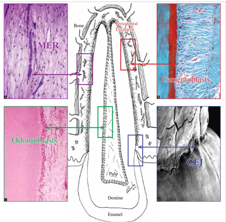
Figure 1 - The four tooth protective structures against resorption: Malassez epithelial rests, (MER), cementoblast cells, odontoblast cells and cementoenamel junc-tion (CEJ). Resorpjunc-tion process is triggered by destrucjunc-tion or local exposure of these structures (A =H.E., 160X; B = T. Mallory, 160X; C = H.E., 40X; D = MEV, 100X).
MER
Cementoblasts
Odontoblasts
Periodontal Ligament
Bone
Pulp
Dentine
Enamel
CEJ
Using this classiication in each clinical case allows a direct and precise diagnosis, immediately linked with its cause, leading to an automatic reasoning of the likely treatment plan with an accurate prognosis.
Root resorptions are grouped as follows:
1. Root resorption by cementoblast cell death with maintenance of Malassez epithelial rests
» Inlammatory root resorptions during orthodon-tic movement.
» Apical Inlammatory root resorption in chronic
periapical lesions.
» Inlammatory root resorption by mild and /or contaminated trauma.
» Inlammatory root resorption by occlusal trauma.
2. Root resorptions by cementoblast and Malassez epi-thelial rests death
» Resorption by replacement in dental trauma. » Resorption by replacement in periodontal ligament
atrophy of unerupted teeth – especially canines.
A
C
B
Orthodonticinsight Consolaro A
© 2013 Dental Press Journal of Orthodontics 9 Dental Press J Orthod. 2013 May-June;18(3):7-9 3. Root Resorption by odontoblast cell death with
maintenance of pulp vitality
» Internal Inlammatory root resorption by dental trauma.
4. Root resorption by direct exposure of dentin to the gingival connective tissue at the cementoenamel junction gaps
» External cervical inlammatory resorption by ac-cidental trauma, especially concussion.
» External cervical inlammatory resorption by trans-operative dental trauma as in impacted ca-nine traction and during intubation in general an-esthetic procedures.
» External cervical inlammatory resorption in as-sociation with internal whitening procedures.
FINAL CONSIDERATIONS
The application of the proposed classification for dental resorption to every clinical case will help the development of a direct diagnosis promptly linked with its cause. This will lead to a treatment plan with a precise prognosis.
1. Consolaro A. Reabsorções dentárias nas especialidades clínicas. 3rd ed. Maringá: Dental Press; 2012.
REFERENCES
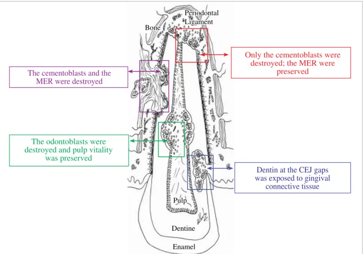
Figure 2 - Destruction or local exposure of the four protective structures of the tooth are the initial phenomena of the four diferent types of resorption pro-cesses leading directly to its cause, treatment plan and prognosis in each clinical case, as represented in this igure.
The cementoblasts and the MER were destroyed
Only the cementoblasts were destroyed; the MER were
preserved
The odontoblasts were destroyed and pulp vitality
was preserved
Periodontal Ligament Bone
Pulp
Dentine
Enamel
Dentin at the CEJ gaps was exposed to gingival