UNIVERSIDADE DE LISBOA
FACULDADE DE MEDICINA
Lancefield group C and G streptococci in human infection:
Molecular typing, virulence and antimicrobial susceptibility
MARCOS DANIEL CAETANO BORGES DE PINHO
DOUTORAMENTO EM CIÊNCIAS E TECNOLOGIAS DA SAÚDE
ESPECIALIDADE MICROBIOLOGIA
UNIVERSIDADE DE LISBOA
FACULDADE DE MEDICINA
Lancefield group C and G streptococci in human infection:
Molecular typing, virulence and antimicrobial susceptibility
MARCOS DANIEL CAETANO BORGES DE PINHO
Tese orientada pelo
PROFESSOR DOUTOR MÁRIO NUNO RAMOS D’ ALMEIDA RAMIREZ
DOUTORAMENTO EM CIÊNCIAS E TECNOLOGIAS DA SAÚDE
ESPECIALIDADE MICROBIOLOGIA
Todas as afirmações efectuadas no presente documento são da exclusiva responsabilidade do seu autor, não cabendo qualquer responsabilidade à Faculdade de Medicina da Universidade de Lisboa pelos conteúdos nele apresentados.
A impressão desta dissertação foi aprovada pelo Conselho
Científico da Faculdade de Medicina de Lisboa em reunião de
18 de Fevereiro de 2014.
My first acknowledgment is to Professor Mário Ramirez, the supervisor of this thesis, for all the guidance provided and the enthusiasm he showed throughout the development of this work. I am also deeply grateful to Professor José Melo Cristino, head of Instituto de Microbiologia, Faculdade de Medicina de Lisboa, for giving me the opportunity to be part of this work team and for the active role he had in all the work included in this thesis. I also wish to thank both for all the knowledge they shared with me throughout the years, and for all the unconditional support which was far beyond the professional issues.
My acknowledgment to Prof. David McMillan, from the Queensland Institute of Medical Research and Griffith Medical Research College, Australia, Prof. Debra Bessen, from the New York Medical College, United States of America, Prof. Constança Pomba, from the Universidade de Lisboa, Portugal, Prof. Silvia Preziuso, from the University of Camerino, Italy, Prof. Antina Lübke Becker and Prof. Lothar Heinz Wieler, from the Freie Universität Berlin, for their collaboration and invaluable contribution to this work, which allowed the purposes of this thesis to be broadened.
I also wish to thank all my colleagues at Instituto de Microbiologia and Unidade de Microbiologia Molecular e Infecção for the nice working environment. A special thanks to Sandra Matos for her help in characterizing some of the bacterial isolates studied. I am also grateful to Ana Friães, Catarina Costa, Sandra Aguiar and João Carriço for their friendship and encouragement.
I thank to my closest family and friends for their care and constant motivation. I express all my gratitude to my parents, whom support and love through all phases of my life have made possible to reach this point. To my sister and brother, Mónica and Miguel, to Bruno, to my grandparents, to Tetyana, Leonid and Olga, thank you for all the support and patience.
Keywords: S. dysgalactiae subsp. equisimilis, S. canis, population, recombination.
Beta-hemolytic, large-colony-forming (diameter greater than 0.5 mm) Lancefield group C and group G streptococci (GCGS) is a group within the Streptococcus genus which includes several species recognized as either colonizers or pathogens in humans and animals. Streptococcus dysgalactiae subsp. equisimilis (SDE), which can express any of these two Lancefield group antigens, is the GCGS species most commonly reported in human infection worldwide and may cause a number of potentially life-threatening infections. SDE is increasingly regarded as an emerging global pathogen and is able to colonize and infect humans, while other GCGS species, such as
Streptococcus canis, are mainly animal pathogens that occasionally infect the human
host. The rising number of human infections reported to be caused by GCGS warrants a better study of their epidemiology, in order to establish the relevance of each species, and clarifying the clonal dynamics and the intra-specific factors influencing the virulence of the strains.
This work aimed at determining the GCGS species responsible for human infection in Portugal and, by assessing the genetic diversity of the isolates recovered, to define the clonal structure of this population. Thus, a special emphasis on molecular typing techniques was given, including those more commonly used to type these streptococci: pulsed-field gel electrophoresis (PFGE), emm typing and multilocus sequence typing (MLST). The application of these techniques to characterize collections of SDE and S.
canis isolates recovered from other geographic locations and, for the latter species, from
animal hosts, allowed to elucidate their respective population structures and provided new insights into the biology and evolution of GCGS.
Initially, the speciation and characterization of GCGS isolates recovered from human infections in Portugal identified the central role of SDE. Not only was there a weak correlation between emm typing and PFGE results, but the polyclonal origin of the SDE population in this region was revealed by each of the methods used that generated multiple partitions. Furthermore, a correlation between emm type and invasive disease potential was suggested.
A more global snapshot of the clonal architecture of this pathogen population was achieved by developing a MLST scheme which was applied to an expanded collection of SDE isolates recovered from distinct continents. An association of Lancefield groups with distinct MLST partitions was found and the high prevalence of a small number of widely distributed MLST sequence types (STs) suggested that a few genetic lineages dominate among SDE causing human infection worldwide. The occurrence of intra-specific and inter-intra-specific recombination with Streptococcus pyogenes (Lancefield group A streptococcus, [GAS]), involving the housekeeping genes used in MLST was detected.
The poor correlation between emm typing and either PFGE or MLST defined groups was illustrated by lineages displaying distinct emm types and the presence of the same
emm type in unrelated genetic backgrounds, as defined by both techniques. These
observations suggested the existence of recombinational replacements involving the
emm locus and question the value of emm typing to accurately ascertain the genetic
relatedness of SDE strains.
The characterization of the antimicrobial susceptibility patterns presented by SDE found a high proportion of levofloxacin-resistant isolates (12%) associated with multiple genetic lineages. Sequence analysis of the quinolone resistance-determining regions of the gyrA and parC genes of representative resistant and susceptible isolates showed that full resistance was associated with mutations in both GyrA and ParC. As observed for the housekeeping genes used in MLST, recombination with GAS DNA in some parC alleles was evident, though this phenomenon was not exclusively associated with resistance.
The final part of the work in this thesis focused on S. canis, the second most frequent GCGS species isolated from human infections in Portugal. A collection of S. canis isolates recovered from infections in both humans and animals, collected in Portugal and abroad, were characterized by employing the same typing methods used for SDE. The S. canis population was polyclonal, and several genetic lineages were shown to possess the ability to infect the human host. The zoonotic nature of S. canis infection was demonstrated, as identical genetic lineages were found infecting house pets and humans, indicating that they constitute a single population. Phylogenetic analysis showed that S. canis was a divergent taxon of SDE and GAS and unveiled the acquisition of genetic material of SDE by S. canis. The presence of emm-like genes was
restricted to a few S. canis isolates and correlated with some MLST-based genetic lineages.
Globally, this thesis contributes to the current knowledge of the molecular epidemiology and evolutionary relationships among members of the two GCGS species studied. The clonal relationships among strains were elucidated and MLST schemes for SDE and S. canis were established, providing useful tools for future studies of their population dynamics. The use of emm typing was shown to be complemented by applying other typing methods and the role of the M protein in SDE virulence was reinforced. The evidence found for recombinational replacements between SDE and GAS in several loci and, to a smaller extent, between SDE and S. canis, indicates that horizontal gene transfer events are important mechanisms driving genetic variability in GCGS populations which may impact key bacterial functions such as virulence and antimicrobial resistance.
Palavras-chave: S. dysgalactiae subsp. equisimilis, S. canis, população,
recombinação.
Os estreptococos beta-hemolíticos formadores de colónias grandes (diâmetro superior a 0.5 mm) dos grupos C e G de Lancefield (GCGS) formam um grupo dentro do género Streptococcus que inclui várias espécies reconhecidas como colonizadoras ou agentes patogénicos no Homem e em animais. Streptococcus dysgalactiae subsp.
equisimilis (SDE), que pode integrar cada um destes dois grupos de Lancefield, é a
espécie de GCGS mais vezes reportada em infecção humana a nível mundial e pode causar diversas infecções potencialmente fatais. SDE é cada vez mais visto como um agente patogénico emergente, com a capacidade de colonizar e infectar o Homem, enquanto as outras espécies de GCGS, tais como Streptococcus canis, são principalmente agentes patogénicos de animais que ocasionalmente infectam o hospedeiro humano. O número crescente de infecções humanas atribuídas aos GCGS exige um estudo reforçado da sua epidemiologia, de modo a estabelecer a relevância de cada espécie e permitindo que a dinâmica clonal e os factores intra-específicos que influenciam a virulência das estirpes sejam clarificados.
Este trabalho teve como objectivo identificar as espécies de GCGS responsáveis por infecção humana em Portugal e, através da avaliação da diversidade genética das estirpes recolhidas, definir a estrutura clonal da população. Deste modo, foi dado especial ênfase às técnicas de tipagem molecular, incluindo aquelas que mais habitualmente são utilizadas para a tipagem destes estreptococos: a electroforese em campo pulsado (PFGE), a tipagem emm e “multilocus sequence typing” (MLST). A aplicação destas técnicas para caracterizar colecções de estirpes de SDE e S. canis isoladas noutras regiões geográficas e, no caso da última espécie, de hospedeiros animais, permitiu elucidar as respectivas estruturas populacionais e percepcionar novos aspectos da biologia e evolução dos GCGS.
Inicialmente, a especiação e caracterização de estirpes de GCGS isoladas de infecções humanas em Portugal mostrou o papel central de SDE. Verificou-se uma fraca correlação entre os resultados obtidos pela tipagem emm e PFGE e a aplicação destas técnicas revelou a origem policlonal da população de SDE nesta região, dado que cada
um dos métodos utilizados gerou múltiplas divisões. Adicionalmente, os resultados sugeriram a existência de uma correlação entre o tipo emm e o potencial de doença invasiva.
Uma imagem mais geral da estrutura apresentada pela população deste agente patogénico foi alcançada através do desenvolvimento de um esquema de MLST, aplicado a um conjunto alargado de estirpes de SDE isoladas em continentes distintos. Esta análise permitiu estabelecer uma associação entre grupos de Lancefield e divisões distintas de MLST. Foi possível observar uma elevada prevalência de um pequeno número de “sequence types” (STs) na população. A ampla distribuição geográfica destes STs sugeriu o predomínio de algumas linhagens genéticas entre as estirpes de SDE que causam infecção humana em todo o mundo. Foi detectada ainda a ocorrência de recombinação intra-específica e inter-específica com Streptococcus pyogenes (estreptococos do grupo A de Lancefield, [GAS]), envolvendo genes “housekeeping” utilizados no MLST.
A fraca correlação entre tipos emm e os grupos definidos por PFGE ou por MLST foi ilustrada pela existência de linhagens com tipos emm distintos e pela presença do mesmo tipo emm em patrimónios genéticos não relacionados, tal como definido pelas duas técnicas de tipagem. Estas observações sugeriram a existência de substituições recombinacionais envolvendo este locus e questionam o valor da tipagem emm para determinar correctamente a relação genética de estirpes de SDE.
A caracterização dos perfis de susceptibilidade aos antimicrobianos apresentados por SDE permitiu detectar uma elevada percentagem de estirpes resistentes à levofloxacina (12%), associada a múltiplas linhagens genéticas. A análise das sequências das regiões determinantes da resistência às quinolonas nos genes gyrA e parC de estirpes resistentes e susceptíveis representativas, mostrou que a aquisição de resistência se encontra associada a alterações dos aminoácidos das proteínas codificadas pelos dois genes. A existência de recombinação com ADN de GAS foi evidente em alguns alelos do gene
parC, como observado para os genes “housekeeping” utilizados no MLST. No entanto,
este acontecimento não foi associado exclusivamente com o desenvolvimento de resistência.
A parte final do trabalho descrito nesta tese focou-se na segunda espécie de GCGS mais frequentemente isolada de infecções humanas em Portugal, S. canis. Uma colecção constituída por estirpes de S. canis isoladas de infecções em seres humanos e animais,
recolhidas em Portugal e no estrangeiro, foi caracterizada recorrendo aos mesmos métodos de tipagem utilizados para SDE. A policlonalidade da população de S. canis foi evidenciada e várias linhagens genéticas mostraram ter a capacidade de infectar o hospedeiro humano. A natureza zoonótica da infecção por S. canis foi demonstrada pois as mesmas linhagens genéticas foram encontradas a infectar animais domésticos e seres humanos, indicando a existência de uma única população. A análise filogenética mostrou que S. canis é um taxon divergente das espécies SDE e GAS e revelou a aquisição de material genético de SDE por S. canis. A presença de genes similares ao gene emm restringiu-se a uma pequena proporção das estirpes de S. canis e correlacionou-se com algumas das linhagens genéticas definidas por MLST.
Globalmente, este trabalho contribuiu para o conhecimento actual da epidemiologia molecular e das relações evolutivas entre os membros das duas espécies de GCGS estudadas. As relações clonais entre as estirpes foram elucidadas e esquemas de MLST foram estabelecidos para SDE e S. canis, constituindo ferramentas úteis para futuros estudos sobre a dinâmica populacional destas espécies. Mostrou-se que a utilização da tipagem emm é complementada pela aplicação de outros métodos de tipagem e a importância da proteína M na virulência de SDE foi reforçada. As indicações encontradas da ocorrência de trocas recombinacionais entre SDE e GAS em vários loci e, em menor extensão, entre SDE e S. canis, indica que os eventos de transferência genética horizontal são mecanismos motrizes da variabilidade genética em populações de GCGS, podendo afectar aspectos fulcrais da biologia destas espécies, como a virulência e a resistência aos antimicrobianos.
The main purpose of the current work was to provide insights into the molecular epidemiology of GCGS causing human infections.
Chapter 1 is a general introduction describing the main aspects of GCGS biology. As the current thesis focuses on human infections by these bacteria, a special emphasis is given to SDE, the species most commonly found and the subject of most of the laboratory work described in this thesis. A few aspects regarding the biology of other GCGS species are also studied, especially those which have also been isolated from the human host. A more detailed description of the typing methodologies used is also found under this section. This chapter concludes by stating the aims of the current work.
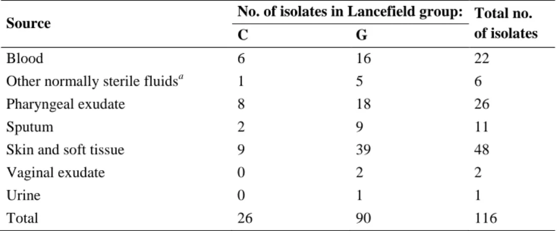
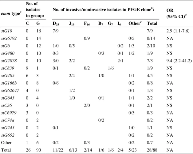
Chapter 2 describes the epidemiology of GCGS recovered from invasive and non-invasive infections in Portugal, and involves the characterization of 116 isolates recovered from 1998 to 2004 in hospital associated laboratories across the country. It starts by identifying the species involved and, since only SDE is detected, it proceeds with a detailed characterization by emm typing and PFGE of the isolates of this species. Also, the association of specific emm types and PFGE defined clones with invasive disease potential is statistically evaluated. The epidemiologic characterization of SDE presented in this chapter is supplemented by the data described in chapters 3 and 4.
Chapter 3 centers on the development of an MLST scheme for the characterization of SDE. In collaboration with groups from Australia and the United States, an epidemiological and genetically diverse collection of isolates was obtained, including 36 isolates recovered in Portugal and representative of several emm types encountered in the work reported in chapter 2. The application of the MLST technique allowed a global view of the SDE population causing infections in humans and to estimate the occurrence of recombination involving the housekeeping genes used in the MLST scheme.
Chapter 4 describes the antimicrobial resistance patterns observed among an expanded collection of SDE human isolates recovered in Portugal (n = 314, recovered from 1998 to 2005), with a special focus on levofloxacin resistance. Prompted by the high resistance rate found and by previous suggestions that members of the S.
dysgalactiae taxon could mediate the appearance of resistance in GAS through
in SDE were investigated and phylogenetic analyses were conducted with the sequence data generated from the resistance-determining regions of the relevant genes.
Chapter 5 focuses on a distinct Lancefield group G species, S. canis, which was found as an infrequent agent of human infection in the last years of the study. This chapter reports the molecular characterization of 85 S. canis isolates employing the same typing techniques used for SDE, as described in the previous chapters. Special emphasis was given to the development of a universally applicable MLST scheme for S.
canis. Given the low number of human infections caused by this species, the collection
was expanded by adding isolates recovered from animal sources, mainly house pets, which allowed the characterization of the S. canis population and provided reinforced evidence of the zoonotic nature of human infection.
Chapter 6 is a general discussion which aims to summarize and articulate the main findings of the thesis. The contribution of this work is contextualized in the current issues on GCGS biology and the working questions that can be pursued in the future are highlighted.
Chapters 2 to 5 can be read independently and are reproductions of the publications listed in the next page. Only minor alterations were made relative to the published version, with the purpose of standardizing text with the other contents of the thesis.
PUBLICATIONS INCLUDED IN THIS THESIS
Chapter 2:
Pinho MD, Melo-Cristino J, Ramirez M. 2006. Clonal relationships between invasive
and noninvasive Lancefield group C and G streptococci and emm-specific differences in invasiveness. J. Clin. Microbiol. 44:841-846.
Chapter 3:
McMillan DJ, Bessen DE, Pinho M, Ford C, Hall GS, Melo-Cristino J, Ramirez M.
2010. Population genetics of Streptococcus dysgalactiae subspecies equisimilis reveals widely dispersed clones and extensive recombination. PLoS One 23;5:e11741.
Chapter 4:
Pinho MD, Melo-Cristino J, Ramirez M. 2010. Fluoroquinolone resistance in Streptococcus dysgalactiae subsp. equisimilis and evidence for a shared global gene
pool with Streptococcus pyogenes. Antimicrob. Agents Chemother. 54:1769-1777.
Chapter 5:
Pinho MD, Matos SC, Pomba C, Lübke-Becker A, Wieler LH, Preziuso S, Melo-Cristino J, Ramirez M. 2013. Multilocus sequence analysis of Streptococcus canis
confirms the zoonotic origin of human infections and reveals genetic exchange with
AW – Adjusted Wallace coefficient CC – clonal complex1
CDC – Centers for Disease Control and Prevention CI – confidence interval
CLSI – Clinical and Laboratory Standards Institute DLV – double-locus-variant1
FCT – fibronectin-binding, collagen-binding and T antigen FQ – fluoroquinolone
GAS – Lancefield group A streptococcus, Streptococcus pyogenes GBS – Lancefield group B streptococcus, Streptococcus agalactiae
GCGS – beta-hemolytic, large-colony-forming (diameter, >0.5 mm) Lancefield group C and group G streptococci2
GCS – Lancefield group C streptococci GGS – Lancefield group G streptococci MIC – minimum inhibitory concentration
MLSB – macrolide, lincosamide and streptogramin B MLST – multilocus sequence typing
NJ – Neighbor joining OR – odds ratio
PFGE – pulsed-field gel electrophoresis
QRDR – quinolone resistance-determining region RDP – Recombination Detection Program
SDE – Streptococcus dysgalactiae subsp. equisimilis SID - Simpson Index of Diversity
SLV – single-locus-variant1 ST – sequence type1
1
These terms are always used in the context of the multilocus sequence typing technique.
2
The designation GCGS used in the current work refers exclusively to large-colony-forming species. Some authors use the GCGS acronym in a broader sense which also includes the small-colony-forming (diameter, <0.5 mm) species belonging to the “Streptococcus milleri” or “Streptococcus anginosus” group. Although some beta-hemolytic “S. milleri” isolates can be found in the same ecologic niches of GCGS and may bear the Lancefield group C and G antigens, they constitute a genetically distinct group, differing from GCGS in the spectrum of clinical manifestations caused, in their pathogenicity and affecting different patient populations.
STSS – streptococcal toxic shock syndrome TLV – triple-locus-variant1
UPGMA – unweighted-pair group method with arithmetic means W – Wallace coefficient
ACKNOWLEDGMENTS ... i SUMMARY ... iii RESUMO ... vii THESIS OUTLINE ... xi ABBREVIATIONS ... xv TABLE OF CONTENTS ... xvii
CHAPTER 1
General Introduction ... 1
INTRODUCTORY NOTE ... 3 1. CLASSIFICATION OF GCGS ... 6 1.1. Morphologic and general characteristics ... 6 1.2. Species delineation and host distribution ... 8 1.2.1. Streptococcus dysgalactiae subsp. equisimilis (SDE) ... 10
1.2.2. Streptococcus canis ... 12
1.2.3. Other GCGS species ... 13 1.3. Laboratory identification ... 15 2. GCGS ECOLOGY AND EPIDEMIOLOGY IN THE HUMAN HOST .
... 18 2.1. Ecological niches and origin of infection... 18 2.2. Infections ... 20 2.3. Clinical and epidemiologic features of GCGS infections ... 23 2.4. Virulence factors ... 24 2.4.1. M and M-like proteins ... 26 2.4.2. Superantigens ... 28
2.4.3. Other factors ... 30 3. ANTIMICROBIAL SUSCEPTIBILITY AND TREATMENT ... 32 4. TYPING METHODOLOGIES ... 35 4.1. Phenotypic methods ... 35 4.2. Genotypic methods ... 36 4.2.1. Pulsed-field gel electrophoresis (PFGE) ... 36 4.2.2. emm typing ... 38
4.2.3. Multilocus sequence typing (MLST) ... 39 4.2.4. Whole-genome sequencing ... 40 5. REFERENCES ... 42 AIMS OF THE THESIS ... 59
CHAPTER 2
Clonal relationships between invasive and noninvasive Lancefield group C and G streptococci and emm-specific differences in invasiveness ... 61
CHAPTER 3
Population genetics of Streptococcus dysgalactiae subsp. equisimilis reveals widely dispersed clones and extensive recombination ... 81
CHAPTER 4
Fluoroquinolone resistance in Streptococcus dysgalactiae subsp. equisimilis and evidence for a shared global gene pool with Streptococcus pyogenes ... 117
CHAPTER 5
Multilocus sequence analysis of Streptococcus canis confirms the zoonotic origin of human infections and reveals genetic exchange with Streptococcus dysgalactiae subsp. equisimilis ... 147
CHAPTER 6
General Discussion ... 187
6. GENERAL DISCUSSION ... 189 6.1. Issues relating to the identification of GCGS species ... 190 6.2. The emm gene: epidemiologic and virulence considerations ... 191 6.3. Improving GCGS typing by applying MLST ... 194 6.4. The relevance of horizontal gene transfer events in GCGS evolution .
... 199 7. Concluding remarks and future perspectives ... 202 8. REFERENCES ... 207
CHAPTER 1
INTRODUCTORY NOTE
Beta-hemolytic, large-colony-forming (diameter greater than 0.5 mm) Lancefield group C and group G streptococci (GCGS) is a group within the Streptococcus genus including several bacterial species which colonize and/or infect humans and animals. Since the original description of streptococci bearing these Lancefield groups by Rebecca Lancefield in the 1930’s, who found them infecting animals (1) and colonizing the vagina of parturient women (2), GCGS were thought to be essentially animal pathogens showing low pathogenicity towards the human host, in which they were present as part of the commensal microbiota (3). Moreover, the low numbers of human infections reported for GCGS, particularly when compared to other beta-hemolytic pathogens, such as Streptococcus pyogenes (Lancefield group A streptococcus [GAS]) or Streptococcus agalactiae (Lancefield group B streptococcus [GBS]), further strengthened that view and contributed to the fact that GCGS infections in humans would be overlooked in subsequent years.
A trend of increasing detection of GCGS in human infections has been noted from the late 1970’s and 80's onwards in various locations around the world (3). Many of the reports published since then have described invasive infections by GCGS (4–7) and these microorganisms were implicated in triggering some of the most severe clinical syndromes caused by beta-hemolytic streptococci (8, 9). Occasionally, increased incidence of GCGS infection has been reported (10–12) and a few studies showed that the disease burden attributable to these bacteria could be similar or even higher than that of GAS or GBS (11–13). The characterization of the genome of GCGS undertaken in the last five years (14–16) confirmed their genetic similarity to other beta-hemolytic streptococci and further showed that these bacteria have a wide array of genes encoding factors that could mediate their virulence. Taken together, these observations led to the recognition of the pathogenic potential of these microorganisms towards humans, and highlighted the need for a better characterization of the GCGS strains causing infection in the human host.
The speciation of GCGS was frequently neglected over the years. Although it was long recognized that the term GCGS included several groups that could be potentially distinguished by their phenotypic traits and host distribution (3), many of the available studies in the literature did not report isolate characteristics other than the Lancefield group. Nowadays, it is recognized that the distinct taxonomic entities included in the
group differ in their significance as human pathogens. A single taxon, Streptococcus
dysgalactiae subsp. equisimilis (SDE), is found established in the human host, being the
only GCGS for which colonization is currently recognized and the most frequently detected in infection in many parts of the world (17–19). Therefore, many authors refer to GCGS isolated from human specimens as SDE (20). The spectrum of infections caused by SDE is similar to that caused by GAS and both bacteria share many proteins that were shown to be virulence factors (6, 15, 20). However, a few other GCGS species may also be found in human infections, as is the case of Streptococcus equi subsp.
zooepidemicus (5) and Streptococcus canis (21). These are primarily found as animal
pathogens and they have been isolated from human infections less frequently than SDE. The current work focuses on the importance of GCGS for human infections and the literature review carried out in this chapter highlights the aspects of the biology of GCGS relevant for the pathogenesis in the human host. Thus, emphasis is given to SDE and to studies which unambiguously identify the isolates included. The comparison between SDE and the other GCGS species is made whenever there are differences that justify it. Particular attention is also given to S. canis, as it was the second most frequent GCGS species isolated in Portugal and part of the work is dedicated to it (Chapter 5). Many studies have focused on the pathogenesis of GCGS in animals, mainly regarding members of the S. equi taxon. Such contributions are not discussed since they are beyond the scope of this thesis, but may be referred to in relevant points.
A few issues should also be noted on the possible limitations of the available GCGS literature. In addition to the studies indicating solely the Lancefield group of the isolates, many authors characterize Lancefield group C and group G strains independently and studies that focus on either one or the other are available. In the context of the human host, it is now clear that most GCGS are SDE and, for this species, there is no evidence supporting a separation between strains bearing Lancefield group C and G antigens based on their clinical relevance. Thus, reports focusing solely on group C or group G SDE strains are included and no separate analysis is made. Finally, in a significant number of studies it is unclear whether the authors discriminate between GCGS and the small-colony-forming “Streptococcus milleri” strains, which can be relevant in particular ecologic niches where the two groups coexist (as is the case of the human oropharynx). The aspects mentioned above limit the value of some studies, since the relative importance of each GCGS species cannot be accurately
assessed, and these aspects were taken into consideration when conducting the literature review.
1. CLASSIFICATION OF GCGS
1.1. Morphologic and general characteristics
As members of the Streptococcus genus, GCGS are facultative anaerobic and catalase negative Gram-positive cocci, with spherical or ovoid cells less than 2 μm in diameter that display chains upon Gram staining (22). The guanine-cytosine content of GCGS species is within the range of 39 to 42%, as evaluated by the currently available genomes (http://www.ncbi.nlm.nih.gov/genome). GCGS are nutritionally fastidious bacteria and their growth in enriched media is enhanced by the addition of blood or serum (22). GCGS have been traditionally classified based on a set of phenotypic characteristics, including the morphology and size of the colonies, the type of hemolysis and the serologic specificity of the cell wall Lancefield group carbohydrate, which determines the classification of streptococcal isolates in this group.
The use of the hemolytic activity to classify streptococci started in the beginning of the twentieth century and is credited to Schottmüller (23). Almost all GCGS strains are beta-hemolytic, i.e. the colonies formed after growth in solid medium containing blood are surrounded by a zone of complete erythrocyte lysis due to the action of an exotoxin (24) (Fig. 1.1). Thus, GCGS are members of the beta-hemolytic or pyogenic group (23), which includes major human pathogens such as GAS and GBS to which GCGS are genetically related (25, 26). Some GCGS strains present variable hemolytic activity, since they can be alpha-hemolytic (partial hemolysis, due to reduction of the hemoglobin in erythrocytes which originates a green colored halo) or gamma-hemolytic (absence of hemolysis) (Fig. 1.1). Such features are described for members of the taxon
Streptococcus dysgalactiae (27, 28) but are not exclusive of GCGS as occasional
non-hemolytic isolates from other beta-non-hemolytic streptococci have been reported (29). Most beta-hemolytic streptococci can be further classified serologically into distinct Lancefield groups, according to the precipitin reaction scheme proposed by Rebecca Lancefield in 1933 (1). Although Hitchcock was the first to describe the presence of a soluble specific substance on the cell wall of these streptococci (30), only after the work of Rebecca Lancefield was the polysaccharidic and group-specific nature of this substance unveiled, which was named carbohydrate C (1). The major constituents of carbohydrate C were identified as rhamnose and one amino sugar (31). Changes in content of this last component originate differences in the antigenic specificity between
bacteria of the various Lancefield groups, which is the basis of this serological technique (31).
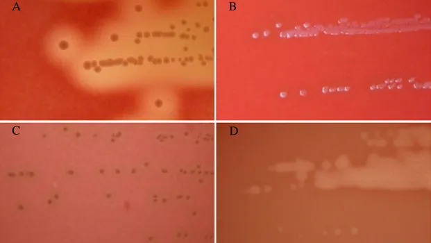
FIGURE 1.1. Colony morphology and hemolytic reaction of
Lancefield group C and G bearing strains
Colonies formed after growth for 24 hours at 37ºC in tryptic soy agar supplemented with 5% (vol/vol) defibrinated sheep blood. (A) Large-colony-forming (>0.5mm) SDE isolate displaying beta-hemolysis; (B) Large-colony-forming gamma-hemolytic (no hemolysis) Streptococcus dysgalactiae subsp. dysgalactiae isolate; (C) Large-colony-forming alfa-hemolytic S. dysgalactiae subsp. dysgalactiae isolate; (D) Small-colony-forming (<0.5mm) “S. milleri” isolate displaying beta-hemolysis.
The Lancefield grouping scheme was a critical contribution to the classification of beta-hemolytic streptococci and has been widely used over the years (23, 25). Although this technique has been particularly useful in identifying both GAS and GBS in the clinical microbiology laboratory, its utilization has some drawbacks which negatively influence the classification of GCGS. It is now recognized that GCGS strains related at the species or subspecies level may have different Lancefield antigens. The most notorious example is found in SDE for which isolates bearing Lancefield group G, C, L and A antigens are known (32, 33). Moreover, distinct species may bear the Lancefield group C or G antigens (25), including certain members of the unrelated “S. milleri” group, which are distinguishable from GCGS by the formation of minute colonies after growth on solid media for 24 hours at 35 to 37ºC (22) (Fig. 1.1).
Contemporaneously with the Lancefield grouping scheme development, it was noted that the serologically defined groups could also be divided by means of physiological
A B
tests (23). The attempted sub-grouping of GCGS into individual species was subsequently based on their serological properties, biochemical testing results and host distribution (3). The GCGS classification obtained by combining these data changed over the years and distinct epithets were used to name the defined groups. More recently, the application of genotypic methods allowed the taxonomy of the group to be clarified (25).
1.2. Species delineation and host distribution
GCGS is a phenotypic and genotypically diverse group comprising several taxonomic entities which have evolved in different hosts. In some situations, this has lead to a niche separation which correlates with the taxonomic divisions accepted at the present time. Seven distinct taxa, including four species, are currently defined (Table 1.1). Further division into subspecies resulted from the genotypic analysis which justified their inclusion under the same epithet (27, 34). GCGS speciation is important because the individual taxa differ in their pathogenic characteristics, may cause distinct infections in the same host, present different infection transmission routes and their prevalence in human infection differs markedly.
Table 1.1 shows the current taxonomic status of GCGS and summarizes key aspects such as host distribution, ecologic behaviour and spectrum of infections caused by the individual taxa. While some GCGS behave as host restricted pathogens, others can be regarded as generalists having the ability to infect a wide range of distinct animal species, including humans. The occurrence in human infection is well established for SDE, S. equi subsp. zooepidemicus and S. canis (25), though their prevalence is quite different. These three taxa display a broad host range and can be found as either colonizers and/or pathogens of many domestic and wild animals (33–35). SDE is by far the most frequent in human infection (17–19) and the only one for which human colonization is recognized (25). Another contrasting feature of SDE is that isolates recovered from human and non-human sources can be distinguished by both phenotypic and genotypic tests (27, 33), a separation not observed for the other two species. S. equi subsp. zooepidemicus and S. canis are mainly animal pathogens but their occurrence in human infection is well documented (5, 36). For the other GCGS listed in Table 1.1, infection of the human host is negligible, as very few confirmed cases of human infection have been published (37–40), the description of poorly characterized isolates
TABLE 1.1. Main characteristics of GCGS species
Speciesa Lancefield
group Host
b Recognized role as
Colonizer/Pathogenc
Commonly reported infections in human/animal hostd
S. dysgalactiae subsp. equisimilis A, C, G, L Human Both SSTI, bacteremia, pharyngitis, STSS
Animal (various) Both Pneumonia, septicemia, arthritis, uterine infections
S. dysgalactiae subsp. dysgalactiae C Animal (cow, fish) Pathogen Bovine mastitis, fish septicemia
S. canis G Animal (various) Both Dermatitis, otitis externa, pneumonia, infective
endocarditis, bacteremia
Human Pathogen Bacteremia, SSTI
S. equi subsp. zooepidemicus C Animal (various) Both Septicemia, pneumonia, arthritis, uterine infections
Human Pathogen Bacteremia, meningitis, nephritis, arthritis, STSS
S. equi subsp. equi C Animal (horse) Pathogen Equine strangles
S. equi subsp. ruminatorum C Animal (various) Pathogen Ovine/caprine mastitis, strangle-like disease in hyenas
Human Pathogen Bacteremia
S. phocae C, F, G, none Animal (seals, fish) Pathogen Systemic infections
a
The taxon S. dysgalactiae subsp. dysgalactiae is not beta-hemolytic (presents gamma or alpha hemolysis). The inclusion in GCGS is justified by the genetic proximity with other species of the group.
b Isolates from a given species are divided in lanes human/animal to differentiate their role in colonization and infection according to host. For species other than SDE, this
separation does not imply that they actually represent distinct populations.
c Refers to the main role. Classification as “pathogen” indicates that role as colonizer or reservoir has not been clearly identified or is restricted to a specific ecological niche. d
questions their true identity or, as is the case of Streptococcus phocae, have not yet been reported.
1.2.1. Streptococcus dysgalactiae subsp. equisimilis (SDE)
The GCGS strains currently included in the S. dysgalactiae taxon were long divided into groups according to their host distribution and phenotypic characteristics (27, 33). At least five groups were recognized over the years, which have been reclassified under the S. dysgalactiae epithet following the genetic evidence for the inclusion in a single species of the non-hemolytic group C S. dysgalactiae strains of bovine origin and the beta-hemolytic strains of Lancefield groups C (S. equisimilis), G and L (34) (Table 1.2).
TABLE 1.2. Subspecies division of S. dysgalactiae strains
Former S. dysgalactiae groupsa
Subspecies proposalb Vandamme et al., 1996 (27)
Vieira et al., 1998 (33)c
Bovine non-hemolytic group C (S. dysgalactiae)
S. dysgalactiae subsp. dysgalactiae
S. dysgalactiae subsp. dysgalactiae Animal beta-hemolytic group L strains
S. dysgalactiae subsp. equisimilis Animal beta-hemolytic group C (S. equisimilis)
Human beta-hemolytic group C (S. equisimilis) S. dysgalactiae subsp. equisimilis
Human beta-hemolytic group G
a
Both terms S. dysgalactiae and S. equisimilis were used in the past. Group L strains have been more frequently isolated from animals than from humans and cannot be distinguished from each other, thus being assigned as animal strains.
b
Dashed lines indicate the separation point according with the two distinct subspecies proposals.
c
The subspecies division proposed by Vieira et al. is the most commonly accepted and is the one adopted in this thesis.
The two subspecies currently defined for S. dysgalactiae were initially established in 1996 by Vandamme et al. (27). Based on whole-cell protein analysis and physiological tests, these authors proposed that all strains isolated from humans should constitute the subspecies equisimilis, while S. dysgalactiae subsp. dysgalactiae would accommodate all strains of animal origin, regardless of the hemolytic activity displayed. However, a study conducted two years later by Vieira et al. (33), based on DNA-DNA hybridization and multilocus enzyme electrophoresis data, showed that the alpha-hemolytic group C strains of bovine origin were a group distinct of all the others and restricted the S.
dysgalactiae subsp. dysgalactiae taxon to these strains. Thus, these authors proposed
segregation by hemolytic reaction but not by host became the most accepted (25). However, the disagreement in the two original studies on the taxonomic position of the animal beta-hemolytic strains and some inconsistencies observed in the 16S rRNA analysis of those strains (41), has led different authors to classify them in distinct manner and at the present time this classification is still a matter of debate (42).
In view of the current taxonomic status, SDE still encompasses a phenotypically diverse set of strains, which may be differentiated by their Lancefield group carbohydrates, biochemical characteristics and by host specificity. Most SDE isolates recovered from human specimens present the Lancefield group G antigen, followed by group C, while Lancefield’s groups L and A are seldom found (17, 43). The occurrence of this last group carbohydrate in SDE strains is of particular significance given their potential erroneous identification as GAS. The first SDE strain bearing the Lancefield group A antigen was identified in 1997 (32), and group A SDE have been isolated and characterized in a few other studies (43, 44). A study from Japan pointed to the dissemination of a single clone in the country (44), a likely indication that this antigen may be restricted to a single or only a few SDE genetic lineages.
SDE is also found as a colonizer and pathogen in a wide variety of animal hosts, including house pets (such as dogs and cats), domesticated cattle (pigs, sheep, cows, horses, chicken) and as an opportunistic pathogen in many other wild species (27, 41, 45). SDE strains isolated from animals are predominantly of Lancefield groups C and L. While group C strains were shown to possess unique phenotypic and genotypic characteristics, allowing their separation from human SDE isolates expressing this Lancefield group, group L strains have been reported to be indistinguishable independently of the host they have been isolated from (33, 46).
The other species of the S. dysgalactiae taxon, S. dysgalactiae subsp. dysgalactiae, is the only non-hemolytic species included among GCGS (25). This taxon now includes alpha- or gamma-hemolytic Lancefield group C strains commonly associated with bovine mastitis and also isolated from the vagina of cows (33), a similar description to the one made by Garvie et al. in 1983 for S. dysgalactiae (47). Over the years, the term
S. dysgalactiae was occasionally used to designate Lancefield group C strains isolated
from animals other than cows (47, 48) and some authors described cases of human infection by S. dysgalactiae subsp. dysgalactiae (49, 50). However, such reports do not give an accurate description of the microbiological characteristics of the isolates which
could unambiguously identify them as S. dysgalactiae subsp. dysgalactiae, especially when the taxonomic issues described above are considered. At present there is no data that consistently supports a role for this species as an agent of infection in humans or other animals, and it is arguable that it might behave as a host restricted pathogen of cows.
In recent years, non-hemolytic Lancefield group C S. dysgalactiae strains were identified infecting several species of cultured fish in Japan and other Asian countries (51, 52), and a case of a woman who developed cellulitis and bacteremia after being stung while cleaning raw fish has been reported from Singapure (38). As suggested by the absence of hemolytic activity and the analysis of the manganese-dependent superoxide dismutase (sodA) gene (53), these isolates seem to belong to the S.
dysgalactiae subsp. dysgalactiae taxon. However, they present unique genotypic
features that distinguish them from the S. dysgalactiae subsp. dysgalactiae type strain isolated from bovine mastitis and other S. dysgalactiae strains isolated from mammals (51). More recently, beta-hemolytic isolates were detected in farmed fish from Brazil (54), making the exact phylogenetic position of these strains still unclear at the moment.
1.2.2. Streptococcus canis
The term Streptococcus canis was used in veterinary clinical microbiology long before the formal establishment of the species by Devriese et al. in 1986 (35). According to these authors, S. canis should include the Lancefield group G strains found causing bovine mastitis and distinct infections in dogs which were biochemically distinguishable from human Lancefield group G streptococci (SDE) (35). S. canis is not only part of the microbiota of cats and dogs (55, 56) but also an important pathogen for these two species and other domestic and wild animals (35, 57). S. canis causes from relatively mild non-invasive infections, such as dermatitis and otitis externa, to severe invasive infections (58–60). S. canis may be transmitted between different animal species living in proximity, as shown by its involvement in outbreaks of clinical and subclinical mastitis with bacterial shedding in milk in cattle herds (61) and in pets living in shelters (59).
The first confirmed report of human infection by S. canis was published in 1997 and described a case of septicemia in a 77-year-old man (62). An increasing number of reports has subsequently identified this species mainly from cases of bacteremia (17, 21,
63) and skin and soft tissue infections (36, 64). The presence of S. canis in human infection is thought to have a zoonotic origin, and in most of the reported cases direct contact with colonized or infected animals was proposed (62, 64). Many studies failed to detect S. canis among GCGS causing human infections (11, 12, 65–67), while others found it at low frequency (17, 68). A single study reported increased incidence of this pathogen among GCGS causing both invasive and non-invasive infections in a hospital in France (36), raising the possibility of increased importance of this pathogen in certain geographic regions. In contrast to SDE, little is known of the relationships between S.
canis strains isolated from distinct hosts and of the characteristics of this population.
The publication of the first S. canis whole-genome sequence showed the close evolutionary relationship to both SDE and GAS, bacteria which share with S. canis many of the genetic determinants that are thought to contribute to the colonization of distinct niches, invasion of specific tissues and evasion of the host immune system (14, 26).
1.2.3. Other GCGS species
Lancefield group C strains found in human infection that do not belong to SDE are part of the Streptococcus equi taxon. The name S. equi was first used in the late 19th century (69), to refer to diplococci causing respiratory infection in horses. Currently, it includes three distinct subspecies which are found as animal pathogens: S. equi subsp.
zooepidemicus (34), S. equi subsp. equi and the recently described S. equi subsp. ruminatorum (70). While S. equi subsp. equi is a strict horse pathogen the other two
infect a broader range of hosts which also includes humans.
S. equi subsp. zooepidemicus is a equine commensal which may opportunistically
cause infections, mainly in the respiratory tract (71). Infections caused by this bacterial species in other animal species, both domestic and wild (72–74), are often severe and include meningitis (74) or hemorrhagic pneumonia (75). In the human host, S. equi subsp. zooepidemicus has been isolated from bacteremia and endocarditis (5, 76), meningitis (77), streptococcal toxic shock syndrome (STSS) (78, 79) and nephritis outbreaks (80, 81). This is the second GCGS species for which more cases of human infection have been reported (5, 77, 81).
S. equi subsp. equi is long known to be the etiological agent of equine strangles, a
characterized by the formation of abscesses in the lymph nodes of the head and neck (82). Although some horses can become carriers of the bacteria in their guttural pouch after the acute phase of the disease, the colonization seems to be restricted to this anatomical region and it is not associated with infections other than strangles. Another contrasting feature to S. equi subsp. zooepidemicus is the host range since it has not been found in other animals. Thus, S. equi subsp. equi is regarded as a highly adapted pathogen of the equine respiratory tract and it is not considered an agent of human infection (25), from which it has been very rarely isolated (4, 37). Genetic analysis of S.
equi subsp. equi isolates suggests that this bacterium is a clone which evolved from S. equi subsp. zooepidemicus to become a horse specific pathogen (83).
The third subspecies of S. equi, S. equi subsp. ruminatorum, was only described in 2004, from ovine and caprine mastitis in Spain (70), and subsequently recognized in hyenas, zebras and wild dogs in Africa (84, 85). Soon after its description, two independent cases of invasive human infection were published (39, 86). The reservoir for this bacterium is still unknown since the niches it may occupy as a colonizer were not identified. Although the literature is still scarce for S. equi subsp. ruminatorum, the apparent distribution by host species seems to indicate that this species may be more similar to S. equi subsp. zooepidemicus than to the restricted host tropism presented by
S. equi subsp. equi.
A very limited number of studies have focused on S. phocae, a bacterium first described by Skaar and colleagues in 1994, as an opportunistic pathogen of seals with a viral infection (87). S. phocae has been later detected infecting several marine mammals (88, 89) and aquaculture salmon in Chile (90). The original study identified strains bearing antigens of Lancefield groups C, F or non-groupable, but subsequent studies reported the occurrence of the Lancefield group G antigen in this species (89, 91). No case of human infection by S. phocae has been reported so far. From the currently available literature on S. phocae it should be noted that although S. phocae isolates present biochemical traits that should allow their distinction from other GCGS species (87), the absence of biochemical profiles specific for this species in databases of currently used identification systems hamper its correct identification. The application of 16S rRNA gene sequencing is the most frequently used method for this species identification (89–91). Thus, infection by this agent may pass unrecognized unless molecular characterization is used.
1.3. Laboratory identification
The most commonly reported procedure for identification of a beta-hemolytic streptococcal isolate as belonging to the GCGS group involves Lancefield group determination, in laboratories performing this serological technique, or identification by means of biochemical testing. However, according to the available literature, in cases of beta-hemolytic pathogens other than GAS and GBS, additional characterization of GCGS isolates, including species level identification and antimicrobial susceptibility testing, may be relatively uncommon (17). The serological identification of these Lancefield groups will imply in first instance the confirmation of a large-colony-forming phenotype. In addition to colony size, the Voges-Proskauer test (acetoin production) will allow the differentiation of GCGS from the small-colony-forming “S.
milleri” strains bearing identical Lancefield groups (25), as only the members of the
latter give a positive reaction in this test.
Most of the GCGS taxa can be distinguished by biochemical testing, including the identification of the subspecies defined for S. dysgalactiae and S. equi (82). Among the carbohydrate fermentation tests allowing species identification, the fermentation of trehalose and sorbitol (22) identify Lancefield group C strains as SDE, S. equi subsp.
zooepidemicus or S. equi subsp. equi (25). On the other hand, the isolation of Lancefield
group G streptococci from a human specimen requires the distinction between SDE and
S. canis, which can be achieved by the positive reactions of beta-glucoronidase activity
and acid production from trehalose for the former, and alfa- and beta-galactosidase activity for the latter (21). SDE isolates which bear the Lancefield group A antigen can be distinguished from GAS by testing pyrrolidonyl arylamidase (or aminopeptidase) activity, since only GAS produces this enzyme (19, 21). Some commercial kits have also proved to be useful in identifying GCGS species, namely SDE (65). However, the phenotypic tests described above not always allow the accurate identification of all taxa currently included in the group.
Other methods have also been applied, including matrix-assisted laser desorption ionization-time of flight mass spectrometry (MALDI-TOF MS), which is being increasedly used in the clinical microbiology laboratory as it provides fast, accurate and cost-effective bacterial identification (92). Few studies have evaluated the capacity of this technique, based on the protein composition of microbial cells, to specifically categorize individual GCGS species. Although one report applying this technique to
beta-hemolytic streptococci showed that species level identification of S. dysgalactiae may be accomplished (92), another one, which used 16S rRNA gene sequencing as the gold standard, reported the misidentification of a SDE isolate (93). Thus, genotypic methods may be the only way to warrant exact identification in many instances.
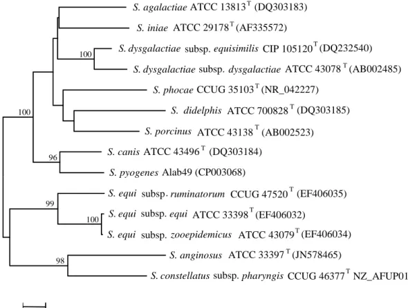
FIGURE 1.2. Neighbor-joining tree of the 16S rRNA gene of GCGS
and other beta-hemolytic species
Branches clustering sequences with greater that 75% bootstrap support (1,000 replicates) are indicated. The tree is drawn to scale, with branch lengths corresponding to the number of base substitutions per site. The species name and strain identification are indicated. GenBank entries whose sequences were used to construct the tree are shown in brackets. Small-colony-forming “S. milleri” species which may also possess Lancefield group C or G antigens are also included. “T” denote type strain.
Several molecular approaches have been employed for the differentiation of GCGS, but only a few were aimed at the routine laboratory identification, while others were directed towards veterinary microbiology (94). A few studies had a special focus on assays allowing the distinction between SDE and GAS (95, 96). Among the sequence based methods developed for streptococci and applied in GCGS speciation, several distinct genes were targeted (21, 41, 42, 53). Among these, sequence analysis of the 16S rRNA gene should be highlighted, because it was important in defining the current
pharyngis (JN578465) (EF406034) equi (EF406032) (AB002523) (DQ303185) (NR_042227) (AB002485) dysgalactiae subsp equisimilis NZ_AFUP01000004 (AF335572) (DQ232540) ATCC 13813 (DQ303183) S. agalactiae T S. ATCC 29178 T S. dysgalactiae . CIP 105120 T S. dysgalactiae subsp . ATCC 43078 T
S. CCUG 35103 T
S. didelphis ATCC 700828 T S. porcinus ATCC 43138 T
S. canis ATCC 43496 T (DQ303184) S. pyogenes Alab49 (CP003068)
S. equi subsp ruminatorum . CCUG 47520 T (EF406035) S. equi subsp . ATCC 33398 T
S. equi subsp . zooepidemicus ATCC 43079 T S. anginosus ATCC 33397 T
S. constellatus subsp . CCUG 46377 T
100 98 99 100 96 100 0.005 phocae iniae
taxonomic status of the genus (25) since it allows the distinction of all the currently defined GCGS species (Fig. 1.2), and it has been the most used method by workers in the field (65).
Disagreements were frequently seen among phylogenies derived from distinct genes (41, 42, 97). For example, 16S rRNA gene analysis places S. canis as the closest relative of GAS (as observed in Fig. 1.2), while analysis based on other housekeeping genes shows a closer relationship between SDE and GAS (97). Multilocus analysis and full genome sequence data have confirmed a close relationship for the three taxa (26, 42), but SDE was identified as most likely sister species of GAS (26).
2. GCGS ECOLOGY AND EPIDEMIOLOGY IN THE HUMAN HOST
The recognition of SDE as part of the human microbiota and the diversity of clinical conditions for which this species is believed to be responsible, makes SDE the most relevant GCGS species in the context of the human host. Although some older literature has limited information on GCGS species identification, more recent studies focusing on the clinical and epidemiologic features of SDE infections produced similar conclusions, confirming previous observations and supporting a major role of this species in the earlier studies.
2.1. Ecological niches and origin of infection
The occurrence of GCGS in human specimens is known since the original description of Lancefield groups C and G bearing strains by Rebecca Lancefield (2). Sites associated with colonization by these organisms include the nasopharynx, the skin, the gastrointestinal and the female genitourinary tracts (2, 98, 99). Although there is a limited number of reports on GCGS carriage in the human host and many did not identify the species, it is generally accepted that SDE is the only GCGS that can consistently colonize humans (100).
Most of the available studies on human GCGS colonization have focused on the pharynx. SDE asymptomatic carriage rates of 2 to 3% have been reported in both children and young adults (98, 101–103). Throat colonization rates have been noted to change over time (101) and others have shown they may be much higher in certain human populations (104–106). A study with school children from India found a SDE colonization rate of almost 10% (106), and in the aboriginal communities of Northern Australia this value may go up to 20% (104, 105).
The origin of the infection has been poorly studied in SDE. Taking into consideration the presence in the human microbiota and the potential separation of human and animal strains within this taxon (33), the most likely source of infection is endogenous. This idea is reinforced by the fact that SDE infections often occur in the skin and upper respiratory tract (19), suggesting that colonization of these areas may have a role in the origin of infection. For example, the presence of these bacteria in toe webs (107) or anal colonization (99) was suggested as reservoirs for erysipelas and cellulitis of the lower limbs. SDE is likely present in the intact skin before the onset of infection and
cutaneous infections, mainly those involving deeper layers of the skin (e.g. cellulitis), may allow SDE to access the bloodstream and cause invasive infections (7, 108). Indeed, studies characterizing SDE bacteremia have repeatedly implied the skin as the primary focus (following not only cellulitis, but also wounds, ulcerations and intravenous drug use) (4, 12, 17, 109). The origin of infection may also be localized to the upper respiratory tract (in neutropenic patients and those with pneumonia), and the genitourinary tract (in women during the peripartum period) (4, 68).
A person-to-person route of transmission has also been demonstrated for SDE. In a case-control study conducted in Finland (67), SDE was more frequently isolated from household members of patients with cellulitis. Molecular typing identified the same SDE strain in the pharynx of patients and members of their household. References can also be found in older literature to healthcare-associated infections associated with both person-to-person transmission and transmission from contaminated environmental sources, with SDE being implicated in outbreaks of skin infections and puerperal sepsis (110) and in post-operative wound infections (111). Less frequently, food borne community outbreaks of pharyngitis attributable to SDE have been detected (110), although this route of transmission is more common for S. equi subsp. zooepidemicus (see below).
The phenotypic and genotypic separation between SDE strains isolated from humans and animals (46) could be interpreted to suggest that zoonotic transmission is not frequent in this taxon. However, no separation between group L strains isolated from human and non-human sources is apparent (33) and a recent report identified the same SDE strain in a child and a dog (112). These observations show that cross-species transmission can occur and question whether animal SDE strains are transmitted to humans, as observed for other GCGS species. It should be noted that infections by such strains may pass essentially unrecognized as neither the genotypic characteristics of such isolates are firmly established, nor is the intra-specific characterization of SDE isolates that could allow such discrimination to be made usually performed.
The other GCGS species are not recognized as agents of colonization in humans. Nevertheless, S. equi subsp. zooepidemicus has been identified in throat swabs of farm workers with no clinical signs of infection at farms experiencing outbreaks caused by this pathogen, and also in convalescent patients several months after the original infection (80). No such evidence have been found for any other of the GCGS species
that are present in animals, but one study detected an unusual high number of S. canis isolates in non-invasive human specimens suggested the occurrence of human colonization by this agent (36). Thus, it is conceivable that transient colonization by S.
equi subsp. zooepidemicus and S. canis may occur in people contacting frequently with
animals.
Human infection by GCGS species other than SDE, as is the case of S. canis, S. equi subsp. zooepidemicus, and more recently, S. equi subsp. ruminatorum, is thought to originate from contact with animals or with their products. For these three species, cases of direct transmission through pre-existing skin lesions, such as patients’ wounds and ulcers, or direct inoculation by bites or scratches, were considered the most likely portal of entry (63, 113, 64, 114, 86). A couple of studies employed typing methods to definitively determine the zoonotic origin of the infection in these patients. One report identified the same S. canis strain in a dog and its owner’s blood cultures following a bite (63), and another one showed the transmission of a S. equi subsp. zooepidemicus strain from a dog to a handler (115). Only S. equi subsp. zooepidemicus infection has been associated with the consumption of unpasteurized milk products, resulting in outbreaks of bacteremia (116) and glomerulonephritis (81). The presence of this bacterium in milk is related to its role as an agent of mastitis in cows and other cattle (80, 117). Although other GCGS species may cause mastitis in animals with bacterial shedding in milk (47, 61, 70), such a route of infection has not been established.
2.2. Infections
GCGS have been associated with a multitude of infections in humans, ranging from mild to life-threatening conditions. Most commonly, infections occur in the skin and in the upper respiratory tract (3, 19) The spectrum of infections caused by this species closely resembles that caused by GAS, and includes numerous skin and soft tissue infections (67), pharyngitis (98), ocular infections (118), bacteremia (119), endocarditis (120), septic arthritis (121), pneumonia (122), meningitis (110), necrotizing fasciitis (123) and streptococcal toxic shock syndrome (STSS) (124). S. canis has been mostly isolated from skin and soft tissue infections and associated bacteremia (21, 36), while S.
equi subsp. zooepidemicus infections are often severe, including septicemia (125),