Related RNAs in Lepidopteran Cells after in Vitro Infection with Hyposoter didymator Virus
Define a New Polydnavirus Gene Family
Anne-Nathalie Volkoff,*,1
Pierre Ce´rutti,* Janick Rocher,* Marc C. P. Ohresser,† Ge´rard Devauchelle,* and Martine Duonor-Ce´rutti*
*Laboratoire de Recherches de Pathologie Compare´e, INRA–CNRS, 30380 St. Christol-les-Ale`s, France; and †University of Algarve, CCMar, Campus de Gambelas, 8000, Faro, Portugal
Received March 4, 1999; returned to author for revision March 29, 1999; accepted July 27, 1999
In the present study, we describe the isolation and the characterization of three different Hyposoter didymator virus (HdV) lepidopteran host-expressed genes, the products of which might interfere with the host physiology during parasitism. In this report, we study the expression of HdV genes in Sf9 cells infected with HdV since results indicate that the Sf9 model mimics to some extent the in vivo model and may be utilized to study expression of HdV genes in lepidopteran host cells. This system allowed us to isolate three HdV-specific cDNAs, termed M24, M27, and M40. cDNA nucleotide sequence analysis demon-strated significant regions of homology. The three cDNAs displayed repeated sequences arranged in tandem array that might have evolved through domain duplication. Similar to other previously described polydnavirus host-expressed genes, two intron positions have been found in the M24 leader region. The cDNAs corresponded to RNAs of 1.5, 1.6, and 2.3 kb that are also detected in parasitized Spodoptera littoralis larvae. They are encoded by different genes likely located on different HdV DNA molecules. Corresponding RNAs are detected early postinfection and remain detectable for at least 10 days postin-fection. They encode secreted glycine- and proline-rich proteins. An antiserum raised against a baculovirus recombinant M24-encoded protein detected similar proteins in the culture medium of infected lepidopteran cells and in parasitized host hemolymph. We propose that the three cloned genes belong to an HdV gene family specifically expressed in parasitized lepidopteran hosts. © 1999 Academic Press
INTRODUCTION
Polydnaviruses (PDVs) are unusual viruses observed in some hymenopteran parasitoids from the ichneu-monid and braconid families. Up to now, PDVs have been described only from parasitic wasps whose preimaginal development occurs inside lepidopteran hosts. PDVs are characterized by polydisperse, double-stranded, super-helical circular DNA genomes (Stoltz et al., 1995) with fewer than 10 and up to more than 25 molecules (Flem-ing, 1992). PDV DNA is integrated into the wasp genome and PDVs are transmitted vertically as proviruses (Flem-ing and Summers, 1991; Gruber et al., 1996; Savary et al., 1997; Xu and Stoltz, 1991). After replication in the repro-ductive tract of infected female wasps, PDVs are inocu-lated into lepidopteran host larva during oviposition along with the proteins produced in the calyx. Once inside the lepidopteran host larva, the viruses do not replicate but specific viral genes are expressed (Sum-mers and Dib-Hajj, 1995).
The PDV genome has been shown to be functionally multipartite (Theilmann and Summers, 1987,1988). Some viral genes are transcribed solely in the hymenopteran
parasitoid or in the lepidopteran host, whereas others are transcribed in both (Webb, 1998). Genes expressed specif-ically in the lepidopteran host, or “host-expressed genes,” have been described from several PDVs associated with braconids (Asgari et al., 1996; Harwood and Beckage, 1994; Hayakawa et al., 1994; Strand et al., 1997) and also from the virus associated with the ichneumonid Campoletis
sono-rensis (Blissard et al., 1987,1989; Theilmann and Summers,
1988; Dib-Hajj et al., 1993; Cui and Webb, 1996).
PDVs associated with parasitic wasps are known to play an essential role in the success of the endoparasitic part of the parasitoid life cycle in their lepidopteran hosts. Indeed, parasitic wasps evade some immune responses of the host insect larvae through expression of PDV genes (Asgari et
al., 1996,1997; Harwood and Beckage, 1994; Lavine and
Beckage, 1995; Shelby et al., 1998; Strand et al., 1997; Yamanaka et al., 1996). In addition, expression of PDV genes causes some cellular and physiological effects in the parasitized host, among them hormonal and develop-ment changes (Beckage et al., 1994; Lawrence and Lanz-rein, 1993; Tanaka et al., 1994). Regarding infection, several tissues of the lepidopteran host are known to be infected. So far, infection has been described in hemocytes (Beck-age, 1998; Strand, 1994; Strand and Pech, 1995) and few reports concern other tissues like the fat body (Shelby and Webb, 1994,1997; Strand et al., 1992). Two modes of action
1To whom reprint request should be addressed. Fax: (33) 04.66.52.46.99. E-mail: volkoff@ensam.inra.fr.
Article ID viro.1999.9929, available online at http://www.idealibrary.com on
0042-6822/99 $30.00
Copyright © 1999 by Academic Press All rights of reproduction in any form reserved.
have been suggested for PDVs: PDV-associated proteins interact directly with host cells and/or virus DNA tran-scribed within the host cell exerts cellular changes through viral gene expression (Asgari et al., 1996; Webb, 1998). The first case is illustrated by the proteins encoded by the Cys-motif genes of the C. sonorensis PDV. Viral proteins bind to and enter host hemocytes, presumably by receptor-mediated endocytosis. This results in disruption of the cy-toskeleton, altering the adhesive properties of the hemo-cytes and thereby inhibiting encapsulation of the wasp egg (Cui et al., 1997; Li and Webb, 1994; Webb, 1998).
Other than these few reports, little is known about the function of viral gene products within the parasitized host. Further analysis of viral gene expression is neces-sary to elucidate the mechanisms by which PDV genes alter lepidopteran host physiology.
In the present study, we describe the isolation and characterization of three different Hyposoter didymator virus (HdV) host-expressed genes the products of which might interfere with the host physiology. This PDV is carried by the ichneumonid H. didymator, a polyphagous parasitoid able to develop in several noctuids, particu-larly in species of Spodoptera (Caballero et al., 1990; Harrington et al., 1993; Tillman and Powell, 1991). We show in this work that Sf9 (Spodoptera frugiperda) cell line is suitable for study of HdV expression in lepidop-teran cells, since HdV expression in Sf9 cells was com-parable to HdV expression in the larva’s hemocytes. Three viral cDNAs generated from HdV-infected Sf9 cells were cloned. Their corresponding RNAs were detected early postinfection and for at least 10 days postinfection. The cDNA nucleotide sequence analysis demonstrated that the RNAs have significant regions of homology. They are encoded by different genes likely located on different HdV DNA molecules. They encode secreted glycine- and proline-rich proteins presenting repeated sequences ar-ranged in a tandem array. We propose that the three cloned genes belong to an HdV gene family specifically expressed in parasitized lepidopteran hosts.
RESULTS
HdV expression in lepidopteran cells: are Sf9 cells suitable for studying viral expression
in lepidopteran host?
The same proteins were detected whether cell lines or hemocytes were infected with HdV. In lepidopteran cell
lines infected with HdV, [35S]methionine incorporation
revealed four proteins of approximately 40, 42, 50, and 70 kDa synthesized de novo in the culture medium (Fig. 1). They were detected 6 h postinfection (p.i.) and continued to be secreted for at least 1 week p.i. (data not shown). These four secreted proteins were observed in the me-dium after infection of all the cell types tested, freshly collected S. littoralis hemocytes (Fig. 1, lane 2), the newly established S. littoralis cells derived from hemocytes
(Fig. 1, lane 6), Sf9 cells (Fig. 1, lane 4), and Hi5 cells (Fig. 1, lane 8), but not in the medium of uninfected cells (Fig. 1, lanes 1, 3, 5, and 7). Furthermore, Sf9 cells infected with HdV that had been previously UV-irradiated did not produce any of the proteins (data not shown), thus sug-gesting they might be encoded by the viral genome. The intracellular protein patterns contained a high number of cellular proteins, and no significant difference was ob-served between infected and uninfected cells whether [35S]methionine labeling or Coomassie blue staining was
used (data not shown).
The main HdV RNA class size detected in HdV-infected Sf9 cells was also detected in parasitized S. littoralis larvae. To analyze HdV-specific RNAs, Northern blot
studies were conducted with RNAs isolated from HdV-infected Sf9 cells and parasitized larvae (Fig. 2). 32
P-labeled total genomic HdV DNA used as a probe hybrid-ized with at least seven RNA class sizes isolated from Sf9 cells infected with HdV. The RNA class sizes ranged from 0.1 to 5.0 kb, the most abundant corresponding to the 1.5- to 1.8-kb band (Fig. 2, lane 1). This band was detected as early as 2 h p.i., thus suggesting that some viral RNAs were transcribed early in infection, and it was observed up to 1 month p.i. (data not shown). The 1.5- to 1.8-kb RNA band was the only band easily observed in samples isolated from parasitized larvae (Fig. 2, lane 2). However, the intensity of the hybridization signal was weak with crude extracts from parasitized larvae
sam-FIG. 1. Proteins secreted by HdV-infected lepidopteran cells. Proteins were detected by autoradiography after [35S]methionine labeling (4-h pulse) followed by 12.5% SDS–PAGE. 30ml of culture medium was loaded per lane. Four cell types were analyzed: Spodoptera littoralis hemocytes (lanes 1 and 2 [H]), Sf9 cell line (lanes 3 and 4 [Sf]), S.
littoralis cell line (lanes 5 and 6 [Sl]), and Trichoplusia ni Hi5 cell line
(lanes 7 and 8 [Tn]). Lanes 1, 3, 5, and 7, uninfected cells (C); lanes 2, 4, 6, and 8, HdV-infected cells, 24 h postinfection (I). Molecular masses in kDa are given for the four proteins detected.
ples compared to that of Sf9 samples. This could prob-ably account for the absence of the other RNA species in the larvae samples. In naturally parasitized S. littoralis, HdV RNAs were detected from 4 h postparasitism (p.p.) to the end of parasitoid development, i.e., 9–10 days p.p. (data not shown). No hybridization was observed with RNA isolated from nonparasitized larvae or uninfected Sf9 cells (data not shown).
Isolation of HdV genes expressed in lepidopteran cells: generation of virus-specific cDNAs
from infected Sf9 cells
Based on the previous results, Sf9 cells were infected for 24 h with HdV to generate a cDNA library. From 720 analyzed clones, 49 were HdV-specific cDNAs, with sizes ranging from 0.8 to 2.5 kb. Thirteen of them were randomly chosen to probe all the HdV-specific cDNAs. This allowed a classification into eight different groups, each representing one or more cross-hybridizing HdV-specific cDNAs (Table 1). Total RNAs from HdV-infected Sf9 cells (24 h p.i.) were then probed with the longest
EcoRI-digested fragment of each group. Northern blot
analysis showed that each probe hybridized with differ-ent RNAs, ranging from 0.8 to more than 3.0 kb (Table 1). In this study, we focused on the group that we referred to as the M group (with 26 cDNA clones; Table 1), which contained 1.5- and 2.3-kb RNAs that hybridized intensely with its specific probe.
The M-group cDNAs were sequenced on both their 59 and their 39 ends. Their alignment (Fragment Assembly; Genetics Computer Group) revealed that most of these cDNAs overlapped with three distinct sequences of 1481, 1636, and 2327 bp, respectively termed M24, M27, and M40.
Characterization of three putative HdV genes expressed in lepidopteran cells: nucleotide and peptide sequence analysis indicated high similarity between the M-group cDNAs M24, M27, and M40 (Fig. 3).
Homologies in untranslated regions. In the 59 untrans-lated region (UTR), M40 was almost identical to M27 (99% identity, compared to 86% with M24); conversely, in the 39UTR, M40 was more related to M24 (98% identity, compared to 79% with M27) (Fig. 3A).
FIG. 2. Northern blot analysis of HdV RNAs. HdV-specific RNAs were detected with32P-labeled BamHI–HindIII-digested genomic viral DNA in total RNA extracted from Sf9 cells 44 h postinfection (lane 1 [Sf]) and in S. littoralis second-instar larvae, 20 h postparasitism (lane 2 [L]). For infected Sf9 cells, detected RNA class sizes are indicated by asterisks or the arrow for the major one. About 20mg total RNA was loaded per lane, exposure was for 24 h. Molecular size markers (kb) are on the left.
TABLE 1
Number and Size of the RNAs Detected by Hybridization with a Specific Probe from Each Characterized HdV cDNA Group
Group
Number of clones
cDNA probe size (EcoRI fragment)
Number of RNAs detected
RNAs with strong hybridization signal S 2 S6: 0.8 kb 3 1.0 kb D 5 D8: 0.9 kb 3 0.8 kb 3.0 kb C 1 C28: 1.6 kb 3 1.0 kb 1.8 kb K 5 K29: 1.7 kb 1 2.0 kb P 6 P30: 1.8 kb 2 1.8 kb 3.6 kb B 2 B36: 1.3 kb 2 1.0 kb 2.0 kb X 1 X41: 1.1 kb 2 1.5 kb M 26 M24: 1.5 kb 4 1.5 kb 2.3 kb
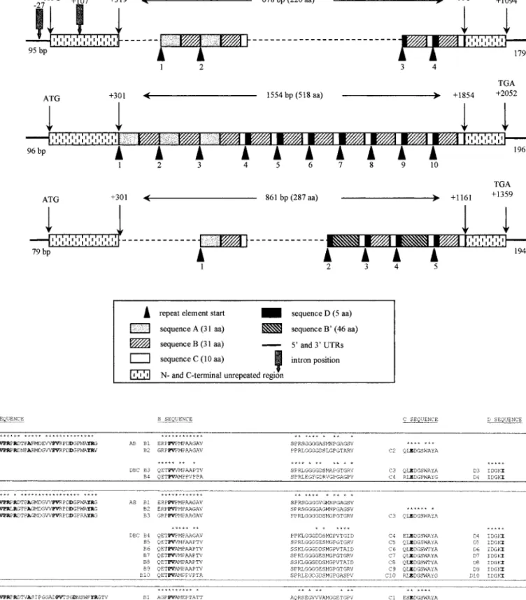
FIG. 3. M-group nucleotide and peptide sequence analysis. (A) Alignment of the nucleotide and predicted peptide sequences of the cDNAs M24, M40, and M27 for the 59UTR, N-terminal and C-terminal coding regions, and 39UTR. Dots indicate identical nucleotides and gaps (2) are introduced to optimize alignment. (B) Positions and sequences of the two introns observed when 59 HindIII-digested ends of M24 and D20 are aligned. Selected restriction site HindIII is in italics and start codon ATG in bold. The region homologous to C. sonorensis cysteine-motif genes is bounded by arrows, and the intron region homologous to M40 is boxed (see text). (C) Schematic representation and alignment of the M-group cDNAs M24, M40, and M27, showing their respective regions of repeated elements, A, B, B9, C, and D. The A of the first ATG is considered 11. (D) Alignments of the repeated sequences A, B and B9, C, and D found in P40 (M24-encoded), P69 (M40-encoded), and P45 (M27-encoded). B sequences from (AB) and (DBC) repeats are analyzed separately. Numeration of repeated sequences indicates the number of the repeat (for example, P40 is A1B1A2B2C2D3B3C3D4B4C4, see
In the 39UTR, a putative polyadenylation signal was identified downstream of the TGA stop codon (170 bp for M24 and M27; 169 bp for M40).
Presence of two introns in the M24-related gene.
Com-parison of M24 cDNA with the HindIII–XbaI fragment generated from the D20 genomic clone, which specifi-cally hybridized with M24, indicated the presence of two introns. These introns were localized 27 bp upstream and 107 bp downstream of the ATG and were 124 and 196 bp long, respectively (Fig. 3B). Flanking exon se-quences were in agreement with eukaryotic consensus splicing sites.
The open reading frames displayed repeated elements arranged in tandem array. Each of the three cDNAs
encoded a single open reading frame (ORF). The se-quences preceding the first ATG were in agreement with the Kozak consensus sequence. The M24, M27, and M40 ORFs were 1094, 1359, and 2052 bp, respectively (Fig. 3C). Corresponding proteins, named P40, P69, and P45, had a predicted molecular mass of 40, 69, and 45 kDa with 397, 452, and 683 amino acids, respectively.
The three ORFs contained a region of imperfectly re-peated elements arranged in tandem array and flanked by nonrepeated regions (Fig. 3C). The coding sequences of these ORFs could be divided into N-terminal nonre-peated, “repeat elements,” and C-terminal nonrepeated regions (Fig. 3A). In the N-terminal nonrepeated region, peptide sequences were 84% identical between P40 and P69, 46% between P40 and P45, and 81% between P69 and P45. In the C-terminal nonrepeated region, se-quences were 95% identical between P40 and P69, 54% between P40 and P45, and 56% between P69 and P45.
Within the repeat-element region, the elements were successively named A, B or B9, C, and D in the order they appeared in the sequence. The repeated region began at amino acid 107 in P40 and was 226 amino acids long. In P45 and P69, the repeated regions began at amino acid 101 and were 287 and 518 amino acids long, respectively. A, B, C, and D sequences were present in all three ORFs, while B9 sequence, related to B sequence, was specific to P45 ORF (Figs. 3C and 3D). A, B, B9, C, and D se-quences were 31, 31, 46, 10, and 5 amino acids long, respectively. A, C, and D amino acid sequences were highly conserved within a same protein, and between the proteins, particularly between P40 and P69 (Fig. 3D). The B element was more variable and, regarding P40 and P69, could be differentiated whether it was part of an (AB) repeat or a (DBC) repeat (Fig. 3D). The P40 repeat region was symbolized by (AB)2-C-(DBC)2, corresponding
to two (AB) repeats, followed by one C repeat and two (DBC) repeats. In the same way, the P69 repeat region was symbolized by (AB)3-C-(DBC)7 and that of P45 by
AB-C-(DB9C-DBC)2. Therefore, the main difference
be-tween P40 and P69 was found in the length of their repeat-element regions, while P45 presented less iden-tity with the other two.
Putative genes related to M-group cDNAs encode re-lated glycine- and proline-rich HdV proteins. All the
pre-dicted proteins P40, P69, and P45 started with a putative signal peptide of 16 amino acids (PSORT analysis; Nakai, 1991). A hydrophobic domain was also present at the C-termini of the proteins (HCA analysis; Caillebaut et al., 1997). These three proteins were glycine (15–16%) and proline (13–14%) rich, particularly in the repeat-element region. No homology was found with any known poten-tial N-glycosylation consensus site. However, the pres-ence of multiple serine and threonine residues might predict some O-glycosylation sites. Prediction of subcel-lular localization of proteins by PSORT analysis (Nakai, 1991) indicated that these proteins have 67% probability of being extracellular. Therefore, the three related gly-cine- and proline-rich HdV proteins are probably se-creted by the host cells.
Comparisons with sequences in GenBank/EMBL and SwissProt databases revealed no significant relationship with previously described polydnavirus gene products or with other proteins. Searches in the available databases did not show significant overall similarity between the repeated sequences A, B, C, and D and other proteins. Localization of M24-, M27-, and M40-related genes on the HdV genome
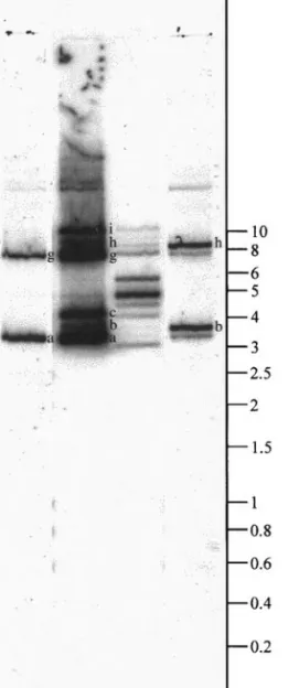
The HdV genome consists of at least 20 DNA mole-cules ranging from 2 to over 20 kb in size (Fig. 4, lane 1; Volkoff et al., 1995). However, in electrophoretically re-solved undigested DNA, several bands may represent different DNAs or conformational differences in the same DNA. Moreover, the pattern may vary from one DNA preparation to the other. Therefore, Southern blot results from undigested genomic HdV DNA should be analyzed with care. To localize M24-, M27-, and M40-related genes on the HdV genome, specific 45-mer oligonucle-otides were used: one specific for the M24 cDNA (cor-responding to nucleotides 149 to 193 in the M24 N-terminal coding region) and one specific for the M27 cDNA (corresponding to nucleotides 653 to 697 in the M27 B9 sequence). Due to the homologies between the sequences, no M40-specific probe could be designed.
M24-specific probe hybridized with the same intensity to two bands, a and g, from undigested HdV DNA (Fig. 4, lane 2), while M24 cDNA hybridized with six bands, namely a, b, c, g, h, and i. M24 also hybridized intensely with a 4.7-kb BamHI–HindIII fragment of HdV DNA (Fig. 4, lane 4). The BamHI restriction fragment of the D20 genomic clone (aligning with M24) was about 5.2 kb and presented a HindIII site. The D20 BamHI–HindIII frag-ment was 4.7 kb size and matched with the 4.7-kb Bam-HI–HindIII fragment of HdV DNA observed in the South-ern blot. Taken together, this indicated that the D20
BamHI restriction fragment might represent an almost
complete HdV molecule of about 5.2 kb containing the
related gene. These results showed that the M24-related gene was more likely present in one HdV mole-cule, bands a and g representing different conforma-tional forms of the same molecule. On the other hand, the M27-specific probe hybridized to the bands termed b and h (Fig. 4, lane 5). Observation of a slight hybridization with bands a and g might be explained by the slight similarity existing between the B9 sequence (correspond-ing to the M27-specific probe) and the M24 B sequence. These results indicated that the genes encoding each of
these two cDNAs were on two different HdV molecules. As with M40, this probe was the only one to hybridize significantly with bands c and i (data not shown), sug-gesting that the M40-related gene was localized on a third molecule.
Synthesis of M24-, M27-, and M40-specific mRNAs during HdV infection of Sf9 cells
and during parasitism
Northern blot analysis showed that the M-group cDNAs hybridized to two or three RNAs isolated from HdV-infected Sf9 cells and from parasitized larvae (Fig. 5), whereas no signal was detected in uninfected cells (Figs. 5A and 5B, lane 7) and in nonparasitized larvae (Figs. 5A and 5B, lanes 1 and 6). In infected Sf9 cells, in parasitized larvae crude extracts, and in parasitized lar-vae hemocytes, M24 hybridized essentially with RNAs of 1.5 and 2.3 kb (Figs. 5A and 3C, lane 5, for M24) while M27 hybridized with a 1.6-kb RNA (Figs. 5B, lanes 7 to 11, and 3C, lane 1). This result was confirmed when highly specific oligonucleotide probes were used (Fig. 5C, lane 5). All cDNAs also slightly hybridized to an approximately 3.0-kb RNA. For M40, results similar to those of M24 were obtained (data not shown), as was expected con-sidering their similarity. Based on the respective sizes of M24 and M40, the 2.3-kb RNA observed on Northern blots might correspond to M40 while the 1.5-kb RNA might correspond to M24.
In HdV-infected Sf9 cells, M-group-specific RNAs were detected from 2 h p.i. (Figs. 5A and 5B, lanes 8) up to 10 days (Fig. 5A, lane IV). The present study indicated that high levels of expression of both transcripts occurred 24 h p.i. in Sf9 cells infected with HdV. No significant decline in their transcription was detectable for at least the first 10 days of infection. In parasitized larvae, tran-scripts were present 2 h p.p. and still detected 5 days p.p. (Figs. 5A and 5B, lanes 8 to 11). RNAs were detected until the end of the parasitoid development (data not shown).
Synthesis of P40 protein in HdV-infected Sf9 cells and in parasitized larvae
An antibody against HdV M24 cDNA-related protein was produced after expression of a histidine-tagged M24 recombinant protein (M24-His) in the baculovirus insect cell system. Protein samples prepared from Sf9 and SlJ cells or from S. littoralis larvae hemolymph were analyzed by Western blot using a polyclonal antiserum raised against this M24-His recombinant protein (Fig. 6). As predicted from sequence analysis, P40 was efficiently secreted in HdV-infected Sf9 cells (Fig. 6A, compare lanes 8 and 11, cell sample contain-ing 2.5-fold more protein). P40 could also be detected in both cells and culture medium from HdV-infected cells and from parasitized larvae (Figs. 6B and 6C). In
FIG. 4. Hybridization pattern of genomic HdV DNA with the M-group cDNAs. Lane 1 shows the pattern of total genomic HdV DNA, observed after hybridization of BamHI–HindIII-digested 32P-labeled viral DNA with undigested viral DNA (2mg per lane, 10 h exposure). Lanes 2 and 5 represent hybridization of M24- and M27-specific probes, respec-tively, with undigested viral DNA (1mg per lane, 48 h exposure). Lanes 3 and 4 represent hybridization of EcoRI-digested32P-labeled M24 with undigested viral DNA and BamHI–HindIII-digested viral DNA, respec-tively (1mg per lane, 48 h exposure). DNA molecular size markers (kb) are indicated at the right. Names of detectable bands (a, b, . . .) are indicated on their right.
infected cell lines, an immunoreactive band was also detected around 70 kDa. As predicted by amino acid sequence identity between P40 and P69, this band might correspond to the P69 protein. Note that for S.
littoralis larvae, an immunoreactive band was detected
around 80 kDa for both parasitized and nonparasitized plasma (Fig. 6C, lanes 4 and 5) that might correspond to a host protein presenting a common epitope with P40. In addition to Western blot analysis, the [35
S]me-thionine incorporation experiments indicated that the radiolabeled prominent 40- and 70-kDa proteins se-creted by several HdV-infected lepidopteran cell lines (Fig. 1) might correspond to P40 and P69.
DISCUSSION
In this paper, expression of some H. didymator polyd-navirus genes was followed in Sf9 cells infected with HdV. Results indicated that the Sf9 model mimics to some extent the in vivo model and may be used to study expression of HdV genes in lepidopteran host cells. This system allowed us to identify three HdV-specific cDNAs, named M24, M27, and M40, corresponding to 1.5-, 1.6-, and 2.3-kb RNAs that were also detected in parasitized
S. littoralis larvae.
The exact number of HdV genes expressed in the lepidopteran host is unknown. It has been described that more PDV genes were specifically host-expressed in
FIG. 5. Northern blot analysis of HdV RNAs related to the M-group cDNAs. (A) Time course of M24 transcription. RNAs were detected with the
EcoRI-digested32P-labeled M24. Lanes 1 to 4 correspond to larvae samples (30mg per lane, 48 h exposure) with lane 1, nonparasitized larvae; lanes 2–4, larvae 2, 24, and 48 h postparasitism, respectively; lanes 5 and 6, hemocytes from fifth-instar larvae (8mg per lane, 48 h exposure) parasitized (lane 5) or not parasitized (lane 6); and lanes 7 to 11, Sf9 cell samples (15mg per lane, 20 h exposure), uninfected cells (lane 7) and cells 2 h (lane 8), 24 h (lane 9), and 24 h treated with RQ1 DNase (lane 10) and 48 h (lane 11) postinfection. Lanes I, II, III, and IV correspond to Sf9 cells 48 h, 6 days, 7 days, and 10 days postinfection, respectively (3mg in lanes I and IV, 4 mg in lanes II and III, 20 h exposure). White asterisks indicate the major RNA species of 1.5 and 2.3 kb in size. The gray asterisks mark the approximately 3.0-kb RNA. (B) Time course of M27 transcription. RNAs detected with the EcoRI-digested32P-labeled M27. Lanes 1 to 11 are the same as in A. White asterisks indicate the major 1.6-kb RNAs. The gray asterisks mark the approximately 3.0-kb RNA. (C) RNAs detected with32P-labeled M24- and M27-specific oligonucleotides. Total RNA from Sf9 cells (30mg per lane, 20 h exposure) were hybridized with M27-specific probe (lanes 1 and 2), M24- and M27-specific probes (lanes 3 and 4), and M24-specific probe (lanes 5 and 6). Lanes 1, 3, and 5 correspond to cells 48 h postinfection and lanes 2, 4, and 6 to uninfected Sf9 cells. Molecular size markers are shown on the right.
FIG. 6. Expression of M-group proteins in HdV-infected cells and parasitized larvae. All cell lines were infected for 72 h and larvae parasitized for 5 days at 27°C. Proteins were detected by immunoblotting with an antibody directed against the recombinant protein NhisP40. (A) Expression in infected Sf9 cells. Total proteins from whole-cell extracts (lanes 1 to 3; 15ml per lane) and culture medium (lanes 4 to 6; 30 ml per lane) were separated on 12.5% SDS–PAGE, transferred onto a nitrocellulose membrane, and reacted with the NhisP40 antibody. Lanes 1 and 4, uninfected cells; lanes 2 and 5, HdV-infected cells; lanes 3 and 6, cells infected with the recombinant baculovirus M24Nhis. As a positive control, approximately 2.5mg of column partially purified recombinant NhisP40 protein was loaded in lane C. (B) Expression in S. littoralis larvae. Proteins were separated on 12.5% SDS–PAGE. Lane 1 represents a positive control with medium from HdV-infected Sf9 cells. P40-related proteins were analyzed in hemocytes (15ml) from parasitized (lane 2) and nonparasitized (lane 3) larvae and in the hemocyte-free hemolymph (30ml) from parasitized (lane 4) and nonparasitized (lane 5) larvae. (C) Expression in S. littoralis cell lines. Proteins were separated on 10% SDS–PAGE. Culture medium (30ml) from uninfected Sf9 cells as a positive control is in lane 1. P40-related proteins were analyzed in the culture medium (30ml) of HdV-infected SlJ2a (lane 2), SlJ2b (lane 3), and Sf9 cells (lane 4). Molecular weights in kDa are indicated on the right. Arrows indicate the immunoreactive bands corresponding to P40 and P69.
ichneumonid than in braconid viruses (Webb, 1998). Two genes were described from the braconid Cotesia
rube-cula PDV (Asgari et al., 1996), while 12 have been
de-tected in the ichneumonid C. sonorensis PDV (Blissard et
al., 1987; Theilmann and Summers, 1988). For the
ichneu-monid-carried HdV, we identified at least eight groups of cross-hybridizing cDNAs expressed in the lepidopteran host. Using different probes from each cDNA group, at least 13 HdV-specific RNAs were detected in HdV-in-fected Sf9 cells. On Northern blots, total genomic HdV DNA hybridized with at least 5 bands. The major band was about 1.5–1.8 kb and probably represented several comigrating RNAs since several cDNAs from the differ-ent groups hybridized to RNAs of this size. This intense band was the only one readily detected in crude extracts of parasitized larvae probably due to the relative smaller amount of viral RNAs in crude extracts from whole in-sects, not all parasitized, compared to cell extracts.
When cDNAs were used as probes, the hybridization signal for larvae was weaker than for Sf9 samples de-spite the fact that two times as much total RNA was loaded for parasitized larvae samples. However, results from hybridization with RNA from host hemocyte isolates were similar to those obtained with Sf9 cells considering that one-third the amount of material was analyzed. This probably indicates that M-group-related genes are pref-erentially expressed in hemocyte tissue. These results also indicated that the M40-related gene is apparently less transcribed than M24- and M27-related genes.
In addition to the 1.5-, 1.6-, and 2.3-kb RNAs, M-group cDNAs hybridized also with a 3.0-kb RNA that might correspond to other related RNAs or to pre-RNAs. The presence of this RNA of greater length suggests that transcription regulation of this gene family should be considered.
The RNAs corresponding to the M-group cDNAs were detected in the lepidopteran host early in infection throughout endoparasite development, as has been re-ported for C. sonorensis PDV Cys-motif genes (Blissard
et al., 1987; Cui and Webb, 1996; Li and Webb, 1994). In
infected Sf9 cells as in parasitized larvae, they were present from 2 h p.i. until at least 10 days p.i. During this period, no significant decline in M-group RNA transcrip-tion was detected. This indicates, together with the fact that Sf9 cells continue to divide, that most of the cells were infected and that viral DNA was maintained and transcribed. In Sf9 cells infected with HdV, M-group-related RNAs are no longer detectable after 1 month p.i.; however, other viral RNAs are still detected (Fig. 2) and are even detected several years after infection (Volkoff et
al., in preparation). This suggests that the viral DNA
corresponding to these RNAs is maintained in infected cells in either an episomal form or, as suggested by Gundersen-Rindal et al. (1998), as an integrated form inside the host genomic DNA.
Results from Southern blots and nucleotide sequence
analysis indicated that RNAs related to the three M-group cDNAs are likely to be encoded by different genes located on at least three distinct viral segments. The M24-related gene is most likely present in one HdV molecule of about 5.2 kb. PDV genome molecules are present in nonequimolar ratio and display restriction length polymorphism (Fleming and Krell, 1993) and the status of the DNA molecules may vary from one species to another (Albrecht et al., 1994; Krell et al., 1982). More-over, PDV genomes have complex homology relation-ships both within and between different DNA molecules (Cui and Webb, 1997; Webb, 1998; Xu and Stoltz, 1993) since members of gene families may be located on multiple segments but also in multiple copies on a single segment (Webb, 1998). In the preparation we analyzed, supercoiled, circular, and linear forms of viral DNA are difficult to differentiate. cDNA bands positive for one probe, a and g (Fig. 4), for example, may represent different DNAs or different conformations of the same DNA. It was therefore difficult to accurately estimate the number and the size of the HdV molecules which were related to the M-group cDNAs. Nonetheless, hybridiza-tion results indicated that genes related to M-group cDNAs might be located on at least three viral DNA molecules. It will now be necessary to determine whether these genes are really present in a single genomic molecule or are distributed on several mole-cules.
Comparison of M24 cDNA with its related genomic sequence indicated the presence of two introns. Splicing of polydnavirus genes was described in C. sonorensis virus (Blissard et al., 1987; Cui and Webb, 1996; Dib-Hajj
et al., 1993) and in Microplitis demolitor virus (Strand et al., 1997). In M24, the first intron was localized in the 59
untranslated region, which has also been described for genes belonging to the C. sonorensis virus host-ex-pressed Cys-motif gene family (Cui and Webb, 1996). Moreover, these C. sonorensis virus genes showed some identity (BLAST analysis) with the M24-related genomic sequence in an intron–exon junction close to the 39 end. This shared gene feature suggests that these genes might be evolutionarily related and that specific regulation might occur through this consensus intron site. Compared to M24, the 59 untranslated region of M40 contained 7 additional nucleotides (Fig. 3B,164 to 170). Alignment of M24-related genomic clone and M40 showed that this extra 7-nucleotide sequence matches almost perfectly the 59 end of the first intron (6 bp iden-tical), suggesting an alternative splicing in this gene’s family.
Further isolation and sequencing of genomic clones containing genes encoding M27- and M40-related RNAs would allow a better characterization of this gene family. Homologies and the presence of imperfect repeated elements arranged in tandem array in the M-group cDNAs suggested that they belong to the same gene
family. The presence of a putative gene family in HdV is consistent with observations done in C. sonorensis PDV (Dib-Hajj et al., 1993; Theilmann and Summers, 1988) and
M. demolitor PDV (Strand et al., 1997). Summers and
Dib-Hajj (1995) suggested that polydnavirus genome segmentation and the existence of gene families in PDVs promote recombination and generation of novel genes. We described in this report three putative genes that belong to a “glycine- and proline-rich” gene family. All these genes encode secreted proteins with similar pre-dicted peptide sequences, suggestive of similar struc-tures and similar function. Their putative ORF contained repeated elements that were symbolized by (AB)2
-C-(DBC)2 for P40, AB-C-(DB9C-DBC)2for P45, and (AB)3
-C-(DBC-DBC)3-DBC for P69. Repeats in the nucleotide
se-quence can increase the likelihood of genetic changes such as mutation or recombination (Gordenin and Resnick, 1998). The presence of related genes with a different number of repeated sequences has been re-ported for M. demolitor PDV (Strand et al., 1997). Variation in the number of repeat units or changes in individual sequences might result from recombination processes or polymerase inadequacy as described for prokaryotic genomes (van Belkum et al., 1998). The similarity be-tween amino acids of the C sequence and the last 14 amino acids of the A sequence was noteworthy. In both P40 and P69, the last B sequence started with a glycine (G) residue (Fig. 8, see P40-B2and P69-B3). In P69 DBC
repeats, similarity was high between B4C4, B6C6, and
B8C8but also between B5C5, B7C7, and B9C9(Fig. 8). This
high degree of identity suggests that, in P69, a duplica-tion of two linked repeat elements (DBC-DBC) might have occurred. The described putative HdV genes might have a common ancestry and possibly be evolved from gene duplication, homologous recombination, or dele-tion events.
The presence of a putative signal peptide suggests that these HdV glycine- and proline-rich M-group en-coded proteins are secreted through cellular mem-branes. Indeed, after infection with HdV particles, the protein P40 is efficiently secreted by S. littoralis hemo-cytes, SlJ cells, and Sf9 cells. Coomassie blue staining allowed its visualization after 72 h p.i., resulting from the accumulation in the culture medium (data not shown). Estimated production level is comparable to that ob-served for recombinant proteins expressed in baculovi-rus expression system. Most of the polydnaviral proteins described in the literature are glycosylated proteins with several N-glycosylation sites (Beckage, 1993; Harwood
et al., 1994; Yamanaka et al., 1996). The HdV gene
prod-ucts described here do not present any potential N-glycosylation site and molecular masses of the observed proteins were close to the predicted molecular masses, suggesting little or no glycosylation.
The M group constitutes a family of related polydna-virus host-expressed genes whose products are
se-creted into the host hemolymph, similar to the Cys-motif gene products described in C. sonorensis PDV (Dib-Hajj
et al., 1993). The M-related proteins, produced in Sf9 and
in hemocytes, were detected from 6 h p.i. until at least 1 week p.i., consistent with the Northern blot results. These results suggest that P40 and P69 are major HdV proteins secreted by lepidopteran cells and in particular by hemocytes. Therefore, they might play an important role in the parasitoid development within the lepidop-teran host. PDVs disrupt several host physiological func-tions (Webb, 1998). The host development (Dover et al., 1988; Strand and Dover, 1991) or the host protein titers (Beckage et al., 1994) might be altered; however, no cloned viral genes have yet been identified as possess-ing one of these functions (Webb, 1998). On the other hand, the host immune system is the known or sus-pected target for several isolated viral genes expressed in the lepidoptera (Shelby et al., 1998). Concerning the M-group HdV proteins, their physiological function re-mained to be elucidated. They are glycine and proline rich but no definite motif could be assigned. The fact that they were secreted by the host hemocytes into the me-dium and that possibly an 80-kDa host protein shares an epitope with the M-group proteins might suggest a role for these proteins in modulating host cellular functions such as interfering with the host immune system.
As only few genes have so far been characterized, it is not known whether all PDVs use similar or unrelated mechanisms to disrupt host defenses or alter host phys-iology. Thus it would be interesting to investigate the interactions between PDV gene products and cellular proteins to identify possible cellular targets for the viral proteins. Functional relationships between P40 and S.
littoralis hemocytes are now being investigated to
ana-lyze the physiological and molecular mechanisms by which the glycine- and proline-rich HdV proteins act on lepidopteran cells and in the parasitized caterpillar.
MATERIALS AND METHODS Insect rearing and cell cultures
H. didymator and its rearing host S. littoralis were
reared as previously reported (Volkoff et al., 1995). Usu-ally S. littoralis larvae were parasitized upon second larval instar for 24 h. Parasitoid larvae hatched 3 days after oviposition, in third-instar host larvae, and reached pupal stage in about 10 days.
The culture medium used to maintain freshly collected hemocytes and cell lines was a TC100 modified medium (Gibco) supplemented with 5% fetal calf serum (Gibco). Hemocytes were collected by bleeding last-instar larvae from a proleg into 500ml of ice-cold anticoagulant buffer (Pech et al., 1994). The hemocytes were pelleted by centrifugation at 250 g, 4°C. Hemocytes were washed twice with phosphate-buffered saline (PBS) and once with cell culture medium, and then were resuspended in
500ml of medium and placed in culture plates. The cell lines used in the different experiments originated from S.
littoralis (SlJ2a, SlJ2b), S. frugiperda (Sf9, ATCC CRL1711)
and Trichoplusia ni (High Five, InVitrogen). The continu-ous S. littoralis cell lines were established from freshly collected hemocytes from one S. littoralis larva (as de-scribed above). After 20 passages, cell population was cloned. Two cloned cell lines were obtained, named SlJ2a and SlJ2b, that had been through more than 40 passages before being considered continuous cell lines. SlJ2a is a clone composed of large cells while SlJ2b is composed essentially of twice smaller cells.
Virus preparation, viral DNA extraction, and infections Polydnavirus were extracted from H. didymator fe-males as described by Beckage et al. (1994). Ovaries were dissected in PBS, placed in 1.5-ml microfuge tubes, and disrupted by several passages through a 23-gauge needle. The resulting suspension was passed through a 0.45-mm filter (cellulose acetate). Filtrate was used either for viral DNA extraction or for cell infection.
For viral DNA extraction, virions were pelleted from the filtrate at 30,000 g for 30 min, 4°C. The pellets were resuspended in extraction buffer (0.1 M Tris–0.1 M Na2EDTA–0.2 M KCl; proteinase K (0.2 mg/ml); Sarcosyl
(0.5%)) and incubated overnight at 50°C. After RNase A treatment (0.3 mg/ml, 2 h at 37°C), DNA was extracted with phenol/chloroform/isoamyl alcohol (25/24/1; v/v) and precipitated with ethanol.
To construct a HdV genomic library, virus was purified from homogenized ovaries on a 20–50% (w/v in PBS) sucrose gradient. Purified viral particles were pelleted and DNA was extracted as described above. Viral DNA was digested with BamHI and fragments generated were cloned into the BamHI site of pUC 19 plasmid (Ce´rutti et
al., in preparation). The library was screened with the
32P-labeled M24. The D20 genomic clone specifically
hybridized with M24 and contained a fragment about 5.2 kb. The HindIII–XbaI fragment of 1737 bp generated from D20 was sequenced and aligned identically to M24.
Infectious medium (IM) was prepared by resuspend-ing a filtrate obtained from 35–50 female ovaries into 12 ml of culture medium. For infections, cells were seeded and infected at the ratio of 1 ml of freshly prepared IM per 106 cells. IM was incubated in the presence of the
cells during the time of the experiment. Cells were di-luted after 4 days (1:2 dilution), then weekly (1:6 dilution). RNA isolation
Total RNA was isolated from uninfected or HdV-in-fected Sf9 cells, and from nonparasitized or parasitized
S. littoralis larvae, at various times postinfection or
post-parasitism. Cells (10 to 123 106) were seeded in 75-cm2
flasks (Falcon): 123 106 cells were seeded for 2-h-p.i.
extraction, whereas 107 cells were seeded for 48-h-p.i.
extraction. They were then infected with freshly prepared IM (ratio of 1 ml IM/106 cells). Parasitism could be
con-trolled only by larvae dissection. Until 3 days p.p., larvae were too small to be dissected, so parasitism rate was estimated as being approximately 50–70% and RNA sam-ples were collected from pooled larvae (25–35 mg of larvae for the 2- and 24-h p.p. samples, 60–65 mg of larvae for the 4- to 5-days p.p. samples). Four to five days p.p., hemolymph and hemocytes were collected from 8–10 larvae, which were then dissected to control the parasitism. As a control, RNA samples were also col-lected from nonparasitized larvae of corresponding age. RNA was extracted with Trizol (Gibco) according to the manufacturer’s protocol. Sf9 cells were washed in PBS and then resuspended in Trizol. Trizol was directly added to hemolymph and hemocytes, and pooled larvae were crushed directly in Trizol. RNA samples were resus-pended in distilled RNase-free water and stored at 280°C. Total RNA concentration was estimated by spec-trophotometry. As a control, some RNA samples were treated with RQ1 DNase (1 U/5mg RNA; Promega). Southern and Northern blot hybridization
Total RNA samples, loaded in a formamide–formalde-hyde buffer (Promega, TB256), and DNA samples were separated by electrophoresis in 1% agarose gels using a Tris–acetate buffer system (TAE). Gels were stained with ethidium bromide before transfer to confirm equal load-ing. They were transferred to charged nylon membranes (Boehringer Mannheim) by capillary blotting using 203 SSC and then fixed by UV irradiation for 3 min. Mem-branes were incubated at 42°C for 3 to 4 h in a prehy-bridization buffer (FPH) containing 50% formamide, 53 Dehnardt’s, 53 SSC, and 100 mg/ml calf thymus DNA (Sigma).
DNA probes (EcoRI-digested cDNA inserts and
HindIII–BamHI-digested HdV DNA) were random prime
(Boehringer Mannheim) labeled with [a-32P]dCTP (3000
Ci/mmol; NEN–Dupont) and hybridized to blots (.2 3 106 cpm/ml). Hybridization was conducted overnight at
42°C in FPH buffer. Membranes were washed twice for 10 min in 23 SSC, 0.1% SDS at room temperature, twice for 15 min in 0.23 SSC, 0.1% SDS at 42°C, and twice for 15 min in 0.13 SSC, 0.1% SDS at 68°C and autoradio-graphed (Kodak Biomax MR).
Two HdV cDNA-specific 45-mer oligonucleotides were synthesized (Eurogentec): one specific for the M24 cDNA (nucleotides 149 to 193 in the M24 N-terminal coding region, 5 9TGCGAATCCGAACTCAGCGTCCTTTACGGCT-TCTTCTCCTCCTCC39) and one specific for the M27 cDNA (nucleotides 653 to 697 in the M27 B91 sequence, 5 9TGGGGCGGGCGGCATTGCATCGCCTCCCCATCTT-GCTGCTGGACC39). Oligonucleotides were phosphory-lated with T4 polynucleotide kinase (Biolabs) in presence of [g-32P]ATP (6000 Ci/mmol; NEN–Dupont) and
ized to Southern and Northern blots (.106cpm/ml).
Pre-hybridization and Pre-hybridization were conducted for 3 h in FPH buffer at 42°C. Membranes were washed twice for 5 min in 23 SSC, 0.1% SDS at room temperature, then twice for 10 min in 0.13 SSC, 0.1% SDS at 42°C and autoradiographed (Kodak Biomax MR).
cDNA cloning and analysis
cDNAs were generated from HdV-infected Sf9 cells 24 h p.i. Total RNA was extracted following Chirgwin’s method (Sambrook et al., 1989). Poly(A)1 mRNA was
isolated with Straight A’s mRNA Isolation System (Nova-gen), according to the manufacturer’s protocol. cDNAs were generated using the Universal Riboclone cDNA Synthesis System (Promega). They were ligated to EcoRI adaptators and cloned into EcoRI-digested dephosphor-ylated pUC18 plasmid vectors. cDNA clones were then transformed on Escherichia coli strain XL1-Blue. To iso-late specific HdV clones, randomly chosen E. coli clones were screened by plaque hybridization (Sambrook et al., 1989) with nick-translated a-32P-labeled HdV genomic
DNA. By Southern blot analysis, cross-hybridization was performed to identify homologous cDNAs. For this pur-pose, 13 cDNAs were randomly chosen and correspond-ing EcoRI inserts were used individually as probes against the entire subset of HdV-specific cDNA clones. The plasmids carrying the cDNAs and derived constructs were sequenced with T7 Sequencing kit (Pharmacia) or with Abi Prism Dye Terminator Cycle Sequencing Ready Reaction kit (Perkin–Elmer) or by Eurogentec, Belgium. Overlapping nucleotide sequence fragments were as-sembled by Fragment Assembly programs, based on the method of Staden (1980), with the Wisconsin Sequence Analysis Package (Version 8 for Unix Server, Genetics Computer Group). Nucleotide and predicted peptide se-quences were aligned manually. Percentages of identity between sequences were calculated excluding the gaps. The sequence data reported here appear in the Gen-Bank database under Accession Nos. AF131648, AF132023, AF132024, and AF156933.
Radiolabeling of proteins
Proteins produced in cells infected with HdV were analyzed by metabolic radiolabeling as reported (Chaabihi et al., 1993). Cells were seeded in 24-well plates (106cells per well) and infected with 1 ml of freshly
prepared IM. At various times postinfection, medium was removed and cells were pulse-labeled with 0.5 ml of medium supplemented with 10mCi of L-[35S]methionine
(Amersham). After 4 h exposure, medium was recovered and cells were lysed in 100ml 1% SDS. Labeled proteins from medium and cell lysate were analyzed by SDS– PAGE in a 12.5% acrylamide gel using the method de-scribed by Laemmli (1970). After electrophoresis,
pro-teins were stained with Coomassie brilliant blue and dried gels were autoradiographed.
Generation of an antiserum against M24-encoded protein
Generation of recombinant baculovirus. A 270-bp
frag-ment encoding the N-terminal part of the gene (from the first ATG to the Eco47III site) was amplified by PCR with the plasmid carrying M24 as template. With the sense primer 5 9GACTGGATCCTACGTAATGAAGATCTTGCTGC-CCTTGATGGTCGC39, two restriction sites, BamHI and
SnaBI, were added upstream of the ATG and a BglII site
was added downstream of the ATG. The antisense primer 5 9CATGAAGCTTCTAGAAGTAGCGCTTCCCTCAC-CCATTTCATTGTC39 allowed us to add XbaI and HindIII sites downstream of the Eco47III site. The 296-bp
BamHI–HindIII fragment was cloned into the
BglII–Hin-dIII sites downstream of the P10 promoter in the bacu-lovirus transfer vector p119 (M. Ogliastro, unpublished data) giving the p119-M24 construct. The 962-bp
Eco47III–XbaI fragment encoding the C-terminal part of
the gene was isolated from the pM24 and introduced into the Eco47III and XbaI sites of the p119-M24 construct. A 26-bp SnaBI–BglII fragment encoding a 6-histidine tag was constructed with two overlapping oligonucleotides, 59GTAATGCATCATCATCACCACCATCATAA39 and 59CAT-TACGTAGTAGTGGTGGTAGTAGTATTCTAG39, and cloned into the SnaBI–BglII site of the p119-M24 (6-his tag intro-duced between the first two amino acids of coding se-quence), giving the p119-M24-Nhis construct. Sf9 cells were cotransfected with viral DNA purified from the mod-ified baculovirus AcSLP10 (Chaabihi et al., 1993) and DNA from recombinant p119-M24-Nhis transfer vector. Recombinant baculoviruses were plaque purified by standard methods (Summers and Smith, 1987).
Purification of the M24-encoded protein. Recombinant
protein was produced by infecting Sf9 cells with the recombinant virus M24-Nhis at a multiplicity of infection of 10 PFU per cell. Cells were collected 4 days postin-fection and purified by NTA-(Ni21) resin-affinity column
chromatography under denaturing conditions according to the manufacturer’s protocol (Qiagen). Fractions con-taining the column-purified protein were concentrated using a Centricon-30K device (Amicon) and proteins were separated by 12.5% SDS–PAGE. M24Nhis protein was purified in acrylamide gel. The gel slice correspond-ing to the protein was used to generate a rabbit poly-clonal antiserum (Eurogentec).
Immunoblotting with the antiserum. To detect the M24
cDNA product among the HdV-encoded proteins, Sf9 and
S. littoralis cells were HdV-infected for 72 h. For the
control M24-Nhis recombinant baculovirus, Sf9 cells were infected as described above for 72 h. Hemolymph and hemocyte samples were collected from nonparasit-ized and parasitnonparasit-ized S. littoralis larvae 4–5 days p.p.
Proteins were resolved on 10 or 12.5% SDS–PAGE and electrotransferred to nitrocellulose membranes using a semidry blotting apparatus. Blots were processed with the polyclonal M24Nhis antiserum (1/100). The protein was detected with anti-rabbit peroxidase-conjugated goat IgG (Sigma) as a secondary antibody (1/1000) fol-lowed by detection with the peroxidase substrate (3-amino-9-ethyl carbozol; Sigma).
ACKNOWLEDGMENTS
We thank R. Bros and B. Limier for kindly providing Spodoptera
littoralis larvae and Professor Santiago-Alvarez and Dr. Vargas for the Hyposoter didymator colony. Thanks to S. Berger for preparing the
buffers. Thanks to L. Croizier for help with sequencing, to F. Cous-serans and M. Lopez for help with GCG software. We are very grateful to S. Blanc, P. Krell, P. Fournier, and K. Shelby for their constructive comments on the manuscript. In addition, special thanks to M. Drucker, M. Ogliastro, and H. Volkoff for the English corrections.
REFERENCES
Albrecht, U., Wyler, T., Pfister-Wilhem, R., Gruber, A., Stettler, P., Heini-ger, P., Kurt, E., Schu¨mperli, D., and Lanzrein, B. (1994). Polydnavirus of the parasitic wasp Chelonus inanitus (Braconidae): Characteriza-tion, genome organization and time point replication. J. Gen. Virol. 75, 3353–3363.
Asgari, S., Hellers, M., and Schmidt, O. (1996). Host haemocyte inacti-vation by an insect parasitoid: Transient expression of a polydnavirus gene. J. Gen. Virol. 77, 2653–2662.
Asgari, S., Schmidt, O., and Theopold, U. (1997). A polydnavirus-en-coded protein of an endoparasitoid wasp is an immune suppressor.
J. Gen. Virol. 78, 3061–3070.
Beckage, N. E. (1993). Games parasites play: The dynamic roles of proteins and peptides in the relationship between parasites and hosts. In “Parasites and Pathogens,” Vol. 1, “Parasites” (N. E. Beck-age, S. N. Thompson, and B. A. Federici, Eds.), pp. 25–57. Academic Press, New York.
Beckage, N. E. (1998). Modulation of immune responses to parasitoids by polydnaviruses. Parasitol. Today 116, S57–S64.
Beckage, N. E., Tan, F. T., Schleifer, K. W., Lane, R. D., and Cherubin, L. L. (1994). Characterization and biological effects of Cotesia congregata polydnavirus on host larvae of the tobacco hornworm, Manduca
sexta. Arch. Insect Biochem. Physiol. 26, 165–195.
van Belkum, A., Scherer, S., van Alphen, L., and Verbrugh, H. (1998). Short-sequence DNA repeats in prokaryotic genomes. Microbiol.
Mol. Biol. Rev. 62, 275–293.
Blissard, G. W., Smith, O. P., and Summers, M. D. (1987). Two related viral genes are located on a single superhelical DNA segment of the multipartite Campoletis sonorensis virus genome. Virology 160, 120– 134.
Blissard, G. W., Theilmann, D. A., and Summers, M. D. (1989). Segment W of Campoletis sonorensis virus: Gene expression, gene products, and organization. Virology 169, 78–89.
Caballero, P., Vargas-Osuna, E., Aldebis, H. K., and Santiago-Alvarez, C. (1990). Parasitas asociados a poblaciones naturales de Spodoptera
littoralis Boisduval y S. exiguae Hb. (Lepidoptera: Noctuidae). Boll. Sanita Vegetal Plagas 16, 91–96.
Caillebaut, I., Labesse, G., Durand, P., Poupon, A., Canard, L., Chomilier, J., Henrissat, B., and Mornon, J. P. (1997). Deciphering protein se-quence information through hydrophobic cluster analysis (HCA): Current status and perspectives. Cell Mol. Life Sci. 53, 621–645. Chaabihi, H., Ogliastro, M. H., Martin, M., Giraud, C., Devauchelle, G.,
and Cerutti, M. (1993). Competition between baculovirus polyhedrin
and p10 gene expression during infection of insect cells. J. Virol. 67, 2664–2671.
Cui, L., Soldevilla, A., and Webb, B. A. (1997). Expression and hemocyte-targeting of a Campoletis sonorensis polydnavirus cysteine-rich gene in Heliothis virescens larvae. Arch. Insect Biochem. Physiol. 36, 251–271.
Cui, L., and Webb, B. A. (1996). Isolation and characterization of a member of the cysteine-rich gene family from Campoletis sonorensis polydnavirus. J. Gen. Virol. 77, 797–809.
Cui, L., and Webb, B. A. (1997). Homologous sequences in the
Campo-letis sonorensis polydnavirus genome are implicated in replication
and nesting of the W segment family. J. Virol. 71, 8504–8513. Dib-Hajj, S. D., Webb, B. A., and Summers, M. D. (1993). Structure and
evolutionary implications of a “cysteine-rich” Campoletis sonorensis polydnavirus gene family. Proc. Natl. Acad. Sci. USA 90, 3765–3769. Dover, B. A., Davies, D. H., and Vinson, S. B. (1988). Dose-dependent influence of Campoletis sonorensis polydnavirus on the development and ecdysteroid titers of last-instar Heliothis virescens larvae. Arch.
Insect Biochem. Physiol. 8, 113–126.
Fleming, J. G. W. (1992). Polydnaviruses: Mutualists and pathogens.
Annu. Rev. Entomol. 37, 401–425.
Fleming, J. G. W., and Krell, P. J. (1993). Polydnavirus genome organi-zation. In “Parasites and Pathogens,” Vol. 1, “Parasites” (N. E. Beck-age, S. N. Thompson, and B. A. Federici, Eds.), pp. 189–225. Aca-demic Press, New York.
Fleming, J. G. W., and Summers, M. D. (1991). Polydnavirus DNA is integrated in the DNA of its parasitoid wasp host. Proc. Natl. Acad.
Sci. USA 88, 9770–9774.
Gordenin, D. A., and Resnick, M. A. (1998). Yeast ARMs (DNA at-risk motifs) can reveal sources of genome instability. Mutat. Res. 400, 45–58.
Gruber, A., Stettler, P., Heiniger, P., Schumperli, D., and Lanzrein, B. (1996). Polydnavirus DNA of the braconid wasp Chelonus inanitus is integrated into the wasp’s genome and excised only in later pupal and adult stages of the female. J. Gen. Virol. 77, 2873–2879. Gundersen-Rindal et al. (1998). In “17th Annual Meeting of the American
Society for Virology.” [Abstract P13–1]
Harrington, S. A., Hutchinson, P., Dutch, M. E., Lawrence, P. J., and Michael, P. J. (1993). An efficient method of mass rearing two intro-duced parasitoids of noctuids (Lepidoptera: Noctuidae). J. Aust.
En-tomol. Soc. 32, 79–80.
Harwood, S. D., and Beckage, N. E. (1994). Purification and character-ization of an early-expressed polydnavirus-induced protein from the hemolymph of Manduca sexta larvae parasitized by Cotesia
congre-gata. Insect Biochem. Molec. Biol. 24, 685–698.
Harwood, S. H., Grosovsky, A. J., Cowles, E. A., Davis, J. W., and Beckage, N. E. (1994). An abundantly expressed hemolymph glyco-protein isolated from newly parasitized Manduca sexta larvae is a polydnavirus gene product. Virology 205, 381–392.
Hayakawa, Y., Yazaki, K., Yamanaka, A., and Tanaka, T. (1994). Expres-sion of polydnavirus genes from the parasitoid wasp Cotesia kariyai in two noctuid hosts. Insect Mol. Biol. 3, 97–103.
Krell, P. J., Summers, M. D., and Vinson, S. B. (1982). Virus with a multipartite superhelical DNA genome from the ichneumonid para-sitoid Campoletis sonorensis. J. Virol. 13, 859–870.
Laemmli, U. K. (1970). Cleavage of structural proteins during the as-sembly of the head bacteriophage T4. Nature 227, 680–685. Lavine, M. D., and Beckage, N. E. (1995). Polydnaviruses: Potent
medi-ators of host insect immune dysfunction. Parasitol. Today 11, 368– 378.
Lawrence, P. O., and Lanzrein, B. (1993). Hormonal interactions be-tween insect endoparasites and their host insects. In “Parasites and Pathogens,” Vol. 1, “Parasites” (N. E. Beckage, S. N. Thompson, and B. A. Federici, Eds.), pp. 59–86. Academic Press, New York. Li, X., and Webb, B. A. (1994). Apparent functional role for a
cysteine-rich polydnavirus protein in suppression of the insect cellular im-mune response. J. Virol. 68, 7482–7489.
Nakai, K. (1991). Predicting various targeting signals in amino acid sequences. Bull. Inst. Chem. Res. Kyoto Univ. 69, 269–291. Pech, L. L., Trudeau, D., and Strand, M. R. (1994). Separation and
behavior in vitro of hemocytes from the moth, Pseudoplusia
inclu-dens. Cell Tissue Res. 277, 159–167.
Sambrook, J., Fritsch, E. F., and Maniatis, T. (1989). “Molecular Cloning: A Laboratory Manual,” 2nd ed. Cold Spring Harbor Laboratory Press, Cold Spring Harbor, NY.
Savary, S., Beckage, N., Tan, F., Periquet, G., and Drezen, J. M. (1997). Excision of the polydnavirus chromosomal integrated EP1 sequence of the parasitoid wasp Cotesia congregata (Braconidae, Microgas-trinae) at potential recombinase binding sites. J. Gen. Virol. 78, 3125–3134.
Shelby, K. S., Cui, L., and Webb, B. A. (1998). Polydnavirus-mediated inhibition of lysozyme gene expression and the antibacterial re-sponse. Insect Mol. Biol. 7, 265–272.
Shelby, K. S., and Webb, B. A. (1994). Polydnavirus infection inhibits synthesis of an insect plasma protein, arylphorin. J. Gen. Virol. 75, 2285–2292.
Shelby, K. S., and Webb, B. A. (1997). Polydnavirus infection inhibits translation of specific growth-associated host proteins. Insect
Bio-chem. Mol. Biol. 27, 263–270.
Staden, R. (1980). A new computer method for the storage and manip-ulation of DNA gel reading data. Nucleic Acids Res. 8, 3673–3694. Stoltz, D. B., Beckage, N. E., Blissard, G. W., Fleming, J. G. W., Krell, P. J.,
Theilmann, D. A., Summers, M. D., and Webb, B. A. (1995). Polydna-viridae. In “Virus Taxonomy: Sixth Report of the International Com-mittee on Taxonomy of Viruses” (F. A. Murphy, C. M. Fauquet, D. H. L. Bishop, S. A. Ghabrial, A. W. Jarvis, G. P. Martelli, M. A. Mayo, and M. D. Summers, Eds.), pp. 143–147. Springer-Verlag, Vienna/New York.
Strand, M. R. (1994). Microplitis demolitor polydnavirus infects and expresses in specific morphotypes of Pseudoplusia includens hae-mocytes. J. Gen. Virol. 75, 3007–3020.
Strand, M. R., and Dover, B. A. (1991). Developmental disruption of
Pseudoplusia includens and Heliothis virescens larvae by the calyx
fluid and venom of Microplitis demolitor. Arch. Insect Biochem.
Physiol. 18, 131–145.
Strand, M. R., McKenzie, D. I., Grassl, V., Dover, B. A., and Aiken, J. M. (1992). Persistence and expression of Microplitis demolitor polydna-virus in Pseudoplusia includens. J. Gen. Virol. 73, 1627–1635. Strand, M. R., and Pech, L. L. (1995). Microplitis demolitor polydnavirus
induces apoptosis of a specific haemocyte morphotype in
Pseudo-plusia includens. J. Gen. Virol. 76, 283–291.
Strand, M. R., Witherell, R. A., and Trudeau, D. (1997). Two Microplitis
demolitor polydnavirus mRNAs expressed in hemocytes of Pseudo-plusia includens contain a common cysteine-rich domain. J. Virol. 71,
2146–2156.
Summers, M. D., and Dib-Hajj, S. D. (1995). Polydnavirus-facilitated endoparasite protection against host immune defenses. Proc. Natl.
Acad. Sci. USA 92, 29–36.
Summers, M. D., and Smith, G. E. (1987). A manual of methods for baculovirus vectors and insect cell culture procedure. Tex. Agric.
Exp. Stn. Bull. 1555.
Tanaka, T., Tagashira, E., and Sakurai, S. (1994). Reduction of testis growth of Pseudaletia separata larvae after parasitization by Cotesia
kariyai. Arch. Insect Biochem. Physiol. 26, 111–122.
Theilmann, D. A., and Summers, M. D. (1987). Physical analysis of the
Campoletis sonorensis virus multipartite genome and identification
of a family of tandemly repeated elements. J. Virol. 61, 2589–2598. Theilmann, D. A., and Summers, M. D. (1988). Identification and
com-parison of Campoletis sonorensis virus transcripts expressed from four genomic segments in the insect hosts Campoletis sonorensis and Heliothis virescens. Virology 167, 329–341.
Tillman, P. G., and Powell, J. E. (1991). Developmental time in relation to temperature for Microplitis croceipes, M. demolitor, Cotesia kazak (Hymenoptera: Braconidae), and Hyposoter didymator (Hymenop-tera: Ichneumonidae), endoparasitoids of the tobacco budworm (Lepidoptera: Noctuidae). Environ. Entomol. 20, 61–64.
Volkoff, A. N., Ravallec, M., Bossy, J. P., Cerutti, P., Rocher, J., Cerutti, M., and Devauchelle, G. (1995). The replication of Hyposoter didymator polydnavirus: Cytopathology of the calyx cells in the parasitoid. Biol.
Cell 83, 1–13.
Webb, B. A. (1998). Polydnavirus biology, genome structure, and evolu-tion. In “The Insect Viruses,” (L. K. Miller and L. A. Ball, Eds.), pp. 105–139. Plenum, New York.
Xu, D., and Stoltz, D. (1991). Evidence for a chromosomal location of polydnavirus DNA in the ichneumonid parasitoid Hyposoter fugitivus.
J. Virol. 65, 6693–6704.
Xu, D., and Stoltz, D. (1993). Polydnavirus genome segment families in the ichneumonid parasitoid Hyposoter fugitivus. J. Virol. 67, 1340– 1349.
Yamanaka, A., Hayakawa, Y., Noda, H., Nakashima, N., and Wa-tanabe, H. (1996). Characterization of polydnavirus-encoded mRNA in parasitized armyworm larvae. Insect Biochem. Mol. Biol. 26, 529–536.
![FIG. 1. Proteins secreted by HdV-infected lepidopteran cells. Proteins were detected by autoradiography after [ 35 S]methionine labeling (4-h pulse) followed by 12.5% SDS–PAGE](https://thumb-eu.123doks.com/thumbv2/123dok_br/18885460.933115/2.921.510.837.105.443/proteins-secreted-infected-lepidopteran-proteins-detected-autoradiography-methionine.webp)
![FIG. 2. Northern blot analysis of HdV RNAs. HdV-specific RNAs were detected with 32 P-labeled BamHI–HindIII-digested genomic viral DNA in total RNA extracted from Sf9 cells 44 h postinfection (lane 1 [Sf]) and in S](https://thumb-eu.123doks.com/thumbv2/123dok_br/18885460.933115/3.921.141.414.109.470/northern-analysis-specific-detected-hindiii-digested-extracted-postinfection.webp)