Universidade de Lisboa
Faculdade de Ciências
Departamento de Biologia Vegetal
Deciphering the genetic makeup of the Bacillus
subtilis Esat-6-like secretion system
Master Thesis
Hugo Manuel Condessa Barreto
Mestrado em Microbiologia Aplicada
2013
Universidade de Lisboa
Faculdade de Ciências
Departamento de Biologia Vegetal
Deciphering the genetic makeup of the Bacillus
subtilis Esat-6-like secretion system
Dissertação orientada por Prof. Dr. Carlos
São-José (FFUL, DMI, CPM-URIA) e Prof. Dr.
Mário Santos (FCUL)
Hugo Manuel Condessa Barreto
Mestrado em Microbiologia Aplicada
Deciphering the genetic makeup of the Bacillus
subtilis Esat-6-like secretion system
Hugo Manuel Condessa Barreto
Master Thesis
2013
This thesis was fully performed at the Center of Molecular
Pathogenesis-Unit of Retroviruses and Associated Infections (CPM-URIA), Faculty of
Pharmacy of the University of Lisbon under the direct supervision of Prof.
Dr. Carlos São-José.
Prof. Dr. Mário Santos was the internal designated supervisor in the
scope of the Master in Applied Microbiology of the Faculty of Sciences of
the University of Lisbon.
i
Acknowledgments
This work was carried out in fulfillment of the Master in Applied Microbiology at the Faculty of Sciences of the University of Lisbon, being performed at Center of Molecular Pathogenesis – Unit of Retroviruses and Associated Infections, (URIA-CPM), Department of Microbiology and Immunology, Faculty of Pharmacy of the University of Lisbon.
I would like to thank all the people that in one way or another contributed to the realization of this work, in particular to:
Prof. Dr. Carlos São-José, for the outstanding orientation, dedication, availability, comprehension, incentive and trust in my work that always trespassed, even when the road seemed closed, giving always good advices and stimulating my scientific curiosity.
Dr. Catarina Baptista, for the availability, guidance, helpful discussions, essential help in all the steps of this work, for the possibility of continue her work and teaching me along the process. Without you this work would be much more difficult.
Prof. Dr. Mário Santos, for the guidance and availability as internal designated supervisor. Prof. Dr. José Moniz Pereira, coordinator of DMI and URIA-CPM, for proving support and excellent conditions to develop my work.
Kazuo Kobayashi, for the kind gift of Bacillus subtilis strains W103, W105 and W128.
Daniel Kearns, for the kind gift of B. subtilis strains DS2415, DS2509, DS2522, DS2569, DS3337, and DS5758. I am also thankful for the helpful discussions.
Sofia Fernandes, for being always available to help me, sharing her knowledge and always making a good atmosphere in the lab.
Nuno Santos, for the excellent friendship and good moments not only in this work but for many years, you rock!
My friends in general, for the support and incentive along this work.
Ana Rita, my girlfriend, for always being supportive and comprehensive along the way. My parents, brother, and rest of the family, for always believing in me and giving me the possibility to follow my dreams.
ii
Abstract
The locus yukEDCByueBC of the Gram-positive model bacterium Bacillus subtilis was recently shown to encode a functional Esat-6-like secretion system (Ess), in natural isolates of this bacterium, with YukE being a typical secretion substrate of the WXG100 superfamily. This genetic locus has been renamed BsEss. Type VII/Ess secretion systems are widespread among bacteria of the phyla Actinobacteria and Firmicutes and in some species they play an important role in bacterial pathogenesis, as in Mycobacterium tuberculosis and Staphylococcus aureus. Thus, the BsEss is viewed as an attractive model to study molecular details of this important secretion pathway. Interestingly, the system is impaired in the classical B. subtilis lab strain 168, although carrying an intact BsEss locus.
There are several reported mutations in the genome of strain 168, when compared to ancestral or undomesticated strains like NCIB 3610 and ATCC 6051, all of them affecting genes involved in Deg-regulated cellular processes. The BsEss is known to be activated by the phosphorylated state of DegU, so in this work we aimed to identify genetic mutations present in strain 168 that could account for the defective operation of the secretion system, and thus to contribute to the understanding of its functioning and regulation. Among the studied mutations, the ones that mostly impaired BsEss operation were those affecting the expression of genes sfp and degQ. Sfp is necessary for surfactin production, a requisite for B. subtilis swarming motility, functioning also as a signaling molecule in biofilm formation. DegQ was shown to enhance phosphotransfer from DegS~P to DegU, being also necessary to robust biofilm development. By complementing these two mutations in strain 168 we could restore YukE secretion to the levels observed in undomesticated strains.
Other genes were included in this study, related to the DegS-DegU regulon. Among these we found swrB, a gene involved in the synthesis of flagella, as being important for YukE export. B. subtilis YeeF, a homologue of the S. aureus Ess-associated protein EsaD, seemed also to be important for BsEss functioning.
Finally, in this work we have further explored previous results that suggested a negative role of BsEss expression in competence development, in an effort to uncover potential cellular functions for this secretion system.
iii
Resumo
Foi recentemente demonstrado que o locus genómico yukEDCByueBC da bactéria Gram-positiva Bacillus subtilis codifica para um sistema de secreção do tipo Esat-6 (Ess). Este sistema é funcional em isolados naturais desta bactéria, sendo YukE um substrato de secreção típico da superfamília WXG100. Este locus foi recentemente renomeado BsEss. Os sistemas de secreção do Tipo VII/Ess são bastante prevalentes em bactérias dos filos Actinobacteria e Firmicutes, sendo que em algumas espécies desempenham um papel importante na patogénese bacteriana, nomeadamente em Mycobacterium tuberculosis e Staphylococcus aureus. Assim, o BsEss é visto como um modelo atractivo para estudar os detalhes moleculares deste importante sistema de secreção.
Curiosamente, este sistema parece inibido na estirpe 168 de B. subtilis, a estirpe laboratorial mais utilizada em trabalhos de investigação, apesar de esta possuir o locus BsEss intacto. São conhecidas várias mutações na estirpe 168 quando comparada com estirpes ancestrais ou não domesticadas, como por exemplo os isolados NCIB 3610 e ATCC 6051. Essas mutações afectam genes envolvidos em processos celulares regulados pelo sistema de dois componentes DegS-DegU. Sabe-se que o BsEss é activado pela forma fosforilada de DegU, pelo que neste trabalho pretendeu-se identificar mutações presentes na estirpe 168 responsáveis pela inibição do BsEss, ao mesmo tempo contribuindo para uma melhor compreensão do seu funcionamento e regulação. Entre as mutações reportadas para a estirpe 168, verificámos que aquelas que mais influenciam negativamente a função do BsEss são as que afectam os genes sfp e degQ. Sfp é necessário para a produção de surfactina, um surfactante essencial para a motilidade de B. subtilis em superfícies sólidas (“swarming”), para além de funcionar também como molécula sinalizadora no desenvolvimento de biofilmes. DegQ aumenta a fosforilação de DegU através de DegS e está também envolvido na formação de biofilmes. Ao complementar ambas a mutações na estirpe 168, observou-se um restauro da secreção de YukE para níveis semelhantes aos das estirpes não domesticadas.
Outros genes foram incluídos neste estudo, relacionados com o regulão DegS-DegU. De entre estes, descobrimos swrB, um gene envolvido na síntese dos flagelos, como sendo importante para a exportação de YukE. A proteína YeeF de B. subtilis, a qual é homóloga à proteína EsaD do Ess de S. aureus, revelou-se também importante para o funcionamento do BsEss.
Finalmente, num esforço para desvendar possíveis funções celulares para este sistema de secreção, foi ainda explorado com mais detalhe resultados anteriores que sugeriam uma influência negativa da expressão de BsEss no desenvolvimento da competência de B. subtilis.
iv
Table of Contents
Acknowledgments ... i Abstract ... ii Resumo ... iii I – Introduction... 1I.1 – Bacterial protein secretion systems ... 1
I.1.1 – The general secretion, twin-arginine and YidC pathways ... 1
I.1.1.1 – The Sec and YidC pathway ... 1
I.1.1.2 – The Tat pathway ... 2
I.1.2.1 – Type I secretion system (T1SS) ... 4
I.1.2.2 – Type II secretion system (T2SS) and type V secretion system (T5SS) ... 4
I.1.2.3 – Type III secretion system (T3SS) ... 5
I.1.2.4 – Type IV secretion system (T4SS) ... 5
I.1.2.5 – Type VI secretion system (T6SS) ... 5
I.2 – The Type VII/ESAT-6 secretion system (T7SS/ESS) ... 6
I.3 – The S. aureus Esat-6-like secretion system (Ess) ... 9
I.4 – The B. subtilis Esat-6-like secretion system (BsEss) ... 10
I.5 – Thesis goals ... 10
II – Results ... 13
II.1 – Why is B. subtilis strain 168 impaired in BsEss functioning? ... 13
II.1.1 – Study of well-known 168 mutations in an undomesticated background ... 13
II.1.2 – Restoring BsEss functionality in B. subtilis strain 168 ... 15
II.2 – Other genes involved in BsEss functioning or regulation ... 16
II.3 – On the track of BsEss cellular function: does BsEss affect competence development? 18 III – Discussion ... 21
IV - Concluding remarks ... 25
V – Materials and Methods ... 27
V.1 – Bacterial strains, plasmids and growth conditions ... 27
V.2 – General recombinant-DNA techniques ... 28
V.3 – Construction of B. subtilis mutants ... 29
V.4 – Production of Protein Extracts and Western Blot Analysis ... 32
V.5 – Study of competence development in B. subtilis ... 33
V.6 – DNA/protein sequencing and Bioinformatics analyses ... 34
1
I – Introduction
Bacteria form a very wide diversity of biotic associations, ranging from biofilms to mutualistic or pathogenic interactions with larger host organisms[1]. Bacterial transport systems are of major interest because of their essential roles in bacterial lifestyles. By mediating transport of molecules across the cell envelope they allow bacteria to perceive, respond and adapt to environmental challenges[2]. They are also fundamental in some processes, such as cell differentiation, horizontal gene transfer, nutrients uptake, motility, intercellular communication and in the establishment of pathogenic interactions with eukaryotic cells, in a context of bacterial infection [1, 3].
In this work we have studied the genetics of a recently discovered protein secretion pathway, the Type VII/Esat-6 secretion system (T7SS/ESS), in the Gram-positive bacterium Bacillus subtilis. The next sections will provide a brief introduction to the best studied bacterial secretion systems and a more detailed description of the key features of T7SS/ESS.
I.1 – Bacterial protein secretion systems
Among the different bacterial transport systems, those involved in protein traffic have been intensively studied. Bacteria use different systems to translocate and insert proteins across and into the cytoplasmic membrane. They can be divided in the widely conserved Sec, Tat and YidC pathways and Type I to Type VII secretion systems, which can transport proteins to different cellular compartments. Gram-negative bacteria contain at least four subcellular locations (cytoplasm, inner membrane, periplasm, and outer membrane), while Gram-positive bacteria contain at least three subcellular locations (cytoplasm, membrane and cell wall)[4]. Proteins can be targeted from their site of synthesis to their correct intracellular destinations or the extracellular space[4].
I.1.1 – The general secretion, twin-arginine and YidC pathways
The general secretion pathway (Sec) and the twin-arginine (Tat) pathways are universal to bacteria and responsible for the secretion of proteins across the single plasma membrane in Gram-positive bacteria and the export of proteins into the periplasm in Gram-negative bacteria
[1]
.
I.1.1.1 – The Sec and YidC pathway
The Sec pathway is essential to cell viability[5] and is a multi-stage reaction that occurs mainly post-translationally, which can be divided in three main steps: protein sorting and
2
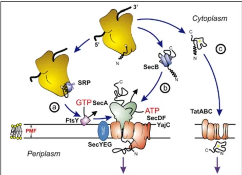
targeting; translocation; release and maturation[6]. The machinery of the Sec pathway recognizes a hydrophobic N-terminal leader sequence on nascent proteins destined for secretion, and translocates proteins in an unfolded state. The nascent proteins are recognized by the ribonucleoprotein signal-recognition particle (SRP) or the SecB chaperone[6]. The proteins recognized by SRP are mainly targeted to the inner membrane. Then, the nascent proteins are targeted to the translocase at the membrane. For SRP, this is achieved by docking to its membrane receptor FtsY[7] and for SecB by docking to the SecA subunit of the
translocase[8] (Fig.1).
In Escherichia coli the Sec translocase is comprised of the SecYEG translocation channel and the accessory components SecA, SecDFYajC, and YidC[4]. The cytoplasmic SecA subunit
hydrolyzes ATP to drive translocation[1], SecDFYajC improves protein translocation and
membrane protein insertion and YidC is required for membrane insertion of certain Sec-dependent substrates and for membrane insertion of Sec-inSec-dependent substrates[4]. In the YidC pathway, YidC functions as a membrane insertase without the Sec machinery[9] (Fig.1).
In comparison to E. coli, the main difference in B. subtilis Sec pathway is the lack of the chaperone SecB and the existence of other two chaperones, GroE and DnaK[10].
I.1.1.2 – The Tat pathway
The machinery of the Tat secretion pathway recognizes a typical twin-arginine motif in the N-terminal domain and translocates the protein in a folded state [11].
In E. coli, the Tat machinery consists of TatA, TatB, TatC and TatE proteins but its variable in other bacteria. Thus, the minimal Tat translocase is composed of one TatA and one TatC subunit[4]. TatA, TatB and TatC are integral membrane proteins that are inserted in the inner membrane in E. coli, forming a “core complex”[12]
(Fig.1). TatC functions in the recognition of
targeted proteins, while TatA is thought to be the major pore-forming subunit[13].
To avoid the translocation of Tat substrates by the Sec pathway, typically the C-terminal region of Tat substrates carries a basic residue, the “Sec-avoidance” motif that, in combination with the twin-arginine motif in the N-region, avoids the translocation of Tat substrates by the Sec pathway[14].
3
Fig. 1 – Schematic overview of the E. coli Sec, Tat and YidC pathways. (a) Co-translational and (b)
post-translational targeting routes of unfolded proteins to the Sec-translocase. (c) Translocation of folded precursor proteins by the Tat translocase. Adapted from [15].
I.1.2 – Type I to Type VI secretion systems
This type of classification evolved from the studies on secretion pathways of Gram-negative bacteria and some authors argue that this classification should apply only to systems that translocate proteins across both the cytoplasmic and outer membranes. However, it is unquestionable that some of these pathways are at least partially conserved in Gram-positive bacteria. Some secreted proteins are exported across the inner and outer membranes in a single step, whereas other are first exported into the periplasmic space via the universal Sec or Tat pathways mentioned above and then translocated across the outer membrane[1] (Fig.2).
Fig. 2 – Summary of Type I to Type VI secretion systems. HM, host membrane; OM, outer membrane;
IM, inner membrane; OMP, outer membrane protein; MFP, membrane fusion protein. ATPases and chaperones are shown in yellow. Adapted from [1].
4
I.1.2.1 – Type I secretion system (T1SS)
The T1SS or ATP-binding cassette (ABC) transporters are heterotrimeric complexes consisting of an inner membrane ABC exporter, a membrane fusion protein and a pore-forming, outer-membrane protein[3] (Fig.2).
In the α-hemolysin (HlyA) secretion of E. coli, the T1SS complex is composed by TolC, HlyD and HlyB. TolC is an integral membrane protein on the outer membrane; HlyD is a membrane fusion protein and HlyB is an inner membrane ABC exporter[16]. Most protein substrates possess a C-terminal signal sequence characterized by loosely conserved secondary structures[17], which is not cleaved off during secretion and must be presented to the ABC exporter in an unfolded state[16].
I.1.2.2 – Type II secretion system (T2SS) and type V secretion system (T5SS)
The T2SS and T5SS are only found in Gram-negative bacteria and take proteins that were exported by the Sec pathway or, in a few cases, the Tat pathway, into the periplasmic space, remove the signal peptides and translocate them in a folded state across the outer membrane[18,
19]
(Fig.2).
The T2SS consists in 12 core components: an outer membrane secretin; a cytoplasmic ATPase; an inner membrane protein; the major and minor pseudopilins; the pre-pseudopilin peptidase/metiltransferase; and a protein that might be involved in substrate recognition and/or secretin interactions[20, 21]. These proteins of the T2SS assemble to form a complex that spans the entire gram-negative cell envelope and is composed by a component in the cytoplasm, an inner membrane sub-complex that reaches into the periplasmic compartment and a secretion pore in the outer membrane[19]. This complex is responsible for the multi-protein assembly in the periplasm and their translocation across the outer membrane[19].
In the T5SS, after the proteins are exported to the periplasm by the Sec or Tat pathway, the secreted proteins are translocated across the outer membrane via a transmembrane pore, formed by a beta barrel[22] and with no energy coupling or accessory factors required for the
translocation[23].
There are three sub-classes of the T5SS machinery: the T5aSS; the T5bSS; the T5cSS. The T5aSS is also named the autotransporter system. The proteins are synthetized with an N-terminal signal peptide and exported to the periplasm via the Sec pathway like T5bSS and T5cSS. The C-terminal domain forms a beta barrel that is required for translocation into the extracellular space[1]. The T5bSS, also known as two-partner secretion pathway[24] consists of pairs of proteins in which one partner carries the beta barrel domain, and the other partner is the secreted protein, needing to interact with each other to be translocated to the extracellular medium[25]. Finally, in the T5cSS the proteins are trimeric and the beta barrel is formed from contributions from all three polypeptides[1] (Fig.2).
5
I.1.2.3 – Type III secretion system (T3SS)
The T3SS are also restricted to Gram-negative bacteria and form complex, supramolecular structures which span the inner membrane, the periplasmic space, the outer membrane, the extracellular space and the host cellular membrane[3] (Fig.2). This machinery is termed the injectisome and delivers effector proteins across the bacterial and host membranes into the cytosol of host cells in a Sec-independent manner[1, 26].
The substrates of this system are divided in two classes: the components of the machinery itself and substrates that are translocated to the target cell[18]. The Type III secretion machinery is highly conserved, consisting of more than 20 proteins, many of them sharing homology to flagellar export factors[27].
The injectisomes are composed of a series of basal rings that span the bacteria inner and outer membranes, connected to an hollow needle, filament or pilus, tipped with a translocation pore that is inserted into the plasma membrane of the target cell[1, 26]. Two oligomeric rings form a ring complex called “basal body” inserted in the inner membrane with an ATPase bounded in the cytoplasmic face of the basal body, energizing the protein translocation[3].
I.1.2.4 – Type IV secretion system (T4SS)
The T4SS are found in Gram-negative and Gram-positive bacteria, and are characterized by its unique ability to translocate nucleic acids and complexes of proteins into plant and animal cells, generally by a contact-dependent mechanism and to receive foreigner DNA (transformation)[1, 3, 18].
T4SS can be divided in three groups: with the ability to mediate the conjugative transfer of plasmid DNA or transposons; with the ability of mediate DNA uptake from and release it into the extracellular medium; with the ability to deliver effector macromolecules into eukaryotic cells during the course of infection[28].
The machinery of T4SS is composed by a substrate receptor, an envelope-spanning translocation channel and an extracellular pilus or surface filament (Fig.2).
I.1.2.5 – Type VI secretion system (T6SS)
The T6SS were first described by Pukatzki in 2006[29], and are characterized by a membrane-penetrating structure, delivering the effector proteins directly into the cytoplasm of host cells, or as a channel for protein translocation without the requirement of hydrophobic N-terminal signal sequences[3, 29, 30].
The machinery of T6SS shows similarities to the T4SS components and a model is proposed to include a cytoplasmic chaperone with ATPase activity, a channel bridging from the inner membrane to the outer membrane and a needle tipped with a pore-forming protein[31] (Fig.2).
6
Recently it has been shown that phage tails and T6SS are structurally, functionally and evolutionary related[32]. It seems that T6SS is a mechanism that can be adapted by individual bacterial species to interact with other prokaryotes and eukaryotes[33].
I.2 – The Type VII/ESAT-6 secretion system (T7SS/ESS)
The T7SS/ESS is a general designation for a protein secretion system with wide distribution among Gram-positive bacteria and some Actinobacteria such as mycobacteria[34]. Apart from a few core elements, the remaining components of this secretion pathway can be completely unrelated among the different bacterial species[35].
The system was first described in Mycobacterium tuberculosis, which has a complex cell envelope that includes a peculiar outer membrane enriched in mycolic acids[36]. For this reason, following the classification scheme in Gram-negative bacteria this secretion pathway was coined as T7SS. Although with some controversy[2, 37] this is the nomenclature currently used in Actinobacteria, whereas in Gram-positive bacteria, which have only a cytoplasmic membrane and a cell wall, the most common adopted designation is Esat-6-like secretion system (ESS) or WXG100 secretion system (WSS)[2, 38] (see below).
Unlike bacterial T3SS or T4SS, secretion of T7SS/ESS substrates in M. tuberculosis occurs in in vitro grown cultures and seems not to require host cell target interaction for translocation of the secreted proteins[39].
I.2.1 – Discovery of the T7SS/ESS in M. tuberculosis
The first clues for a new T7SS/ESS came from the in silico analysis of the M. tuberculosis virulence effectors ESAT-6 (early secreted antigen target, 6 kDa) and CFP-10 (culture filtrate protein, 10 kDa), encoded respectively by esxA and esxB genes in the region of difference 1 (RD1)[35]. These effectors are known to be secreted without a recognizable secretion signal[40, 41] and interact with each other forming a tight 1:1 heterodimer[42, 43].
The RD1 region, which contributes to the pathogenesis of M. tuberculosis[44], is deleted in the vaccine strain Mycobacterium bovis (BCG), being this one of the reasons for the attenuated virulence of BCG[45]. When the complete RD1 region was reintroduced in the BCG strain the secretion of ESAT-6 and CFP-10 was restored[46], revealing the first evidences for a new secretory system.
The ESAT-6 and CFP-10 proteins are important T-cell antigenic targets[47] and belong to the
WXG100 superfamily, whose members were initially defined as sharing a central tryptophan-variable-glycine (WXG) motif, a protein length of ~100 residues[35, 48] and the tendency to cluster with genes for membrane proteins, ATPases and/or chaperones[40, 41, 48]. M. tuberculosis has up to five T7SS/ESS, termed ESX-1 to ESX-5, being the ESX-1 the best studied and the responsible for the secretion of ESAT-6 and CFP-10[49].
7
There are other substrates of the T7SS/ESS of M. tuberculosis, such as the PE and PPE proteins that form heterodimeric complexes[50] and are associated with virulence[51] like ESAT-6
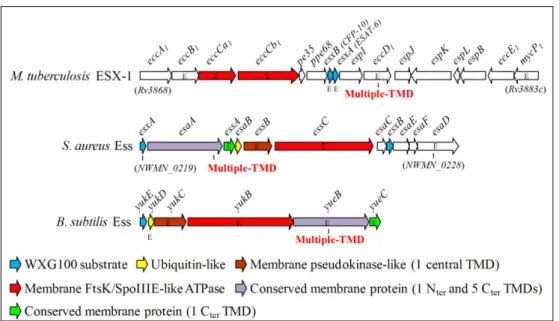
and CFP-10. Distantly related gene clusters were also found in the genome of pathogenic and non-pathogenic Gram-positive bacteria such as Staphylococcus aureus and B. subtilis[1, 35] (Fig.3).
Fig. 3 – Schematic representation of the gene clusters encoding core components and substrates of T7SS-like systems in M. tuberculosis (ESX-1), S. aureus (Ess) and B. subtilis (BsEss). The
nomenclature of ESX-1 genes is that propose by Bitter et al.[37]. For simplicity, the genes located immediately or far upstream of M. tuberculosis eccA1 are not represented. Genes described as essential or important for secretion of cognate WXG100 proteins, or of other specific substrates, are marked with the letter “E”. Genes coding for products sharing conserved domains are depicted with the same color code, whereas those specific of each system are colored in white. Features of conserved gene products are indicated below. TMD, transmembrane domain.[52]
I.2.2 – Molecular composition of T7SS/ESS
In M. tuberculosis, the five T7SS/ESS share a number of highly conserved components, named Ecc (ESX-conserved-component)[37]. Five of these components are predicted to be membrane proteins and could form a translocation channel. Two other components show homology to proteins with known function: MycP, a protease that belongs to the subtilisin protein family; and EccC which is a member of the FtsK/SpoIIIE family of ATPases[53].
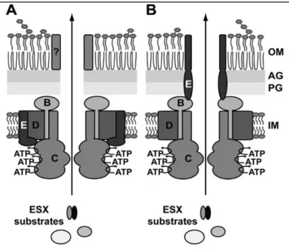
In M. tuberculosis, the membrane complex is thought to be composed of the membrane proteins EccB, EccC, EccD and EccE, that likely form a translocation channel[49]. This complex has a size of ~1500 kDa and might consist of six copies of EccB, EccC and EccD and three copies of EccE, being EccE only absent in the ESX-4[49]. The six copies of the ESX-5 EccCare in accordance to members of the FtsK/SpoIIIE family which forms hexamers[54]. Recently, it has been proposed two models for the topology of the T7SS/ESS membrane complex (Fig.4).
8
Fig. 4 – The two proposed models for the T7SS/ESS membrane channel. EccBCDE are embedded in
the cytosolic membrane were they assemble into a large membrane complex. The three nucleotide-binding domains of EccC are likely involved in energizing translocation of substrates through the translocation channel. In (A) is a two-step process and in (B) a one-step process. IM, inner membrane; PG, peptidoglycan; AG arabinogalactan; OM, mycobacterial outer membrane.[49]
I.2.3 – Mechanism of action of T7SS/ESS
Although the substrates and the membrane complex have been studied intensively, the mechanism of action is still unclear. It is known that all the substrates in ESX-1 are mutually dependent upon each other for secretion[55] and in mycobacteria they seem to be preferentially
secreted as heterodimers[56].
It has been identified a CFP-10 secretion signal in its C-terminus[57], more specifically a YxxxD/E motif that appears to be shared by all known classes of T7SS/ESS substrates in mycobacteria, except for PPE and ESAT-6-like. These last proteins are secreted as a complex with a motif-containing substrate and can be used to predict putative unknown T7SS/ESS substrates, although the signal does not discriminate among the various ESX pathways[56].
Recently it has been added the function of EspG, a protein that may maintain the PE/PPE proteins in a stable conformation[58], possibly directing them to the membrane-embedded secretion machinery where the YxxxD/E motif is potentially recognized by EccE and then translocated across the mycomembrane.
I.2.4 – Role on pathogenic interaction with eukaryotic host
As it was mentioned above, ESAT-6 and CFP-10 act as virulence effectors of M. tuberculosis, inducing a strong T-cell response during infection[47]. Among all the different proposed functions for ESAT-6, CFP-10 and ESX-1, the most frequently reported observations
9
points towards a function linked to lysis of cells and/or membranes[39, 44], induced apoptosis, translocation to the cytosol and phagosomal rupture in the host cell[59, 60, 61]. The immune responses induced by ESX-1 antigens are generated at both the innate and adaptive levels to limit mycobacterial replication, and an ESAT-6-specific T-cell response is obtained only when ESAT-6 and CFP-10 are expressed and secreted[39].
It is known that ESAT-6 plays a crucial role in M. tuberculosis infection[62], namely by interacting and disintegrating the host cell membrane[63, 64], by modulating the activation of the macrophage[65] and interacting with the host signaling pathway[62]. It is also known that the RD1 is involved in the translocation of M. tuberculosis from the phagosome into cytoplasm of the host cell at later stages of infection[66], which results in the induction of cytotoxicity and in the end, host cell death[61].
Altogether, this data shows that the T7SS/ESS of M.tuberculosis is clearly involved in pathogenicity and host-cell infection.
I.2.5 – DNA transfer mediated by T7SS/ESS
The T7SS of Mycobacterium smegmatis, which is homologous to the M. tuberculosis ESX-1, is involved in DNA transfer by a conjugation-like mechanism[67]. Interestingly, T7SS has opposite impact in donor and recipient strains: donor secretion negatively regulates transfer whereas in recipient is essential[68]. Such opposed effects cannot be explained by the nature of the secreted substrates (ESAT-6/CFP-10 homologues) as these are identical in donor and recipient strains[68]. Thus, secretion by T7SS and DNA transference appear to have a close interplay in M. segmatis, suggesting that this relation can also occur in other bacteria that possess Esat-6-like secretions systems.
I.3 – The S. aureus Esat-6-like secretion system (Ess)
As mentioned before, S. aureus possesses a gene cluster, the ess locus (Fig.3), encoding an Esat-6-like secretion system with some elements displaying homology to those of T7SS/ESS of M. tuberculosis.
The Ess proved to be functional and its WXG100 substrates required for the pathogenicity of S. aureus[38]. The component EssC is a member of the FtsK/SpoIIIE family, whose function is essential for secretion of EsxA and EsxB, which in their turn are two secreted substrates of the WXG100 superfamily[38]. EsxA and EsxB depend on each other to be secreted. In addition, EsaC is a polypeptide that is also secreted by the S. aureus Ess[69].
Recently it has been shown that EsaD and EssB are involved in the secretion of Ess substrates[70, 71]. EsaD is a polypeptide located in the staphylococcal membrane, is part of the Ess and supports the secretion of EsxA, being therefore involved in the pathogenesis of staphylococcal infections[70]. EsaD also shows homology to the B. subtilis YeeF[70]. EssB is a
10
membrane protein, required for EsxA secretion and an essential component of the Ess translocon. It probably interacts with itself and other machinery components[71]. EssB is homologous to B. subtilis YukC[71]. The Ess was proved to interfere with host immune responses and to favor the establishment of persistent infections[69].
I.4 – The B. subtilis Esat-6-like secretion system (BsEss)
The Gram-positive non-pathogenic model bacteria B. subtilis has a gene cluster, the yuk locus that is positively regulated by phosphorylated DegU[72] and which carries some
components homologous to elements of the M. tuberculosis T7SS and S. aureus Ess[48].
This B. subtilis locus is composed by an FtsK/SpoIIIE-like ATPase encoded by yukB, an ubiquitin-like protein YukD[73], the YukC protein with a pseudokinase-like fold[74], the membrane proteins YueB and YueC and the putative WXG100 substrate YukE. YueB was previously known as the receptor essential for phage SPP1 infection[75]. Recently, YukE was shown to be secreted as a homodimer, being its export absolutely dependent on elements of the Yuk cluster and on the phosphorylated form of the response regulator DegU[52].
In this work the yuk locus was renamed BsEss (B. subtilis Esat-6-like secretion system). The only conserved elements among the T7SS/ESS of M. tuberculosis, S. aureus and B. subtilis are the FtsK/SpoIIIE-like ATPase (YukB in B. subtilis) and the WXG100 substates (YukE in B. subtilis)[38, 48]. In addition to these elements, S. aureus and B. subtilis share esaA/yueB, essA/yueC; esaB/yukD and essB/yukC[52] (Fig.3).
BsEss operation was demonstrated in the undomesticated strain B. subtilis ATCC 6051 when entering the stationary growth phase. Genes of the BsEss locus were required for YukE stable production, secretion and accumulation in culture supernatants and their activation depended on high levels of phosphorylated DegU[52]. It is known that the classic lab strain B. subtilis 168 has accumulated several mutations during is domestication that seem to diminish DegU phosphorylation or its action as transcriptional activator[72, 76], thus possibly affecting the expression of the BsEss locus. In strain ATCC 6051, where YukE is secreted[52], these mutations are not present[77].
I.5 – Thesis goals
The T7SS/ESS secretion systems are widespread among bacteria of the phyla Actinobacteria and Firmicutes and in some species they play an important role in bacterial pathogenesis. Due to the amenability and availability of tools for the genetic manipulation of the B. subtilis lab strain 168 and its derivatives, the BsEss could be an attractive model to study molecular details of this important secretion pathway.
However, as mentioned above, secretion of YukE is strongly diminished in this domesticated strain. Strain 168 is known to have mutations affecting genes epsC, sfp, swrA and degQ[77, 78, 79],
11
all of them involved in Deg-regulated cellular processes like swarming and biofilm formation[72,
79, 80, 81, 82]
. In addition, strain 168 is also cured from a plasmid that has strong influence in biofilm architecture of ancestral strains such as NCIB 3610[79]. This plasmid was recently shown to be responsible for the low competence for transformation of undomesticated strains[83].
In this work, we aimed to study if genetic mutations reported in B. subtilis 168 could account for the defective operation of BsEss in this domesticated strain, at the same time contributing to the understanding of the functioning and regulation of this secretion pathway. The effect of other genes such as swrB, aprE, degR and yeeF, the latter of which is homologue to S. aureus EsaD, shown to support Ess functioning[70], was also studied. As an attempt to uncover cellular functions of the BsEss (none is presently known) we have also explored in this work previous results that linked the BsEss cluster to the development of competence for DNA uptake (C. Baptista, unpublished).
13
II – Results
II.1
– Why is B. subtilis strain 168 impaired in BsEss
functioning?
II.1.1
– Study of well-known 168 mutations in an undomesticated
background
As previously mentioned, the genome of the B. subtilis reference strain 168 (GenBank NC_000964) carries mutations affecting genes epsC, swrA, sfp and degQ, when compared to undomesticated strains such as NCIB 3610 (GenBank NZ_CM000488) and ATCC 6051 (GenBank NC_020507)[77, 79].
Mutations in swrA and sfp correspond to frameshift mutations[79, 82], whereas that impairing epsC is a missense mutation[79]. The mutation affecting degQ lies in the -10 box of the promoter sequence, thus decreasing its transcription[79, 81]. These mutations impair swarming and biofilm development, which are cellular processes under the control of one major cell-fate regulator, DegU[72, 84]. In addition, the 3610 strain carries an 84-kb plasmid encoding functions that stimulate and repress biofilm formation and competence development, respectively[79, 83].
Our lab as recently confirmed that the BsEss is part of the DegS-DegU regulon, being its expression and functioning highly enhanced by the phosphorylated form of DegU, DegUP[52]. The same study showed that strain 168 secreted very low levels of YukE when compared to strain 6051, suggesting a deficient activation and/or functioning of the BsEss in the domesticated strain. Given the link between the 168 mutations referred above and the Deg regulon, we have decided to study the involvement of the respective genes on BsEss functioning. Gene epsC was not included in this study since the mutation reported in strain 168 seems to impair a specific step of exopolysaccharide biosynthesis[79], and thus we have considered it unlikely to be involved in BsEss operation.
We have started by checking YukE production and secretion in the stationary growth phase of strains derived from the 6051 or 3610 backgrounds, which carried mutations affecting the genes altered in strain 168. In terms of their chromosomal sequences strains 3610 and 6051 are very similar[78] and in our lab conditions the level of yukE accumulation in culture
supernatants is indistinguishable between the two ancestral strains (not shown). The different mutant strains used in this study are described in the Materials and Methods section.
Culture samples taken 2 hours after entry in the stationary phase were processed for precipitation of total proteins present in cell-free supernatants. After solubilization of the precipitates, total protein was quantified by the Bradford method, which seemed to give overestimates of protein content, probably due to the colored PRMM precipitates (see
14
methods). The precipitates were analyzed by SDS-PAGE and Coomassie blue staining to monitor the quality and relative protein quantities before transfer for Western blot analyses (see methods).
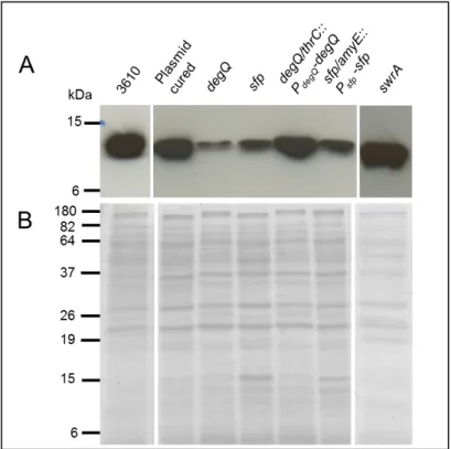
YukE secretion in a 3610 derivative lacking the 84-kb plasmid (strain DS2569) seemed similar to that observed in the WT strain (Fig.5). This result indicates that the absence of this plasmid in strain 168 should not be responsible for the malfunction of the BsEss. In contrast, a degQ knockout mutant of strain 6051 (strain W105) resulted in a marked decrease of YukE secretion (Fig.5). Complementation of the degQ mutant by inserting in the dispensable thrC locus a WT copy of degQ under the control of its native promoter (strain HCB1, see methods) restored high levels of YukE secretion (Fig.5).
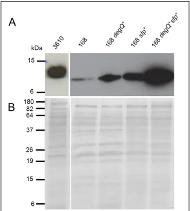
Fig. 5 – Immunodetection of YukE in supernatant precipitates of different B. subtilis mutants derived from undomesticated strains 6051 or 3610. A. For details of strains and mutations see section
Material and Methods. 3610 (wild-type strain NCIB 3610); Plasmid cured (strain DS2569 ); degQ (strain W105); sfp (strain DS3337); swrA (strain DS2415); degQ/thrC::PdegQ-degQ (strain HCB1); sfp/amyE::Psfp-sfp (strain HCB2). Each lane was loaded with 20µg total protein. B. Coomassie blue-stained gel to show
even protein loading.
In absence of swrA (strain DS2415) YukE secretion seemed to be unaffected (Fig.5), suggesting that the swrA mutation described in strain 168 should not be affecting BsEss operation. The sfp mutation (strain DS3337) seemed to cause significant reduction of the YukE signal (Fig.5). However, when we tried to complement the sfp mutation by inserting Psfp-sfp in
the amyE locus of strain DS3337 (HCB2, see methods) we did not obtain the expected increase in YukE secretion (Fig.5). At this point the results were inconclusive concerning the real contribution of sfp to the BsEss pathway (see discussion).
15
In summary, by introducing in undomesticated backgrounds genetic modifications that mimic those known to occur in strain 168 we have concluded that the 84-kb plasmid and gene swrA have no major role in BsEss functioning. On the contrary, mutations affecting degQ and eventually sfp seem to produce an impact on BsEss operation, as judged by the observed reduction in YukE secretion.
II.1.2 – Restoring BsEss functionality in B. subtilis strain 168
The results from the previous section suggested that the mutations known to affect degQ and sfp expression in strain 168 could be responsible for the defective BsEss functioning in this genetic background. We have confirmed by sequencing that strain 168 from our collection carried the reported mutations (not shown). To check the role of these mutations in the BsEss defective phenotype of strain 168 we have tested if their complementation resulted in an increase of YukE secretion. Complementation of the degQ mutation was carried out by inserting the wild-type cassette PdegQ-degQ in the thrC locus of strain 168, resulting in the derivative
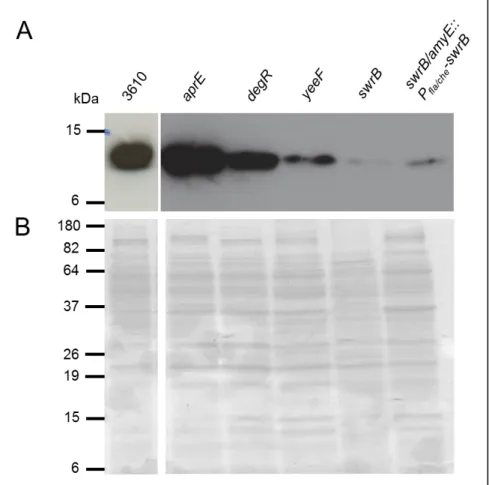
HCB3 (see methods). This strain showed a clear increase in YukE secretion when compared to the parental strain 168, yet YukE signal was still much smaller than that observed in an undomesticated strain (Fig.6). This result suggested that increasing degQ expression in strain 168 was not sufficient to fully restore BsEss operation and/or YukE accumulation in culture supernatants.
Fig. 6 – Immunodetection of YukE in supernatant precipitates of different B. subtilis mutants derived from the domesticated strain 168. For details of strains and mutations see section Material and
Methods. 3610 (wild-type strain NCIB 3610); 168 (L16601 strain); 168 degQ+ (HCB3 strain); 168 sfp+ (HCB4 strain); 168 degQ+sfp+ (HCB5 strain). Each lane was loaded with 20µg total protein. B. Coomassie blue-stained gel to show even protein loading.
16
A similar result was obtained with a 168 derivative (HCB4) carrying Psfp-sfp in the amyE
locus (Fig.6). Interestingly, a double-complemented 168 strain (HCB5) showed levels of YukE secretion comparable to those observed in ancestral strains (Fig.6), indicating that the correction of these two mutations are necessary and sufficient to fully restore the BsEss functioning in strain 168. In summary, the impairment of BsEss in strain 168 seems to be due to the mutations reported in the promoter region of degQ and in sfp.
II.2
– Other genes involved in BsEss functioning or
regulation
Several studies on other T7SS/ESS systems and on the Deg regulon suggested that other B. subtilis genes could be involved in the operation and/or regulation of the BsEss. Among these we have selected to study genes swrB, degR, aprE and yeeF. When growing in rich liquid media, B. subtilis populations are majorly composed of swimming (motile) cells thanks to the activity of SwrA that stimulates the fla/che operon. In addition to be involved in flagella synthesis and chemotaxis functions, the fla/che operon also encodes σD
and its activator SwrB, which are responsible for stabilizing the motility state by driving the expression of other genes[85]. It is also known that swrB is required for swarming motility, probably by stimulating the assembly and number of cell surface flagella[82]. DegR expression is driven by σD[86], and DegR has a positive effect on phosphorylated DegU by stabilizing their phosphorylated state[87]. The gene product of aprE is the extracellular protease subtilisin[88], which is representative of a large subset of serine proteases (PFAM family Peptidase_S8, PF00082). Expression of aprE is stimulated by phosphorylated DegU, which binds to the aprE promoter[89, 90]. Interestingly, MycP1, a subtilisin-related mycosin has been shown to have a role in M. tuberculosis ESX-1 functioning, probably by cleaving specific domains of ESX-1 components[91, 92].
Given the link of all these genes to the DegS-DegU regulon, which as shown before is essential to the activation of the BsEss[52], we have probed secretion of YukE in an undomesticated strain individually affected in these genes. In an aprE mutant (strain W103) secretion of YukE seemed to be unaffected when compared to strain 3610, whereas in a degR mutant (strain W128) it was only slightly reduced (Fig.7). These results suggested that YukE and DegR are not crucial for BsEss function. Somewhat surprisingly (see discussion), YukE secretion was much reduced in a swrB deletion mutant (strain DS2509), suggesting a role for this gene in BsEss operation and/or YukE extracellular accumulation. Unfortunately, YukE secretion levels could not be restored in a complementation strain (DS2522) carrying native swrB, under the control of the operon fla/che promoter, in the amyE locus of the parental DS2509 (Fig.7), a result that claims for additional confirmatory studies.
17
Fig. 7 – Immunodetection of YukE in supernatant precipitates B. subtilis mutants derived from ancestral strains. 3610 (wild-type NCIB 3610 strain); aprE (W103 strain); degR (W128 strain); yeeF
(HCB6 strain); swrB (DS2509 strain); swrB/amyE::Pfla/che-swrB (DS2522 strain). Each lane was loaded with
20µg total protein. B. Coomassie blue-stained gel to show even protein loading.
Recently, it has been shown that EsaD, an homologue of B. subtilis yeeF, is required for normal secretion of EsxA by the S. aureus Ess[70]. Therefore, we have decided to test if yeeF disruption would produce some impact on YukE sectretion. yeeF was disrupted through integration of a pMutin4 derivative. The advantage of this strategy is that, in principle, only the target gene is affected. This is achieved by two main properties of the vector[93]: i) vector encoded transcriptional terminators block the transcription initiated upstream of the integration site, and ii) the vector-borne, IPTG-inducible promoter Pspac allows expression downstream of
the inactivated gene, thus bypassing polar effects in case of operon structures. In the undomesticated strain with yeeF disrupted (HCB6; see methods) the culture samples were obtained in the presence of IPTG, to avoid polar effects. YukE secretion in this strain was reduced (Fig.7), suggesting that yeeF has a similar role to his homologue EsaD. For a full confirmation of these results, the complementation of this strain with yeeF expressed ectopically shall be made in future works.
18
II.3
– On the track of BsEss cellular function: does BsEss
affect competence development?
At this moment there is no known cellular function for the BsEss. In this work we have decided to follow a lead from preliminary results that seemed to implicate the BsEss cluster in a reduction of natural competence for DNA transformation (C. Baptista, unpublished). This lead came from a global analysis that aimed to study the effect of BsEss deletion in general cellular processes like competence development, sporulation and secretion of degradative enzymes. Interestingly, these results were obtained in the lab strain 168, where BsEss expression/functioning seem to be repressed when compared to undomesticated strains (see above). We should note that competence development is very difficult to induce in undomesticated strains in lab conditions[77, 83], making reliable quantitative measurements of competence efficiency very difficult to perform. An additional motivation to study the effect of BsEss in competence development was the fact that DegU is also known to be involved in the regulation of competence genes[94], in addition to the master competence regulator ComK[95, 96].
As the regulation of BsEss and of competence genes involves DegU, BsEss functioning
could somehow interfere with the development of competence. Although impaired in YukE secretion in LB medium, strain 168 is still able to transcribe BsEss genes at different levels, depending on growth conditions[75, 97].
We have studied development of competence in the control strain L16601.amy::CM, which is a 168 derivative with a chloramphenicol cassette in the amyE locus, and in strain L.Del6, which carries a deletion covering the promoter region of the BsEss cluster located upstream yukE and downstream genes until yueB[98]. Construction of this deletion mutant implied the insertion of a chloramphenicol resistance cassette in gene yukF, which lies immediately upstream and is divergently transcribed relatively to yukE. To control a putative effect derived from yukF inactivation, a 168 derivative strain carrying yukF disrupted by a chloranphenicol cassette (L.cat86) was also tested for transformation efficiency.
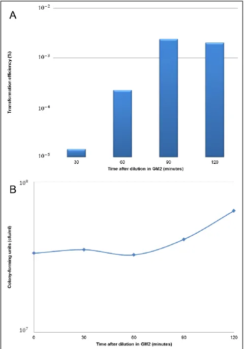
The protocol followed for developing B. subtilis competence was essentially that described by Yasbin et al.[99] (see methods). Literature is a bit ambiguous about the time necessary for optimal competence development after culture dilution in GM2 medium and about the minimal quantity of transforming DNA for maximal efficiency of transformation[99, 100, 101, 102]. We have thus, in a first step, conducted experiments to optimize the protocol in order to have the highest transformation efficiency possible. The results showed a peak of transformation efficiency 90 minutes after dilution and incubation in GM2 (Fig.8A), which roughly corresponded to the time when cultures re-gained exponential growth (Fig.8B).
19
Fig. 8 – B. subtilis transformation efficiency in GM2. GM1 cultures that had been in stationary growth
phase for 90 min were 10-fold diluted in GM2 and assayed for their efficiency of transformation at different time points post-dilution. A. Strain 168 showed the highest transformation efficiency 90 minutes after dilution in GM2. B. Strain 168 re-entered in exponential growth phase after 90 minutes in GM2.
Next we determined the efficiency of transformation for three concentrations of transforming DNA (1, 5 and 25 g/ml). With 5 µg/ml of DNA the number of transformants per total cell number was clearly higher than with 1 µg/ml, but only slightly lower than with 25 µg/ml of DNA (not shown). Therefore, in all subsequent experiments we have used for transformation 5 µg/ml of DNA and 90 minutes of incubation in GM2.
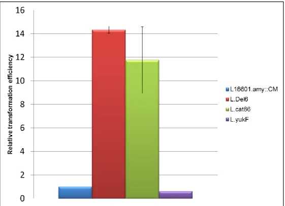
Strain L.Del6 showed transformation efficiency about fourteen times higher than the control strain (Fig.9), suggesting that the deletion of the BsEss cluster and/or the disruption of yukF play a role in the development of competence. In an attempt to clarify this question, we tested
20
strain L.cat86, which supposedly only carried yuKF disrupted by a chloramphenicol resistance cassette. Surprisingly, the results showed a transformation efficiency similar to that of strain L.Del6 (Fig.9). According to Baptista[98], the construction of strain L.cat86 implied several amplifications of the sequences covering the 5’ end of yukF, the BsEss promoter region, yukE, yukD and the 5’ end of yukC. Although this strain showed the expected DNA structure and behaved like the parental 168 in terms of SPP1 infection[98], the presence of eventual point mutations in BsEss genes, due to amplification errors, was never ruled out. So, for clarifying the role of yukF in the transformation efficiency, a strain with yukF disrupted by single crossover plasmid integration (L.yukF, see methods), and therefore more reliable than L.cat86, was equally tested. The results showed transformation efficiency similar to the control strain, in clear contrast to the results observed for L.cat86.
Fig. 9 – Relative transformation efficiency of 168 mutant strains. The efficiency of transformation is
expressed as a ratio of that observed in control strain L16601.amy::CM. The data was obtained with at least three independent experiments. The minimal number of transformants was 40/µg of DNA.
In summary, the results were contradictory in terms of the effect of disrupting yukF in competence development, making it difficult to draw any conclusion about the results obtained with the BsEss deletion strain, which also carried inactivated yukF. We are currently constructing a strain only deleted for the BsEss operon in order to elucidate these results.
21
III – Discussion
Recently, it was proved that the Esat-6-like secretion system of B. subtilis, encoded by the BsEss gene cluster, was functional in an undomesticated strain and dependent of high levels of phosphorylated DegU[52]. In this work we sought to understand the genetic bases for the deficient functioning of the BsEss in the domesticated B. subtilis lab strain 168. In addition to the advantages of having a fully operational BsEss in strain 168, which is much more amenable to genetic manipulation when compared to ancestral strains, with this work we also wanted to get further insight on the genetic circuits involved in the regulation and/or molecular mode of action of this secretion system.
B. subtilis 168 has accumulated during its domestication several mutations that seem to diminish DegU phosphorylation or its action as a transcriptional activator, namely mutations blocking degQ and swrA expression. Other differences between strain 168 and the 6051 or 3610 ancestral strains include mutations blocking sfp and epsC and the absence of a plasmid in strain 168. The mutation in epsC seems to specifically affect the production of an exopolysaccharide important for biofilm formation[79], and thus we have decided not to include it in our study.
Based on the literature, mutations affecting degQ and swrA would be the ones more probable to affect the function of BsEss. DegQ stimulates production of DegUP, a major activator of BsEss and it also enhance its own transcription and of yukC[72]. Expression of degQ is in its turn modulated by the two-component regulatory system ComP-ComA, and also to some extent by DegS-DegU, upon different nutritional stimuli[103]. DegQ is required to swarming motility and biofilm formation[79, 82]. SwrA had been previously described as a positive modulator of DegU function as transcriptional activator[76]. DegQ and SwrA seem to be functionally “linked” as mutations in their genes affect the same cellular phenomena such as swarming and biofilm formation[79, 82]. In our genetic analysis we have confirmed the expected positive contribution of degQ to BsEss operation, both in an undomesticated background and in strain 168, but not for SwrA.
Intriguingly, we found that an in frame deletion of swrB, a gene belonging to the SwrA-stimulated operon fla/che, resulted in a drastic decrease of YukE secretion. Unfortunately, ectopic expression of a native swrB was not successful in reverting the YukE secretion phenotype, which calls for a re-evaluation of these results. In any case, if we confirm the effect of swrB mutation, the apparent contradictory swrA/swrB results can be explained if we assume that: i) swrB is still expressed to some level in swrA mutants and ii) the range of SwrB upregulated genes may extend beyond those involved in stabilizing motility[82, 85]. In fact, it appears that SwrA is required to express swrB and the upstream gene sigD above a certain threshold in a subpopulation of B. subtilis cultures, being swrB still expressed at considerable levels in a swrA-independent manner in the other fraction of the population (D. Kearns, personal communication).
22
Another gene that seems important to BsEss functioning is sfp. Its inactivation in an undomesticated strain resulted in an apparent decrease of YukE secretion, whereas its ectopic expression in stain 168, which is defective for sfp, resulted in a clear increase of extracellular YukE. Sfp is necessary for the production of the lipopeptide antibiotic surfactin[104]. Surfactin acts also as biosurfactant to reduce solid surface tension, which is an essential requisite for swarming motility[82, 105, 106, 107]. The mechanism by which sfp function stimulates BsEss is presently unknown. In addition of being an essential component of surfactin synthesis, sfp was also suggested to be involved in the regulation of the surfactant biosynthesis at the transcriptional level[104]. Moreover, surfactin itself seems to have the ability to function as modulator of gene expressing as it happens in biofilm formation, in which surfactin acts as an autoinducer or a quorum-sensing signal leading to the derepression of genes involved in matrix synthesis[108]. It will be interesting to test whether the simple addition of commercially available surfactin to media is sufficient to stimulate YukE secretion.
Disruption of gene yeeF inhibited YukE secretion, similarly to what happens when its homologue, the S. aureus Ess protein EsaD is disrupted[70]. EsaD is located in the staphylococcal membrane and is proposed to contribute to the selection of secretion substrates and/or interact with the Ess secretion machine, supporting the secretion of EsxA[70]. A similar role can be envisaged for YeeF. Further studies with ectopically expressed yeeF in HCB6 are still necessary to confirm its role in BsEss.
Other genes involved in Deg-regulated processes also tested in this work were aprE and degR. Their individual inactivation produced no major impairment in YukE secretion, despite a slight and somewhat expectable reduction observed with the degR mutant.
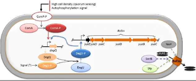
Overall, our genetic analysis indicates that correction of mutations affecting degQ and sfp in strain 168 are required, and should be sufficient, to restore YukE secretion to levels similar to those observed in ancestral strains. Other genes that seem to be involved in the activation and/or functioning of the BsEss are swrB and yeeF. These results, allied to the already known role of phosphorylated DegU in the activation of this system[52] are summarized in Figure 10.
In another part of this work we aimed to uncover a possible cellular function of this secretion system, based on the previous results that linked the BsEss cluster to the development of competence (C. Baptista, unpublished). At least in M. smegmatis the T7SS equivalent to the M. tuberculosis ESX-1 has been previously shown to be involved in DNA transfer[68]. By testing this hypothesis in the 168 strain, it was observed a ~14-fold increase of the transformation efficiency in a strain deleted of genes yukE to yueB of the BsEss cluster (L.Del6). As result of the construction strategy, this strain also carried yukF, the gene immediately upstream of yukE, disrupted by a cat86 cassette (confers chloramphenicol resistance), an so we had to rule out this mutation as being involved in the observed phenotype. Unfortunately, we obtained opposite results when using two strains that supposedly only carried yukF disrupted: L.cat86 behaved essentially as L.Del6, whereas strain L.yukF displayed competence levels similar to control strain L16601.amy::CM. Since the steps used to construct L.cat86 might have inadvertently introduced point mutations in some genes of the BsEss cluster (see section II.3), we tend to
23
believe that results with strain L.yukF are the trustable ones, that is, that simple yukF disruption produces no effect on competence development. In that scenario, which of course needs further confirmation, it is the lack of BsEss that is the responsible for the increased competence in L.del6. When yukF is inactivated in an undomesticated background by the same strategy used to generate L.yukF, no effect on YukE secretion is observed (C. Baptista, unpublished).
The development of competence is related to sporulation, both processes sharing a lot of regulatory and essential genes[109]. Interestingly, recently yukF was renamed adeR and shown to be required for normal sporulation of B. subtilis, being a transcriptional activator that mediates ald expression, also required for normal sporulation, in response to alanine availability[110]. Our results allied with these recent findings suggest that yukF and the BsEss cluster play a role in sporulation and competence, respectively. Further studies with a more “clean” strain only deleted for BsEss will be conducted in a near future.
Fig. 10 – Model for activation and regulation of B. subtilis BsEss. Note that the mechanism by which
Sfp, SwrB and YeeF exert their positive contribution to YukE secretion is completely unknown (dashed arrows).
25
IV - Concluding remarks
One major goal of this work was to elucidate the genetics underlying the defective BsEss operation in B. subtilis lab strain 168. We believe this was achieved as we found that mutations affecting degQ and sfp are sufficient to explain the defective YukE secretion in this strain. This is a valuable knowledge as it will permit to construct a marker-less derivative of strain 168 with fully functional BsEss. Such derivative might be advantageous to further dissect BsEss molecular details in a more amenable genetic background. As result of this work, two additional genes were identified, swrB and yeeF, which seem to contribute positively to BsEss functioning. Although the real contribution of these genes still requires formal confirmation by gene complementation assays, we are convinced that these preliminary findings will lead to new insights on this secretion system.
Unfortunately, we could not also obtain definitive conclusions about the interplay between BsEss and B. subtilis competence, but our interpretation of the results tend to favor the previously raised hypothesis that BsEss expression negatively affects competence development. This is a topic that certainly deserves further investigation.
27
V – Materials and Methods
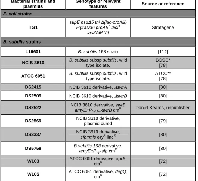
V.1 – Bacterial strains, plasmids and growth conditions
The bacterial strains and plasmids used in this work are listed in Table 1. E. coli and B. subtilis strains were usually grown in Luria-Bertani (LB) medium[111] with aeration at 37ºC,
except for E. coli transformants harboring pMutin4 derivatives, which were selected and propagated at 30ºC. Agar was added to LB medium at a final concentration of 1.5% (wt/vol) for bottom plates. E. coli strains carrying vectors or recombinant plasmids were grown in the presence of ampicillin (100µg/ml), while erythromycin (1µg/ml), chloramphenicol (5µg/ml), spectinomycin (100µg/ml) or lincomycin (200µg/ml) were used for the selection of B. subtilis transformants. B. subtilis transformants expressing β-galactosidase were selected in X-Gal (5-bromo-4-chloro-3-indolyl-β-D-galactopyranoside) suplemented (0.02%, wt/vol) LB plates.
Table 1 – Strains and plasmids used in this work.
Bacterial strains and plasmids
Genotype or relevant
features Source or reference
E. coli strains
TG1
supE hsdΔ5 thi Δ(lac-proAB) F’[traD36 proAB+ lacIq lacZΔM15] Stratagene B. subtilis strains L16601 B. subtilis 168 strain [112]
NCIB 3610 B. subtilis subsp subtilis, wild
type isolate.
BGSC* [78]
ATCC 6051 B. subtilis subsp subtilis, wild
type isolate.
ATCC** [78]
DS2415 NCIB 3610 derivative, swrA [80]
DS2509 NCIB 3610 derivative, swrB [80]
DS2522 NCIB 3610 derivative, swrB amyE::Pfla/che-swrB cm
R Daniel Kearns, unpublished
DS2569 NCIB 3610 derivative, plasmid cured [79] DS3337 NCIB 3610 derivative, sfp::mls eryR lincR [80] DS5758 B.subtilis 168 derivative, amyE::Psfp-sfp cm R [80]
W103 ATCC 6051 derivative, aprE;
cmR [72]
W105 ATCC 6051 derivative, degQ;