Antiparasitic potential
of natural marine
compounds
Gisela Almeida Castro
Biologia e Gestão da Qualidade da Água
Departamento de Biologia 2016
Orientador
Maria do Rosário Martins, Investigador CIIMAR-UP; Professora-Adjunta, ESS-IPP
Co-orientador
Todas as correções determinadas pelo júri, e só essas, foram efetuadas. O Presidente do Júri,
Acknowledgements
To Professor Doctor Rosário Martins and Sandra Pereira for their availability, great support, guidance, patience, friendliness and help that was essential to this work. I will be always thankful for all the effort.
I would like to thank also to Professor Vitor Vasconcelos, Agostinho Cruz and Piedade Barros for the access to the BEE laboratory at CIIMAR, and laboratories of Pharmacy and Morfological Sciences at the Health School respectively.
To Professor Natividade Vieira for all patience and help over the years of the Masters.
To professors Rita Ferraz, Ana Oliveira and Cláudia Pinho for the availability, the good mood that turned the experimental part easier and to provide their equipment to perform part of this work.
To Dr Maria José Borrego from Laboratório Nacional de Referência das Infeções Sexualmente Transmissíveis, Instituto Nacional Saúde Dr Ricardo Jorge, for kindly providind the Trichomonas vaginalis strain INSA 157654. Thank you for your important collaboration. To Prof. Doctor Maria José Alves from Escola Superior de Saúde, Instituto Politécnico de Bragança, for the availability demonstrated when help was asked concerning the Trichomonas culture conditions.
To Prof. Doctor Gabriela Santos Gomes from Instituto de Higiene e Medicina Tropical, Lisboa, and to Prof Doctor Ana Tomás from I3S, Porto, for kindly providing the Leishmania infantum isolates MON-1. Thank you for the fruitful collaboration.
To Doctor Aldo for the availability and the patience in the statistical analysis. To Sara Freitas for the help in the initial part of this work.
To my friends, for the support, for the moments of relaxation and fun, friendship, and for keeping me motivated.
To my family, my parents and my sister who made this possible. Thanks for your patience, support in good and bad times, dedication and the moments of joy. I will be forever grateful. To all these and others that were not mentioned, thank you very much!
This research was partially supported by FCT – Foundation for Science and Technology under the project UID/Multi/04423/2013 and by the Structured Program of R&D&I INNOVMAR - Innovation and Sustainability in the Management and Exploitation of Marine Resources (reference NORTE-01-0145-FEDER-000035, Research Line NOVELMAR), funded by the Northern Regional Operational Program (NORTE2020) through the European Regional Development Fund (ERDF).
Abstract
Leishmaniasis is a neglected disease that affects mainly the poorest and most vulnerable populations in developing countries. Currently, high rates of clinical failures have been reported associated with the classical treatments like miltefosine. Giardiasis is the most common parasitic diarrheal disease affecting humans. The first-line nitroimidazoles drugs were found to have relevant side effects and drug resistance to compounds have been reported. Trichomoniasis is a sexual transmitted parasitosis that increases the risk of HIV transmission and leads to adverse outcomes of pregnancy. Treatment options are in this case reduced to nitroimidazole compounds. For all the described parasitosis the search for new medicines seems mandatory.
Marine organisms such as cyanobacteria and sponges are recognized as source of compounds promising for drug discovery. In Portugal, the Interdisciplinary Centre of Marine and Environmental Research (CIIMAR) hosts a cyanobacteria culture collection (LEGE culture collection) composed manly by strains isolated from the Portuguese coast. At the Department of Chemistry Giacomo Ciamician, Alma Mater Studiorum at the University of Bologna, Italy, studies concerning the antiparasitic potential of natural based compounds isolated from organisms such as sponges have been performed. Considering the bioactive potential of cyanobacteria and compounds of the University of Bologna, allied with the need for new drugs against major human parasites, we aimed with this work to study the potential of marine cyanobacteria and natural based compounds as antiparasitic for visceral leishmaniasis (Leishmania infantum), giardiasis (Giardia duodenalis) and trichomoniasis (Trichomonas vaginalis).
Cyanobacteria extracts obtained from lyophilized biomass, extracts from the LEGE culture collection and marine natural based compounds from the University of Bologna, were tested for toxicity against L. infantum promastigotes, and Giardia duodenalis and. Trichomonas vaginalis trophozoites. The 3- (4,5 dimethylthiazol-2-yl) - 2,5-diphenyl tetrazolium bromide-212 (MTT) assay was applied in order to verify an antiparasitic effect. The results showed that Leishmania infantum was affected by the cyanobacteria extracts of the strains LEGE06099, LEGE06102 and LEGE07167, Giardia duodenalis was inhibited by the strains LEGE06108 and LEGE07175 and no significant toxic effects were registered to Trichomonas vaginalis. The cyanobacteria strains tested included the picoplanktonic forms of the genera Cyanobium, Synechocystis and Synechococcus and the filamentous forms Leptolyngbya. Although cyanobacteria have already being studied in antiparasitic studies, there are no references concerning those genera. In this sense, this study represents a contribution to broaden the range of cyanobacteria from which new antiparasitic compounds might be isolated. Natural based compounds were only tested for Giadia duodenalis and Trichomonas vaginalis. For these parasites only a slight reduction in viability was registered with compound AQ1812f27-28 in
Giardia duodenalis and compounds AQ1897f26-33 and AQ1906f33-42 for Trichomonas vaginalis.
The results obtained in the present study although preliminary, allow us to confirm the interest of these picoplanktonic and filamentous cyanobacteria genera as a source of antiparasitic compounds. Further studies using other viability assays are needed to confirm their potential antiparasitic effect.
Key words: Giardia duodenalis, Leishmania infantum, marine cyanobacteria, marine natural based compounds, MTT, Trichomonas vaginalis.
Resumo
A leishmaniose é uma parasitose que afeta principalmente as populações mais pobres dos países em desenvolvimento. Atualmente, têm sido verificadas falhas com os tratamentos clássicos como a miltefosina. A giardíase é a doença parasítica diarreica que mais afeta o Homem. Efeitos secundários relevantes e resistência a drogas comuns como os nitroimidazóis têm sido descritos. A tricomoníase é uma parasitose de transmissão sexual que aumenta o risco de contágio por HIV e conduz a resultados adversos na gravidez. As opções de tratamento estão neste caso, limitados aos nitroimidazóis. Para todas estas parasitoses a pesquisa e desenvolvimento de novas terapias mostra-se fundamental.
Os organismos marinhos, tais como cianobactérias e esponjas são reconhecidos como fontes promissoras de compostos com atividade terapêutica. Em Portugal, o Centro Interdisciplinar de Investigação Marinha e Ambiental (CIIMAR) possui uma coleção de estirpes de cianobactérias maioritariamente isoladas do litoral português (coleção de culturas LEGE). No Departamento de Química Giacomo Ciamician, Alma Mater Studiorum da Universidade de Bolonha, Itália, estudos sobre o potencial antiparasitário de compostos baseados em produtos naturais isolados de organismos como esponjas têm sido realizadas. Considerando o potencial bioativo de cianobactérias e compostos da Universidade de Bolonha, aliado com a necessidade de novas drogas contra parasitoses comuns em humanos, foi objetivo deste trabalho estudar o potencial de cianobactérias marinhas e compostos baseados em produtos naturais como antiparasíticos para a leishmaniose visceral (Leishmania infantum), giardíase (Giardia duodenalis) e tricomoníase (Trichomonas vaginalis).
Extratos de cianobactérias obtidos a partir de biomassa liofilizada, extratos da coleção cultura LEGE e compostos da Universidade de Bolonha, foram testados quanto à sua toxicidade contra promastigotas de Leishmania infantum e trofozoítos de Giardia duodenalis e Trichomonas vaginalis. O ensaio de MTT foi aplicado a fim de verificar um efeito antiparasítico. Os resultados mostraram que Leishmania infantum foi a mais afetada pelos extratos de cianobactérias, neste caso pelas estirpes LEGE06099, LEGE06102 e LEGE07167, seguida, de Giardia duodenalis pelas estirpes LEGE06108 e LEGE07175. Para Trichomonas vaginalis não foi registado efeito tóxico para nenhum dos produtos testados. As estirpes de cianobactérias incluídas no estudo pertencem a formas picoplanctônicas dos géneros Cyanobium, Synechocystis e Synechococcus e a formas filamentosas do género Leptolyngbya. Apesar de existirem estudos relativos ao potencial antiparasítico de cianobactérias, não há referências aos géneros mencionados. Os compostos baseados em produtos naturais foram apenas testados para Giadia duodenalis e Trichomonas vaginalis. Foi registado uma pequena
redução na viabilidade com o composto AQ1812f27-28 na Giardia duodenalis e para a Trichomonas vaginalis com os compostos AQ1897f26-33 andAQ1906f33-42.
Os resultados obtidos neste estudo, embora preliminares, permite-nos confirmar o interesse destas cianobactérias filamentosas e picoplânctonicas, como uma fonte de compostos antiparasíticos. Mais estudos usando outros testes de viabilidade são necessários para confirmar o potencial efeito antiparasitico.
Palavras chave: cianobactérias marinhas, compostos marinhos naturais, Giardia duodenalis, Leishmania infantum, MTT, Trichomonas vaginalis.
Table of Contents
Acknowledgements ... iii Abstract ... iv Resumo ... vi List of Tables ... x List of Figures ... xiList of Abbreviations ... xiii
1. Introduction ... 1
1.1 Cyanobacteria ... 1
1.1.1 Cyanobacteria Bioactive compounds ... 2
1.2 Parasitology ... 3
1.2.1 Leishmaniasis ... 3
1.2.2 Giardiasis ... 5
1.2.3 Trichomoniasis ... 6
2. Objectives ... 8
3. Materials and Methods ... 10
3.1 Literature review ... 10
3.2 Cyanobacteria strains ... 10
3.3 Cyanobacteria Culture, lyophilisation and extraction ... 12
3.4 Marine natural based compounds ... 13
3.5 Parasites ... 18
3.5.1 Leishmania infantum ... 18
3.5.1.2 Growth curve determination ... 18
3.5.1.3 Viability MTT assay ... 19
3.5.2 Giardia duodenalis ... 19
3.5.2.1 Parasite density optimization... 19
3.5.2.2 Viability MTT assay ... 20
3.5.3 Trichomonas vaginalis ... 20
3.5.3.1 Growth curve determination ... 20
3.5.3.2 Viability MTT assay ... 21
3.6. Statistical analysis ... 21
4. Results and Discussion ... 23
4.1Antiparasitic potential of Cyanobacteria ... 23
4.2.1 Leishmania ... 31
4.2.1.1 Culture Optimization ... 31
4.2.1.2. Viability assays ... 31
4.2.2 Giardia ... 33
4.2.2.1 Parasite density optimization... 33
4.2.2.2 Viability assays ... 34 4.2.3 Trichomonas ... 39 4.2.3.1 Culture Optimization ... 39 4.2.3.2 Viability assays ... 40 5. Conclusion ... 44 6. References ... 45
List of Tables
Table I- Cyanobacteria strains cultured for extract preparation………..…11
Table II – Marine cyanobacteria extracts of LEGE cyanobacteria culture collection included in this study……….….11 Table III - Natural based compounds that were evaluated as potential antiparasitic with their chemical basic structure……….…..14 Tabela IV – Natural based compounds that were evaluated as potential antiparasitic……….…..17 Tabela V – Parasites included in this study and summary of culture condition….………18
Table VI- Review of cyanobacteria compounds tested against Leishmania parasites…………25
Table VII - Review of cyanobacteria compounds tested against Plasmodium falciparum parasites………27 Table VIII- Review of cyanobacteria compounds tested against Trypanosoma cruzi and Toxoplasma gondii parasites………....29
List of Figures
Figure 1 – Left: Culture of cyanobacteria after the inoculation (beginning of the culture). Right: culture of cyanobacteria after fifteen days, where the exponential stage was reached………..………...12 Figure 2- Growth curve of Leishmania infantum………31
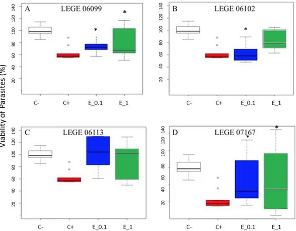
Figure 3- Effect of cyanobacterial crude extracts (LEGE0699, LEGE06112, LEGE06113, LEGE07167) on the viability of Leishmania infantum (“C-“ negative control, “C+” positive control, “E_0.1” 0.1 µg/mL of cyanobacterial crude extract and “E_1”1 µg/mL of cyanobaterial crudeextract)………...33 Figure 4- Percentage of Viability of Giardia duodenalis exposed to LEGE06104, LEGE06020, LEGE00038, LEGE00041, LEGE06361, LEGE11428 ……….………35 Figure 5 - Effect of cyanobacterial crude extracts (LEGE06108, LEGE06113, LEGE07167, LEGE07175) on the viability of Giardia duodenalis (“C-“ negative control, “C+” positive control, “E_0.1” 0.1 µg/mL of cyanobaterial crude extract and “E_1” means 1 µg/mL of cyanobaterial crude extract)……….36 Figure 6- Effect of marine natural based compounds from sponges(A- “AQ1972f39”; B- “AQ1974f13”; C- “LCL173R”; D- “LCL168R”; E- “LCL169R”; F- “DS-23”; G- “DS-139”; H- “DS-130”, I-“DS-131”) on the viability of Giardia duodenalis (“C-“ negative control, “C+” positive control and “E_1” 1 µg/mL)……….………37 Figure 7- Effect of marine natural based compounds from sponges (A- “DS-24”; B- “DS-215”; C- “DS-217”; D- “DS-221”; E- “AQ1963f50-51”; F- “AQ1964f43-51”; G- “AQ1865f33-38”; H- “AQ1851f23-25”; I- “AQ1829f13-16”) on the viability of Giardia duodenalis (“C-“ negative
control, “C+” positive control and “E_1” 1
µg/mL).……….………38 Figure 8- Effect of marine natural based compounds from sponges (A- “AQ1812f27-28”; B- “AQ1872f20-24”; C- “AQ1893f44-46”; D- “AQ1897f26-33”; E- “AQ1906f33-42”; F- “AQ1880f48-55”; G- “AQ1866f26-30”; H- “AQ1846f18”) on the viability of Giardia duodenalis (“C-“ negative control, “C+” positive control and “E_1” 1 µg/mL)………...……….………..39 Figure 9- Growth curve of Trichomonas vaginalis………..40
Figure 10- Effect of cyanobacterial crude extracts (LEGE0699, LEGE06102, LEGE06113, LEGE06194, LEGE 07163, LEGE 07167) on the viability of Trichomonas vaginalis (“C-“ negative control, “C+” positive control and “E_1” 1 µg/mL of sponge compounds)…………..41 Figure 11- Effect of marine natural based compounds from sponges(A- “AQ1972f39”; B- “AQ1974f13”; C- “LCL173R”; D- “LCL168R”; E- “LCL169R”; F- “DS-23”; G- “DS-139”; H- “DS-130”, I- “DS-131”) on the viability of Trichomonas vaginalis (“C-“ negative control, “C+” positive control and “E_1” 1 µg/mL).……….…..………41 Figure 12- Effect of marine natural based compounds from sponges (A- 24”; B- “DS-215”; C- “Ds-217”; D- “DS-221”; E- “DS-222”; F- “AQ1963f50-51”; G- “AQ1964f43-51”; H- “AQ1865f33-38”; I- “AQ1851f23-25”) on the viability of Trichomonas vaginalis (“C-“ negative control, “C+” positive control and “E_1” 1 µg/mL).……..………..…….……..42 Figura 13- Effect of marine natural based compounds from sponges (A- “AQ1829f13-16”; B- “AQ1812f27-28”; C- “AQ1872f20-24”; D- “AQ1893f44-46”; E- “AQ1897f26-33”; F- “AQ1906f33-42”; G- “AQ1880f48-55”; H- “AQ1866f26-30”, I- “AQ1846f18” ) on the viability of Trichomonas vaginalis (“C-“ negative control, “C+”positive control and “E_1” 1 µg/mL)...43
List of Abbreviations
AIDS - Acquired Immune Deficiency Syndrome BBE- Blue Biothecnology and Ecotoxicology groupCIIMAR- Interdisciplinary Centre of Marine and Environmental Reasearch DMSO- Dimethilsulfoxide
FBS- Fetal Bovine Serum
HIV- Human Immunodeficiency Virus HPV- Human Papiloma Virus
MTT - 3- (4,5 dimethylthiazol-2-yl) - 2,5-diphenyl tetrazolium bromide-212 STD - Sexually Transmitted Disease
VL - Visceral Leishmaniasis WHO - World Health Organization
1. Introduction
The planet Earth is covered by 70% of water in which 96% are oceans. Considered the “mother of origin life”, oceans are the source of a wide range of bioactive natural products that are mainly produced by organisms such as sponges (37%), algae (9%) and echinoderms (6%) (Gerwick and Moore 2012). Marine organisms, through evolution, had to develop characteristics to cope with the changes in the environment, namely extreme conditions of temperature and pressure. Many of these adaptions resulted in the production of compounds that revealed interesting activities in the pharmacological field namely in deadly diseases such as cancer, acquired immune deficiency syndrome (AIDS) and arthritis (Imhoff et al., 2011). In fact, several marine compounds were approved by FDA (Food and Drug Admnistration) and are currently available to the public as drugs. As an example, Cytosar-U® (1969) or Halaven® (2010) are a trademark where the bioactive compound was found in sponges and now are used to treat leukemia and metastatic breast cancer, respectively; or even Adcetris®(2011) in which the compound was discovered from a symbiotic relationship between a mollusk and a cyanobacterium and is now a medicine to Hodgkin's disease.
1.1 Cyanobacteria
Cyanobacteria, also known as cyanophyta, are a phylum from the Eubacteria Domain (Castenholz et al., 2001).These photosynthetic prokaryotes captures sunlight using clorophyll a and phycocyanin, a bluish pigment that was responsible for the name of these group of organisms (Whitton and Potts, 2007). Through their photosynthetic activities, 2 billion years ago, cyanobacteria played a major role in creating the atmosphere as we know nowadays. In fact, they were responsible for the accumulation of oxygen in the early atmosphere, which lead to our oxygenic environment (Hartman, 1998).
Cyanobacteria are found in a wide range of habitats, from terrestrial to aquatic ones, and even in environments with extreme conditions such as extreme temperatures (Wieland, 2000). Due to a long evolution period and adaptation to different environmental conditions, cyanobacteria developed mechanisms of adaptation that include the production of several secondary metabolites.
In the marine environment cyanobacteria occur as free-living organisms in open ocean or near shores, as benthic forms along the maritime coasts and as symbiotic forms with animals such as sponges (Paerl et al., 2000). In this environment, cyanobacteria play an important ecological role thought the involvement in the primary production and in nitrogen fixation (Carpenter et al., 2004) and by offering food and shelter to many aquatic organisms (Paerl et al., 2000).
1.1.1 Cyanobacteria Bioactive compounds
Secondary metabolites are chemicals produced by an organism in which a role in primary functions such as growth or reproduction has not been found (Campàs et al., 2010). Even though they are not absolutely required for the survival of the organism, in cyanobacteria they are responsible for many functions such as nutrient storage (Istvánovics et al. 1993, Kaplan et al., 1988) defense (Nogueira et al. 2004) and allelopathy (Leão et al., 2009).
The high biodiversity of cyanobacteria in nature leads to a high diversity in a chemical level, which in turn gives to various and different secondary metabolites that can be potential bioactive compounds. From this group of organisms a variety of bioactive secondary compounds, that includes alkaloides, peptides, lipopetides, amino acids and fatty acids is described (Burja et al., 2001), being cyanobacteria considered a good source of compounds for biotechnology aplications, namely pharmaceutical and therapeutic application (Dahm et al., 2006; Dixit et al., 2013; Tan 2013). In fact, in the pharmacology field, compounds isolated from cyanobacteria were found to have antibacterial (Jaki et al., 2000), antifungal (T. Shishido et al. 2015), antiviral (Patterson et al., 1994), anticancer ( Gerwick et al., 1994), immunosuppressive (Koehn et al., 1992), algicide (Papke et al., 1997) antiparasitic (Burja et al., 2001) and antiplasmodial (Papendorf et al., 1998) potential. In this sense, in the last few years, cyanobacteria have been considered one of the most promising sources of natural compounds (Dixit et al., 2013).
1.2 Parasitology
All over the world, the diseases caused by parasites are a public health problem affecting about one billion people (WHO, 2015), with most of them being also neglected tropical diseases (Molyneux et al., 2016). Despite their main prevalence in tropical and subtropical regions, recent epidemiological studies about leishmaniasis have shown an increasing prevalence in Europe largely caused by an augment in international travel, difficulty eradicating leishmanial infection in AIDS patients, and the use of immunosuppressive medications (Torpiano et al., 2015).
In spite of the progress that has been made in the prevention, control and elimination of human parasitic diseases, there are however some limitations concerning the therapeutics namely due to parasite resistance and adverse effects on patients.
Three main parasitosis affecting humans worldwide are visceral leishmaniasis, giardiasis and trichomoniasis.
Concerning leishmaniasis, it is a parasitic disease with clinical presentations that vary from asymptomatic infection to cutaneous, mucocutaneous or visceral disease. 1.3 million new cases occur annually: 300 000 are visceral and 1 million are cutaneous or mucocutaneous (WHO, 2015). The estimated number of deaths from visceral leishmaniasis ranges from 20 000 to 50 000 annually (Alvar et al., 2012).
Giardiasis is the most common non-bacterial and non-viral diarrheal diseases affecting humans worldwide (Escobedo et al., 2015). Because of its link with poverty and the potential for dissemination through food and water supplies, giardiasis was included in the Neglected Disease Initiative, estimating that 280 million people are infected each year.
Trichomoniasis is a sexual transmitted parasitosis that increases the risk of human immunodeficiency virus (HIV) transmission and leads to adverse outcomes of pregnancy. In an approach for 2005, a total number of 248 million new cases cases of trichomoniasis were estimated in adults between the ages of 15 and 49 (WHO, 2011).
1.2.1 Leishmaniasis
Leishmaniasis continues to pose a major public health problem worldwide. It is a zoonotic, infectious and non-contagious disease caused by different species of protozoan parasites of the genus Leishmania (Class Kinetoplastida, Family Trypanosomatidae) and transmitted by the bite of a female phlebotomine sandfly. This disease presents itself in three
forms: mucosal (infection of macrophages in the naso-oropharyngeal mucosa), cutaneous (infection of macrophages in the dermis) and visceral (infection of macrophages throughout the reticuloendothelial system). It is recorded each year, nearly 2 million new cases of leishmaniasis in the world. The most severe form of the disease is the visceral leishmaniasis (VL), being 85-90% of untreated cases fatal (Chappuis et al., 2007).
The specie Leishmania infantum is the etiologic agent of VL and has two forms of life: the amastigote stage, oval or spherical with no free flagellum, and the promastigote stage, elongated with a free flagellum.
Leishmania infantum is a digenetic parasite that completes his life cycle in two hosts: phlebotomine sandflies (intermediate host), that hosts the promastigote stage, and a mammal (definitive host) where the amastigote stage of the parasite develops.
The life cycle of the parasite begins when the intermediate host, the female sandfly (vector), bites the definitive host (vertebrate) already infected, ingesting the parasites in an amastigote form. Within the vector, the amastigotes migrate to the phlebotomine intestine where they differentiate into infective promastigotes and multiply (Bates, 2007). In the next blood meal, the infected females inoculated into the vertebrate host the promastigotes forms of the parasite, which in turn are rapidly phagocytosed by macrophages. After phagocytosis, parasite differentiate into amastigotes and proliferate to the point of causing the rupture of the infected macrophage and causing the release of amastigotes in vertebrate host's body (Almeida-Souza et al. 2016; Chan et al., 2005). The infected macrophages migrate to the subcutaneous tissues, to the liver, spleen, bone marrow and lymph nodes, leading to the development of cutaneous and V, respectively. Thus, the development of the disease depends on the parasite species's efficiency with respect to the amastigote differentiation as well as the immune response of the host (Barral et al. 1991; Grimaldi et al., 1996).
Visceral leishmaniasis is a disease with worldwide distribution, with 90% of human cases occuring in India, Sudan, Bangladesh, Nepal and Brazil. (Molyneux et al., 2016; WHO, 2015). The number of cases of leishmaniasis are increasing worldwide. Some reasons are the lack of vaccines, difficulties in controlling the vectors and increasing the number of resistance to chemotherapy of parasites. The human VL caused by L infantum is a major public health problem in areas where canine leishmaniasis is endemic and dogs are the main reservoir of infection. The disease affects the vital organs of the body and is characterized by irregular attacks of fever, weight loss, anemia, and enlargement of spleen and liver (WHO, 2015). Malnutrition has been recognized as a risk factor and may explain why the disease is more prevalent among children in poor countries than in developed countries, despite high prevalence rates in canine populations. The human disease is also prevalent in immunosuppressed
individuals; with HIV patients being the predominant risk group in southern Europe (Monge-Maillo et al., 2014).
According to WHO, leishmaniasis is among the parasitic diseases that mostly disturb the public health, by the frequency, and especially the therapeutic difficulties and also the clinical sequels that may result (WHO, 2004).
Although therapy historically has relied on antimonials, the latest treatment options include amphotericin B, liposomal, paromomycin and miltefosine (Sinha et al,. 2014). Combinations of drugs showed positive results and can be a short term solution to delay or prevent the onset of resistance, increased efficiency, and shorten the treatment course (Alvar et al., 2006; Sundar et al., 2011). Miltefosine is a recognized oral agent to treat this parasitosis, but with limitations to the safety of use, presenting relevant toxicity and a high frequency of side effects (Coelho et al., 2014).
1.2.2 Giardiasis
Giardiasis occurs worldwide, with a distribution more prevalent in developing countries where hygiene conditions are poor, favoring its spread in warm climates (WHO, 2012). This is also a zoonotic disease caused by the infectious protozoan parasite Giardia duodenalis (also known as Giardia intestinalis or Giardia lamblia, Class Zoomastigophora, Family Hexamitidae) considered the major diarrheal diseases found worldwide. Its transmission is fecal-oral route and is due to ingestion of water and food contaminated with protozoa in the form of cysts (infective form), or from person to person (hand contact with contaminated feces, anal sex). Giardiasis is the most common infection caused by protozoa in children throughout
the world (Coles et al., 2009; Gardner et al., 2001).
Giardia (G. duodenalis, G. intestinalis) is a flagellate protozoan that survives under two distinct morphological forms - cyst and trophozoite. These organism prefer humid locals, with water at a temperature of 4-10ºC. Cysts can remain viable for more than a few months (deRegnier et al., 1989); although the boiling point and instantaneous freezing result in inactivation (Meyer et al., 1980). Although humans are the primary reservoir of the parasite, several animals transport different species, which can be transmitted to humans. Giardia have been isolated from various animal species, and by 1950 these isolates were considered highly specific hosts as to be named according to the host species, e.g. G. bovis, G. felis, among others. However, only three distinct species managed to survive being G. duodenalis the species that infect humans and many other mammals (Filice, 1952).
The Giardia duodenalis life cycle is composed of the two forms of the parasite transmitted by feces: cyst (infective form) and trophozoite. Cysts are ingested by fecal-oral route or through consumption of food and / or contaminated water. When the cysts are ingested, digestive enzymes leads to the release and excystation of trophozoites (each cyst producing two trophozoites). Trophozoites proliferation leads to acute giardiasis, with an incubation period of 1 to 14 days (mean 7 days) and usually lasts 1 to 3 weeks. Symptoms include diarrhea, abdominal pain, flatulence, nausea and vomiting. In chronic giardiasis symptoms are recurrent and can lead to debilitation. Several studies have shown that chronic infection of Giardia during childhood can contribute to calorie protein malnutrition, vitamin A deficiency, iron deficiency anemia, zinc deficiency and poor cognitive and educational performance (Halliez et al., 2013). In relation to treatment, the first-line drugs are nitroimidazoles, with the prototype, metronidazole, being the most common drug used worldwide (Miyamoto et al., 2013). Alternatives to these drugs include paromomycin, quinacrine, and furazolidone (Escobedo et al., 2007). However some of these compounds have relevant side effects and single and multi-drug resistance to compounds, including metronidazole have also been reported (Alves et al., 2011) .
1.2.3 Trichomoniasis
Trichomoniasis is a sexually transmitted disease (STD) caused by the infectious protozoan parasite Trichomonas vaginalis (Class Zoomastigophorae, Family Trichomonadidae). Trichomoniasis is the most common STD non-viral in the world, with 170 million new cases occurring annually (Menezes et al., 2016).
Trichomonas vaginalis is a flagellate protozoan (10-23μm) of rounded shape that during its life cycle only possess the trophozoite stage. It has four flagella that produce motion and a fifth that can help with direction. Its high motility contributes to the pathogenicity. The Trichomonas vaginalis is in lower genital tract of women while most affected men hosts the parasite inside the penis. Trichomonas multiplies by binary division and does not have the form of cyst, thus do not survive well in the external environment. The parasite causes damage to the vaginal epithelium, leading to the formation of microscopic ulcers that increase the risk of contamination by other sexually transmitted diseases, including HIV, Human Papilloma Vírus (HPV), herpes, gonorrhea and chlamydia.
This parasite occurs worldwide, prevailed more among people with multiple sexual partners or other sexually transmitted diseases. Trichomoniasis is a sexual transmitted parasitosis so it is important that infected sexual partners perform treatment simultaneously.
Treatment options are in this case reduced to nitroimidazole compounds (Alves et al., 2011), which highlight the need to search for new therapies.
2. Objectives
It is imperative the search for new antiparasitic compounds, due to the great difficulty of finding drugs that ensure an efficient therapeutic action and are less toxic to the host, so different natural products are being investigated, including cyanobacteria and sponges compounds.
The Blue Biotechnology and Ecotoxicology group (BBE) at the Interdisciplinary Centre of Marine and Environmental Reasearch (CIIMAR) has been involved in the study of the bioactive potential of cyanobacteria isolated from the portuguese coast. From the cyanobacteria culture collection several strains have been studied and compounds have been isolated. As a result of these studies different bioactivities were described such as anticancer (Costa et al., 2014; Freitas et al., 2016a; Freitas et al., 2016b), antimicrobial (Costa et al., 2014; Martins et al., 2008) and antiviric (Lopes et al., 2011). Also the antiparasitic potential has been studied to a less extent, being a compound described as potential antimalarian (Compound under a patent process). Also, the Department of Chemistry Giacomo Ciamician, Alma Mater Studiorum at the University of Bologna, Italy has been performing studies concerning the antiparasitic potencial effect of natural based compounds isolated from organisms such as sponges (Cerisoli et al., 2016b; Persico et al., 2011; Monari et al., 2015). In this sense, considering the bioactive potential of the cyanobacteria strains of the LEGE culture collection, and compounds of the University of Bologna, allied with the need for new drugs against major human parasites, the main aim of this thesis was:
- to study the potential of marine cyanobacteria isolated from the portuguese coast and natural based compounds as antiparasitic for visceral leishmaniasis (Leishmania infantum), giardiasis (Giardia duodenalis) and trichomoniasis (Trichomonas vaginalis).
1. To perform a literature review concerning cyanobacteria antiparasitic activity;
2. To culture cyanobacteria strains in order to prepare extracts from freeze dried biomass; 3. To perform culture optimization of the parasites Leishmania infantum, Giardia
duodenalis and Trichomonas vaginalis, in vitro;
4. To perform a toxicological assay in vitro ( 3- (4,5 dimethylthiazol-2-yl) - 2,5-diphenyl tetrazolium bromide-212 - MTT assay) to evaluate the antiparasitic potential effect of extracts and compounds.
3. Materials and Methods
3.1 Literature review
In order to compile information concerning the antiparasitic potential of cyanobacteria a search for scientific papers and reviews was performed by using the databases ScienceDirect and PubMed, until June 2016. The search was conducted by using “cyanobacterial
antiparasitic compounds, “antiprotozoal compounds, “cyanobacterial bioactive compounds, “cyanobacterial antileishmanial compounds, “cyanobacterial antimalarial compounds” as example of key words.
3.2 Cyanobacteria strains
In this work eight marine cyanobacteria strains were cultured under laboratory conditions in order to obtain biomass (Table I). From this strains crude extracts were prepared. This cyanobacteria strains were selected according to bioactive potential already described in previous studies. Also crude extracts from cyanobacteria strains that are being studied in ongoing studies for bioactivities were included in the antiparasitic assays (Table II).
Table I. Cyanobacteria strains cultured for extract preparation.
Strain Species/ Genera Order Origin Culture
medium LEGE
06102 Leptolyngbya halophile Oscillatoriales
S. Bartolomeu do
Mar Z8 + NaCl
LEGE
07167 Leptolyngbya fragilis Oscillatoriales Lavadores Z8 + NaCl LEGE
06113 Cyanobium Chroococcales Aguda
Z8 + NaCl
LEGE
07175 Cyanobium Chroococcales Martinhal Z8 + NaCl
LEGE
06099 Synechocystis salina Chroococcales Moledo Z8 + NaCl
LEGE
06108 Synechocystis salina Chroococcales
S. Bartolomeu do
Mar Z8 + NaCl
LEGE
07163 Pseudanabaena persicina Oscillatoriales Z8 + NaCl
LEGE
06194 Pseudanabaena sp. Oscillatoriales Z8 + NaCl
Table II – Marine cyanobacteria extracts of LEGE cyanobacteria culture collection included in this study.
Strain Species/ Genera Order Origin
LEGE 06014 Leptolyngbya sp. Oscillatoriales Arellho
LEGE 06020 Leptolyngbya sp. Oscillatoriales Coxos
LEGE 00038 Synechocystis sp. Chroococcales Mindelo
LEGE 00041 Synechocystis sp. Chroococcales Espinho
LEGE 06361 Leptolyngbya sp. Oscillatoriales Unknown
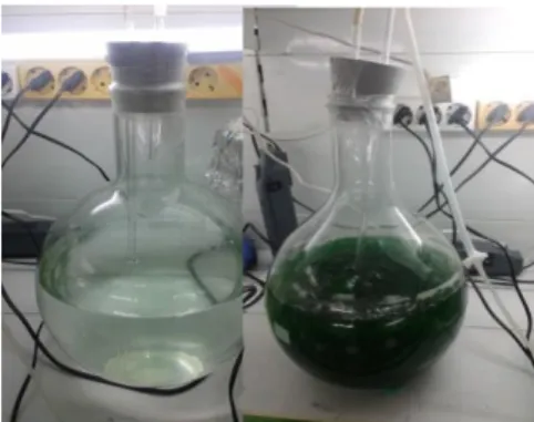
Figure 1 – Culture of cyanobacteria. Left: after the inoculation (beginning of the culture). Right: after fifteen days.
3.3 Cyanobacteria Culture, lyophilisation and extraction
In order to obtain sufficient biomass for the antiparasistic assays, six cyanobacteria strains (Table I) were cultured under laboratory conditions.
Cyanobacteria strains were cultured following the procedures already optimized in the LEGE laboratory. Briefly, cyanobacteria strains were incubated in the laminar flow chamber in six liter glass flasks containing 4 liters of nutrient Z8medium (with 20g/L NaCl) (Kotai 1972) and a 500 mL of cyanobacteria stock culture. Cultures were maintained under an aeration systems with controlled temperature conditions (25°C) and luminance (light cycles / dark 14h / 10h).
When cultures reached the exponential growth stage, after approximately 30 days of the inoculation, visually recognized by the high concentration of biomass (Figure 1), the culture was harvested by centrifugation.
Cultures were centrifuged at 4,500 rpm, 4°C, 10 minutes. The supernatant was removed and the pellet washed with sterile distilled water and centrifuged again. The supernatant was removed one second time, and the pellet was placed in a flask collector to freeze at -20°.
Before lyophilisation, the concentrated biomass was placed at -80ºC. The frozen biomass was lyophilized for 4 days at -48ºC and then stored in a refrigerator. A stock culture of all cyanobacteria was maintained during this work in order to obtain biomass for the different bioassays. These cultures were performed by the inoculation process mentioned above, by removing a small sample of the culture and placing it in flasks with 100 ml of Z8 medium (20g/L NaCl).
From the cultured cyanobacteria strains a crude extract was prepared by also following the procedure already optimized in the LEGE laboratory. Briefly, 1g of lyophilized material of each strain was extracted for 10 minutes with 50mL of a dichloromethane: methanol (2:1) solution by stirring periodically. The solution was them placed into a Bücher funnel, under
vacuum for filtration. At the end of the filtration the residual mass was collected and extracted for another 10 minutes. After extraction the solvents were evaporated in the rotary evaporator at -7 °C. The flask was then washed with a mixture of isooctane: ethanol (1:1, v/v) to dissolve the pellet, using ultrasounds, and transferred to a previously weighted 22 mL clear glass vial. The mixture was evaporated using N2. The glass vial was weighted again and the total mass of the
crude extract calculated. The dry extract was dissolved in DMSO for the antiparasitic assays. Cyanobacteria extracts were tested against parasites at a final concentration of 1 µg/mL and 0,1 µg/mL with DMSO at a final concentration of 1%.
3.4 Marine natural based compounds
In addition to the cyanobacterial natural extracts it was also evaluated the antiparasitic potential of several natural based compounds provided by Arianna Quintavalla, researcher at the Department of Chemistry Giacomo Ciamician, Alma Mater Studiorum, University of Bologna, Italy.
All these compounds are synthetic but their design was based on natural compounds. Indeed they have in common structural motifs, some of them with a marine origin, that have already shown bioactivity against protozoan and helminthic parasites (Persico et al., 2011); Trost et al, 2013).
The compounds tested in this study are listed in Table III and IV and their chemical basic structure belongs to:
-Peroxide derivatives (Persico et al., 2011; Sonawane et al., 2015a; Sonawane et al., 2015b) (Table III);
-Spiro oxindole derivatives (Monari et al., 2015a; Cerisoli et al., 2016a) (Table
III)
Compounds solubilized in DMSO were tested against Giardia duodenalis and Trichomonas vaginalis at a final concentration of 1 µg/mL as described in 3.5 and 3.6. Leishmania infantum assays were not performed as a demand of the authors.
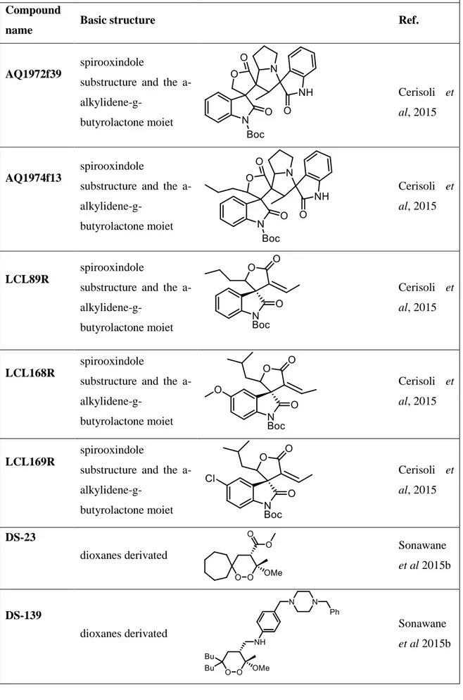
Table III- Natural based compounds that were evaluated as potential antiparasitic with their chemical basic structure Compound
name Basic structure Ref.
AQ1972f39
spirooxindole
substructure and the a- alkylidene-g-butyrolactone moiet Cerisoli et al, 2015 AQ1974f13 spirooxindole
substructure and the a- alkylidene-g-butyrolactone moiet Cerisoli et al, 2015 LCL89R spirooxindole
substructure and the a- alkylidene-g-butyrolactone moiet Cerisoli et al, 2015 LCL168R spirooxindole
substructure and the a- alkylidene-g-butyrolactone moiet Cerisoli et al, 2015 LCL169R spirooxindole
substructure and the a-
alkylidene-g-butyrolactone moiet
Cerisoli et al, 2015
DS-23
dioxanes derivated Sonawane
et al 2015b
DS-139
dioxanes derivated Sonawane
DS-130
dioxanes derivated Sonawane
et al 2015b
DS-131
dioxanes derivated Sonawane
et al 2015b
DS-24
dioxanes derivated Persico et
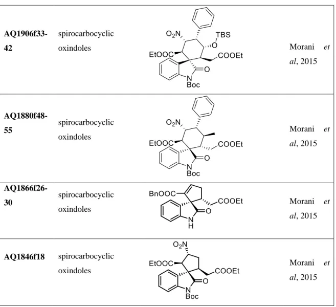
al, 2011 DS-215 Endoperoxide derivated (artemisina bone) Sonawane et al 2015a Ds-217 Endoperoxide derivated (artemisina bone) Sonawane et al 2015a DS-221 Endoperoxide derivated (artemisina bone) Sonawane et al 2015a DS-222 Endoperoxide derivated (artemisina bone) Sonawane et al 2015a AQ1963f50-51 spirocarbocyclic oxindoles Morani et al, 2015
AQ1964f43-51 spirocarbocyclic oxindoles Morani et al, 2015 AQ1865f33-38 spirocarbocyclic oxindoles Morani et al, 2015 AQ1851f23-25 spirocarbocyclic oxindoles Morani et al, 2015 AQ1829f13-16 spirocarbocyclic oxindoles Morani et al, 2015 AQ1812f27-28 spirocarbocyclic oxindoles Morani et al, 2015 AQ1872f20-24 spirocarbocyclic oxindoles Morani et al, 2015 AQ1893f44-46 spirocarbocyclic oxindoles Morani et al, 2015 AQ1897f26-33 spirocarbocyclic oxindoles Morani et al, 2015
AQ1906f33-42 spirocarbocyclic oxindoles Morani et al, 2015 AQ1880f48-55 spirocarbocyclic oxindoles Morani et al, 2015 AQ1866f26-30 spirocarbocyclic oxindoles Morani et al, 2015 AQ1846f18 spirocarbocyclic oxindoles Morani et al, 2015
Table IV – Natural based compounds that were evaluated as potential antiparasitic. Basic structure
molecule Compound reference
Peroxide derivates DS-23; DS-24; DS-130; DS-131; DS-139; DS-215; Ds-217; DS-221; DS-222 Spiro oxindole derivates
AQ1812f27-28; AQ1829f13-16; AQ1846f18; AQ1851f23-25; AQ1865f33-38; AQ1866f26-30; AQ1872f20-24; AQ1880f48-55;
AQ1893f44-46; AQ1897f26-33; AQ1906f33-42; AQ1963f50-51; AQ1964f43-51;
3.5 Parasites
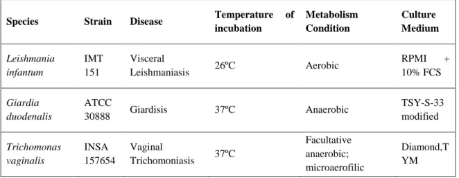
The parasites included in this study were Leishmania infantum, Giardia duodenalis and Trichomonas vaginalis. Information concerning the strain reference, culture medium and incubation temperature is presented in Table V. All parasites were stored at -80ºC in complete culture medium with 10% DMSO.
Table V – Parasites included in this study and summary of culture condition
Species Strain Disease Temperature of
incubation Metabolism Condition Culture Medium Leishmania infantum IMT 151 Visceral Leishmaniasis 26ºC Aerobic RPMI + 10% FCS Giardia duodenalis ATCC 30888 Giardisis 37ºC Anaerobic TSY-S-33 modified Trichomonas vaginalis INSA 157654 Vaginal Trichomoniasis 37ºC Facultative anaerobic; microaerofilic Diamond,T YM
3.5.1 Leishmania infantum
For culturing and parasites maintenance, an 1 ml aliquot of promastigotes of L. infantum IMT 151 was poured into 9 mL RPMI GlutaMAX-I medium (Gibco, Life Tecnologies) medium supplemented with 10% fetal bovine serum, 50U/ml penicilin, 50 µg/mL streptomycin and 25mM HEPES soidum salt (RPMI complete medium), pH7,4 in a 25cm2 flask (Orange Scientific's advanced, high-quality Tissue Culture). Parasites were maintained at 26ºC.
3.5.1.2
Growth curve determination
The characterization of parasite growth in vitro is fundamental to evaluate the compounds effect on parasite cultures. For growth curve determination, promastigotes of L. infantum IMT 151 were incubated at 5 x 10⁶ parasites/ml in RPMI complete medium supplemented with fetal bovine serum (10%) (Keister, 1983), at 26ºC. During 5 days, a daily quantification of parasite density was performed, in triplicate, by counting promastigotes in a Neubawer chamber and an estimative of viability was obtained by Trypan blue exclusion. Trypan blue is a blue dye often used as a quick test to evaluate the cellular viability. This dye enters the cell when cell membrane is damaged. Thus, after adding trypan blue to a cellular
suspension (1:1), viable cells do not stain while not viable cells acquire a blue color. This test was performed during cell counting for the determination of the growth curve and for the antiparasitic assays, to have a direct assumption of the viability of the parasites. Culture exponential phase was defined after growth curve determination.
3.5.1.3 Viability MTT assay
The potential antiparasitic effect for Leishmania infantum was tested only with cyanobacteria strains referred on Table I. For viability assays Leishmania infantum promastigotes at a density of 1,5x10⁶ parasites/mL were cultured in 1 mL RPMI complete medium in 1,5ml eppendorfs. Cyanobacteria extracts and natural based compounds were added and incubated for 72 hours at 26 ºC. After incubation the viability assay using the 3- (4,5 dimethylthiazol-2-yl) - 2,5-diphenyl tetrazolium bromide-212 (MTT) was carried out. These MTT assay conditions were a result of an optimization procedure also performed in this thesis.
The MTT assay (Mosmann, 1983) is a colorimetric assay that relies on the cellular reduction of tetrazolium salt by mitochondrial dehydrogenases in viable cells. In solution, the MTT salt has a yellow color but, after reduction by mitochondrial dehydrogenases present in metabolically active cells yields formazan crystals purple. Subsequently, these crystals after dissolved with DMSO, absorb in the visible region, allowing spectrophotometrically quantitate the number of viable cells. Since formazan crystals are slightly soluble in the RPMI medium, it was necessary to remove this medium before incubation with the MTT. After incubation time with the cyanobacteria extracts, eppendorfs were centrifuge in order to concentrate the parasites. The culture medium was carefully removed and changed to a solution of PBS-Glicose (1:1). MTT was them added to each eppendorf to a final concentration of 0.02mg/mL and incubated for 4 hours. After the MTT incubation, eppendorfs were centrifuged again and the PBS-Glicose aspired carefully. The formazan crystals were them solubilized with 100% DMSO and the absorbance was read at 550nm. Each assay was run in quadruplicate and three experiments were performed independently. As a positive control amphotericin B was tested at a concentration of 0.1 µM. The negative control consisted in culture medium with 1% DMSO.
3.5.2 Giardia duodenalis
3.5.2.1 Parasite density optimization
An 1 ml aliquot containing trophozoites of Giardia duodenalis ATCC 30888 was poured into a Nunclon tube with TYI-S-33 modified medium (Keister, 1983) supplemented
with 50U/ml penicilin, 50 µg/mL streptomycin and culture at 37ºC. After incubation for 48h, trophozoites were detached from the substracte by cooling on ice for 20 minutes and subcultured. Culture tubes were full completed with culture medium, in order to obtain an anaerobic environment.
For determination of the initial parasite density for viability tests, preliminary MTT assays were performed in 96 well plate with 300 µL medium/well and using three initial different concentrations, 2x10⁴parasites/mL, 5x10⁴parasites/mL and 1x10⁵ parasites/mL. After an incubation time of 24 hours, cells were incubated for 4 hours with MTT at a final concentration of 0.05mg/mL. After the incubation the medium was aspired, formazan crystals were solubilized with 100%DMSO and the absorbance was read at 550nm. These MTT assay conditions were a result of an optimization procedure also performed in this thesis.
3.5.2.2 Viability MTT assay
Viability assays using Giardia duodenalis trophozoites were performed for all the cyanobacteria extracts and the natural based compounds presented above, at a density of 5x10⁴ parasite/mL were seeded in a 96-wells plate, in triplicate. After 3 hours of cell adhesion to the plate, cyanobacteria extracts and natural based compounds were added and incubated for 24 hours at 37 ºC. After the incubation time, the culture medium was exchanged to PBS-Glicose (1:1), since the TSY-S-33 medium spontaneously reacts with the MTT. MTT was added to a final concentration of 0.05mg/mL and incubated for 4 hours. After the incubation the medium was aspired, formazan crystals were solubilized with 100% DMSO and the absorbance was read at 550nm. These assays were performed three times independently. The positive control consisted in 1% of metronidazole at a concentration of 0,856g/L. The negative control consisted in culture medium with 1% DMSO. During incubation the plates were maintained in a low-oxygen environment achieved through a system of AnaerocultC (Merck) and by using an anaerobic jar. For cell counting the trypan blue assay was performed in order to infer about parasites viability before the MTT assay.
3.5.3 Trichomonas vaginalis
3.5.3.1
Growth curve determination
Growth curve determination of Trichomonas vaginalis trophozoites was performed by incubating the parasites in Diamond-TYM medium (Green et al., 2012), at 37ºC, in microaerophilic conditions. During 3 days, a daily quantification of parasite density was performed, in triplicate, by counting trophozoites in a neubawer chamber and an estimative of viability was obtained by trypan blue exclusion.
3.5.3.2 Viability MTT assay
The antiparasitic effect on Trichomonas vaginalis was tested with the cyanobacteria strains referred in Table I and with the natural based compounds referred in Table III. For viability assay Trichomonas vaginalis trophozoites at a density of 2,8x10⁵ parasites/mL were seeded in eppendorfs, in triplicate. Cyanobateria extracts and natural based compounds were added and incubated for 24hours at 37ºC. After incubation, eppendorfs were centrifuge at 1200 rpm for 5 minutes at 10ºC. Culture medium was carefully exchanged to PBS-Glicose (1:1) with careful not to aspirate the trophozoites pellet. Parasites were then treated with 0.02mg/mL MTT final concentration, for 4 hours. After incubation with MTT, the eppendorfs were centrifuged and the medium was aspired carefully. Formazan crystals were solubilized with 100% DMSO and the absorbance was read at 550nm. These assays were performed three times independently. The positive control consisted in 1% of metronidazole at a concentration of 0,856g/L. The negative control consisted in culture medium with 1 % DMSO. These MTT assay conditions were a result of an optimization procedure also performed in this thesis.
3.6. Statistical analysis
Differences in the effect of cyanobacterial extract on parasite viability were analyzed with a general linear model, using parasite viability as dependent variable and treatment (levels: concentration of cyanobacterial extract, positive control and negative control) as fixed factor. The assumption of normality of model residuals was tested with a Shapiro-Wilks test. If residuals were not normal they were transformed using the Box-Cox function. If this transformation did not produce the intended effect, data would be analyzed with a generalized linear model, assuming that residuals follow a Gamma distribution.
Since the data were obtained from 3 independent assays with identical design, we also considered if the inclusion of 'assay' as a random factor in the model would significantly improve our model. For every single data set (parasite cyanobacterial extract) the AIC value of a mixed effects model including 'assay' as random factor was compared to the AIC value of an equivalent fixed effects model, and the model with the lowest value was chosen.
After obtaining the suitable model for each case, we tested the effect of treatment with analysis of variance (for general linear models with only fixed effects) or analysis of deviance (for generalized linear models with only fixed effects or for any mixed effects models). If the effect of treatment was found to be statistically significant (p<0,05) we performed a post-hoc Tukey test for all the pairwise comparisons between treatment levels (Positive and negative control, and cyanobacterial extract).
All these analysis were performed using the packages stats, MASS and lme4(Bates et al., 2014) from the software R version 2.15.2 (R Core Team 2015).
4. Results and Discussion
4.1Antiparasitic potential of Cyanobacteria
In order to compile the information concerning studies related to the antiparasitic effects of cyanobacteria compounds, an extensive search for scientific papers was performed. From this search, papers concerning effects against Plasmodium falciparum (malaria), Leishmania species (leishmaniasis), Trypamosoma and Toxoplasma were found. For more easly present data, results concerning the different parasites are presented separately (Tables VI, VII and VIII). In Table VI it is presented the cyanobacteria compounds that were tested as antileishmanial. The cyanobacteria compounds that were tested as antiplasmodium by using the parasites that cause malaria, the Plasmodium falciparum, are presented at Table VII while the cyanobacteria compounds that were tested against Trypanossoma cruzi and Toxoplasma gondii are organized in Table VIII.
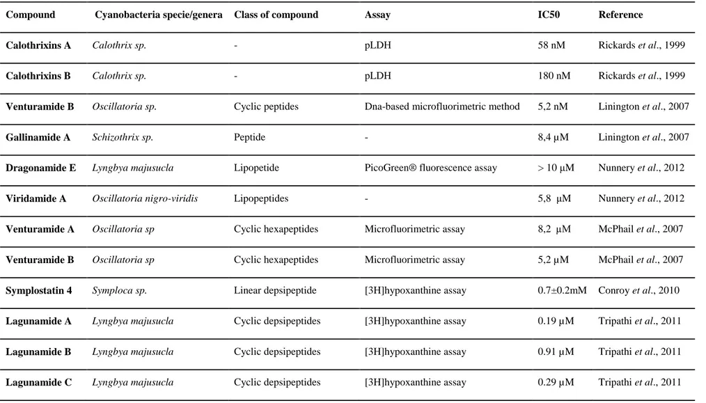
Most of the papers found are related to the parasite Plasmodium falciparum, the agent of the more severe form of malaria infection (Table VI). According to the 2014 WHO Malaria report (WHO, 2014) it is estimated that 3.2 billion people are at risk of being infected with malaria and 1.2 billion are at high risk (>1 in 1000 chance of getting malaria in a year). In 2013, 198 million cases of malaria were reported, with around 584 000 deaths. The treatment for malaria is still based on parasite chemotherapy with drugs such as quinine, mefloquine and artemisinin. However its poor tolerability and poor compliance, associated to an increasing resistance of the parasite against these drugs (Elfawal et al., 2015) has forced the research for effective new antiplasmodial agents.
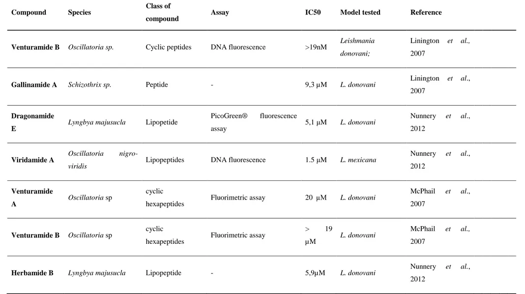
Concerning the potential of cyanobacteria against Leishmania parasites, we found Leishmania donovani as the most evaluated species (Table VII). Leishmaniasis is also a deadly parasitic diseases with 200 000–400 000 people contracting the infection every year in developing countries. Miltefosine is a recognized oral agent to treat this parasitosis, but with limitations to the safety of use, presenting relevant toxicity and a high frequency of side effects (Coelho let al. 2014) and thus, the research for effective drugs remains essential.
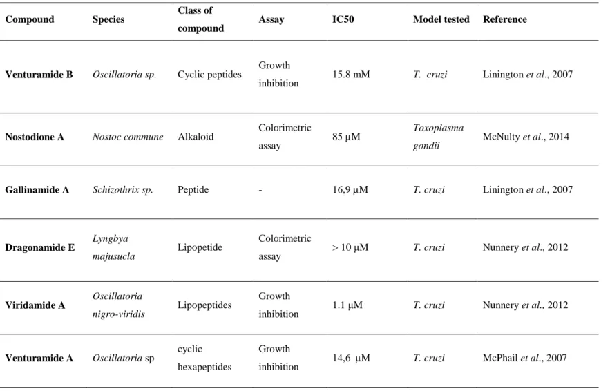
Studies involving Trypanossoma and Toxoplasma species were also found (Table VIII) namely with the specie Trypamossoma cruzi. Trypanossoma cruzi is the causing agent the human american trypanosomiasis, also known as chagas desease. It is estimated that about 6 million to 7 million people worldwide are infected with Trypansosoma cruzi. The Chagas disease is found mainly in Latin American countries (WHO, 2016). Although drugs such benznidazole and also nifurtimox are effective they cause severe side effects and the cost of treatment f remains substantial. Also there is no vaccine against this disease, which leads to the search for new therapeutical agents (WHO, 2016).
Looking through the Tables VI, VII and VIII it is notorious that most of the studies are related to filamentous cyanobacteria, namely of the genera Lyngbya and Oscillatoria. In fact, the majority of studies concerning marine cyanobacteria as producers of bioactive compounds with pharmacological potential, involved cyanobacteria that growth in high densities along the maritime shores of tropical and subtropical regions being the genera Lyngbya, Microcoleus, Oscillatoria, Schizothrix, Symploca and Trichodesmium the most referenced ones (Gerwick et al., 2008). Due to the massive growth of these cyanobacteria in natural conditions, little effort is applying in getting biomass. On the contrary there are numerous other genera that have not been studied in such detail, mainly because under natural conditions they occur in low densities. In this group it is included the picoplanktonic forms of the genera Cyanobium, Synechocystis and Synechococcus and the filamentous forms Leptolyngbya included in this study (Tables I and II) In this sense, this study represents a contribution to broaden the range of cyanobacteria from which new bioactive compounds might be isolated.
Table VI- Review of Cyanobacteria species/compounds tested against Leishmania parasites
Compound Species Class of
compound Assay IC50 Model tested Reference
Venturamide B Oscillatoria sp. Cyclic peptides DNA fluorescence >19nM Leishmania
donovani;
Linington et al., 2007
Gallinamide A Schizothrix sp. Peptide - 9,3 µM L. donovani Linington et al.,
2007
Dragonamide E
Lyngbya majusucla Lipopetide PicoGreen® fluorescence
assay 5,1 μM L. donovani
Nunnery et al.,
2012
Viridamide A Oscillatoria
nigro-viridis Lipopeptides DNA fluorescence 1.5 μM L. mexicana
Nunnery et al., 2012 Venturamide A Oscillatoria sp cyclic
hexapeptides Fluorimetric assay 20 µM L. donovani
McPhail et al.,
2007
Venturamide B Oscillatoria sp cyclic
hexapeptides Fluorimetric assay
> 19
µM L. donovani
McPhail et al.,
2007
Herbamide B Lyngbya majusucla Lipopeptide - 5,9µM L. donovani Nunnery et al.,
2012
Coibacin A Oscillatoria sp Polyketide pLDH 2.4 μM L. donovani Marcy J et al., 2012
Almiramides B L. majuscula Lipopeptides DNA fluorescence 2.4 μM L. donovani Sanchez et al., 2010
Almiramides C L. majuscula Lipopeptides DNA fluorescence 1.9 μM L. donovani Sanchez et al., 2010
Table VII- Review of Cyanobacteria species/compounds tested against Plasmodium falciparum parasites
Compound Cyanobacteria specie/genera Class of compound Assay IC50 Reference
Calothrixins A Calothrix sp. - pLDH 58 nM Rickards et al., 1999
Calothrixins B Calothrix sp. - pLDH 180 nM Rickards et al., 1999
Venturamide B Oscillatoria sp. Cyclic peptides Dna-based microfluorimetric method 5,2 nM Linington et al., 2007
Gallinamide A Schizothrix sp. Peptide - 8,4 µM Linington et al., 2007
Dragonamide E Lyngbya majusucla Lipopetide PicoGreen® fluorescence assay > 10 μM Nunnery et al., 2012
Viridamide A Oscillatoria nigro-viridis Lipopeptides - 5,8 μM Nunnery et al., 2012
Venturamide A Oscillatoria sp Cyclic hexapeptides Microfluorimetric assay 8,2 µM McPhail et al., 2007
Venturamide B Oscillatoria sp Cyclic hexapeptides Microfluorimetric assay 5,2 µM McPhail et al., 2007
Symplostatin 4 Symploca sp. Linear depsipeptide [3H]hypoxanthine assay 0.7±0.2mM Conroy et al., 2010
Lagunamide A Lyngbya majusucla Cyclic depsipeptides [3H]hypoxanthine assay 0.19 µM Tripathi et al., 2011
Lagunamide B Lyngbya majusucla Cyclic depsipeptides [3H]hypoxanthine assay 0.91 µM Tripathi et al., 2011
Tumonic acids I Blennothrix cantharidomum Acylproline derivate Inhibition of quorum sensing activities 2 µM Clark et al., 2008
Carmabin A Lyngbya majusucla Linear alkynoic lipopeptides Fluorometric method 4,3 µM McPhail et al., 2007
Dragomabin Lyngbya majusucla Linear alkynoic lipopeptides Fluorometric method 6 µM McPhail et al., 2007
Dragonamides A Lyngbya majusucla Linear alkynoic lipopeptides Fluorometric method 7,7 µM McPhail et al., 2007
Table VIII - Review of Cyanobacteria species/compounds tested against Trypanosoma cruzi and Toxoplasma gondii parasites.
Compound Species Class of
compound Assay IC50 Model tested Reference
Venturamide B Oscillatoria sp. Cyclic peptides Growth
inhibition 15.8 mM T. cruzi Linington et al., 2007
Nostodione A Nostoc commune Alkaloid Colorimetric
assay 85 µM
Toxoplasma
gondii McNulty et al., 2014
Gallinamide A Schizothrix sp. Peptide - 16,9 µM T. cruzi Linington et al., 2007
Dragonamide E
Lyngbya
majusucla Lipopetide
Colorimetric
assay > 10 μM T. cruzi Nunnery et al., 2012
Viridamide A Oscillatoria
nigro-viridis Lipopeptides
Growth
inhibition 1.1 μM T. cruzi Nunnery et al., 2012
Venturamide A Oscillatoria sp cyclic
hexapeptides
Growth
Venturamide B Oscillatoria sp cyclic
hexapeptides
Growth
4.2 Antiparasitic activity
The antiparasitic activity was evaluated using the MTT assay. Since it is a colorimetric assay that relies on the cellular reduction of the tetrazolium salt in viable cells, it will be imply that higher metabolic activity indicate higher parasite viability, which in turn can be interpreted as an increase in parasites proliferation.
4.2.1 Leishmania
4.2.1.1 Culture Optimization
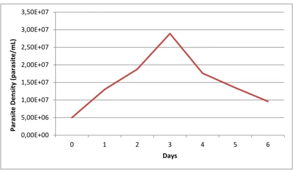
The conditions for the viability assay using Leishmania parasites, such as initial parasite concentration and incubation time, was based on the growth curve of promastigotes. Exponential phase occurred during the 3 first days of culture, so the exposition of the parasites to cyanobacteria extracts and natural based compounds was performed during 72hours.
Figure 2 – Growth curve of Leishmania infantum
4.2.1.2. Viability assays
The search for new treatments against leishmaniasis has increased due to high frequency of drug resistance registered in endemics areas, side effects, and complications caused by coinfection with HIV(Almeida-Souza et al., 2016). Classic drugs used to treat leishmaniasis, such as antimonials and amphotericin B are considered toxic to some patients, justifying the rational search for new anti-leishmanial compounds (Yamamoto et al., 2015).
0,00E+00 5,00E+06 1,00E+07 1,50E+07 2,00E+07 2,50E+07 3,00E+07 3,50E+07 0 1 2 3 4 5 6 Par asi te D e n si ty (p ar asi te /m L) Days