ABSTRACT
The study of the human microbiome—and, more recently, that of the respiratory system—by means of sophisticated molecular biology techniques, has revealed the immense diversity of microbial colonization in humans, in human health, and in various diseases. Apparently, contrary to what has been believed, there can be nonpathogenic colonization of the lungs by microorganisms such as bacteria, fungi, and viruses. Although this physiological lung microbiome presents low colony density, it presents high diversity. However, some pathological conditions lead to a loss of that diversity, with increasing concentrations of some bacterial genera, to the detriment of others. Although we possess qualitative knowledge of the bacteria present in the lungs in different states of health or disease, that knowledge has advanced to an understanding of the interaction of this microbiota with the local and systemic immune systems, through which it modulates the immune response. Given this intrinsic relationship between the microbiota and the lungs, studies have put forth new concepts about the pathophysiological mechanisms of homeostasis in the respiratory system and the potential dysbiosis in some diseases, such as cystic fi brosis, COPD, asthma, and interstitial lung disease. This departure from the paradigm regarding knowledge of the lung microbiota has made it imperative to improve understanding of the role of the microbiome, in order to identify possible therapeutic targets and to develop innovative clinical approaches. Through this new leap of knowledge, the results of preliminary studies could translate to benefi ts for our patients.
Keywords: Microbiota; Microbiology; Immune system.
The pulmonary microbiome: challenges of a
new paradigm
André Nathan Costa1,a, Felipe Marques da Costa1,b, Silvia Vidal Campos1,c,
Roberta Karla Salles1,d, Rodrigo Abensur Athanazio1,e
Correspondence to:
André Nathan Costa. Rua Dr. Enéas Carvalho de Aguiar, 44, CEP 05403-000, São Paulo, SP, Brasil. Tel./Fax: 55 11 2661-5695. E-mail: [email protected]
Financial support: None. INTRODUCTION
“The lungs of healthy humans are sterile sites, unlike the upper airways where there are commensal microorganisms—they live in homeostasis with the human body”.(1) Although the respiratory system has a surface area greater than 70 m2—which is the size of a tennis court—and is in direct contact with the environment, the concept above pervaded knowledge of the respiratory
system until the early 21st century, when the fi rst studies based on molecular techniques for the identifi cation of
bacterial DNA revealed the presence of genetic material from microorganisms in the lower respiratory tract.(1,2) Much of this delay in knowledge of the lung microbiota
is due to the diffi culty in characterizing the human
lung environment by means of conventional culture techniques based on bacterial growth in material collected by bronchoalveolar lavage.(2,3) This occurs because the bacterial load of the lungs is lower than that of other sites in the human body, such as the gastrointestinal and genitourinary tracts. In addition, there has always been intense debate over possible contamination of material collected from the lower airways with microorganisms from the upper airways, which resulted in exclusion
of the lungs from early studies mapping the human microbiome.(2,4-7)
After the overcoming of these initial obstacles in the study of the lung microbiome, science has advanced to an understanding of the interaction of this microbiota with the local and systemic immune systems, through which it modulates the immune response in the context of health and of various respiratory diseases. The characterization of the lung microbiome therefore has the potential to provide new concepts about the pathophysiological mechanisms of homeostasis in the respiratory system and the loss of this balance, known as dysbiosis, in some
diseases such as cystic fi brosis (CF), COPD, asthma, and
interstitial lung diseases.(8-11)
It is highly likely that the microbiome and its
changes have a direct infl uence on the natural history
of respiratory diseases, as well as there is certainly a change in the microbiota resulting from antibiotic treatment of infectious respiratory tract diseases. In addition, increasing knowledge of the lung microbiome has brought about a discussion of a possible distinction between those bacterial species that are pathogens and those that behave as commensals in the composition of our physiological microbiome.(12-15)
1. Divisão de Pneumologia, Instituto do Coração – InCor – Hospital das Clínicas, Faculdade de Medicina, Universidade de São Paulo, São Paulo (SP) Brasil. a. http://orcid.org/0000-0002-8025-6940 b. http://orcid.org/0000-0001-5118-2033 c. http://orcid.org/0000-0003-3297-5948 d. http://orcid.org/0000-0003-3074-445X e. http://orcid.org/0000-0002-9399-5275
Submitted: 12 June 2017. Accepted: 16 July 2017.
To better tread this new path that opens up to pulmonology, some concepts are important. Microbiota, microbiome, metagenome, and 16S rRNA are terms
that pervade studies in this fi eld, and mastery of
these terms facilitates the understanding of this new
dimension of knowledge (Chart 1). With regard to
technique, analysis of the bacterial microbiome is based
on the identifi cation and sequencing of variable regions
of the 16S gene encoding bacterial rRNA. Given that this gene is not present in mammals, the confounding bias with human DNA is nonexistant.(1,4,8) Finally, the 16S DNA sequence contains nine variable regions
that can be identifi ed by various techniques, the most
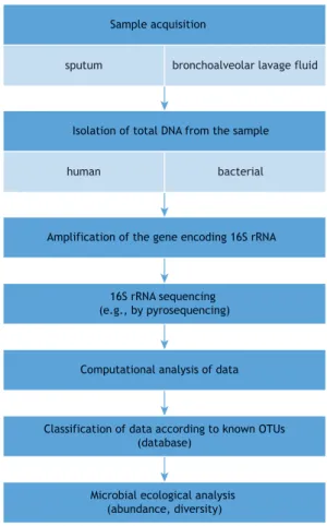
commonly used being pyrosequencing, phylogenetic microarrays, and terminal restriction fragment length polymorphism.(1,3,4) Figure 1 summarizes the sequence of events leading to the recognition of the microbiome at a given site.
Although the results of published studies differ a little,
Proteobacteria, Firmicutes, and Bacteroidetes are the most commonly identifi ed bacterial phyla in healthy individuals. With regard to genus, Streptococcus, Prevotella, Fusobacteria, and Veillonella predominate, with potential pathogens, such as Haemophilus and Neisseria, contributing a small fraction. However, those studies are based on case series involving a small number of healthy subjects and few centers around the world.(2,3,16)
Being as important as qualitative knowledge of the
bacteria present in the lungs, describing the richness of organisms and the coexistence of various species is essential. Although the healthy lung microbiome presents low colony density, it presents high diversity; however, some pathological conditions lead to a loss of that diversity, with increasing concentrations of some bacterial genera, to the detriment of others.(15,17,18) This
could imply the development of specifi c therapies, to
the detriment of broad-spectrum antibiotic therapies, with great potential to cause more imbalance in an already dysbiotic microbiome.(19) Following this line of
reasoning, the anti-infl ammatory effects of macrolides,
which are considered to be immunomodulators and are used for prolonged periods in diseases such as
bronchiectasis, bronchiolitis obliterans, and COPD, have been revisited, since these anti-infl ammatory
properties appear to be related to changes in the
lung microbiota and in the microbial metabolites, with subsequent downregulation of alveolar macrophage function.(13)
Finally, as far as the microbial population in the
respiratory system is concerned, early studies failed to address two important components of the lung ecosystem: viruses and fungi. The lung mycobiome
and virome, especially in some conditions, such as CF
and lung transplantation, appear to have particular importance.(20) Viral and fungal identifi cation uses the same technique as that used in bacterial microbiota analysis, but with detection of 18S rRNA in the case
Chart 1. Glossary of nomenclature and defi nitions used in the routine evaluation of the human microbiome.
Microbiota All of the microorganisms of a given region or habitat
Microbiome The collection of microorganisms, their genes, and their environmental interactions Metagenome Genetic information of the microbiota, obtained from genetic sequencing that is
analyzed, organized, and identifi ed through computational tools, using databases of
previously known sequences
16S rRNA Component of the prokaryotic 30S subunit. It encodes DNA 16S genes, used to obtain phylogenetic data
Operational taxonomic unit Operational defi nition of a species or group of species, used when only DNA sequence
data are available
Dysbiosis An imbalance in the composition of the microbiota of a given niche, related to changes in local conditions
Figure 1. Sequence of events leading to the recognition of the microbiome at a given site. OTU: operational taxonomic unit.
sputum bronchoalveolar lavage fluid Sample acquisition
Isolation of total DNA from the sample
human bacterial
Amplification of the gene encoding 16S rRNA
16S rRNA sequencing (e.g., by pyrosequencing)
Computational analysis of data
Classification of data according to known OTUs (database)
of fungi and with nucleic acid sequencing and PCR in
the case of viruses.(21)
THE LUNG MICROBIOME AND ITS RELATIONSHIP WITH THE GASTROINTESTINAL TRACT
The theory that the facial sinuses were the major determinants of the microbiological changes found in the lower respiratory tract persisted for a long time. The glottis was then considered to be an effective structure in protecting the lungs from the gastrointestinal tract. However, the microbiome of the lower respiratory tract is now recognized to be similar to that of the oropharynx, leading to the concept that microbial migration from this region is the major determinant
of the lung microbiome in healthy individuals.(22)
Microaspiration appears to play a key role in shaping the lung microbiome, although other bacteria that are present in the lower respiratory tract, such as those of the genera Prevotella, Veillonella, and Streptococcus, have their origin attributable to inhalation through the upper airways.(23,24) The interrelationship of these systems and the local determinants of the lung
microbiome are presented in Figures 2 and 3.
THE MICROBIOTA AND IMMUNE MODULATION
Commensal interactions between microorganisms
and humans throughout evolution, as well as the relevance of the luminal ecosystem (genitourinary and gastrointestinal tracts) in immune modulation, have emerged as a new paradigm. This ecosystem is separated from the interior of the host by a thin layer of epithelial cells that acts as an interface between the host and the environment, and this epithelium is equipped with cilia, microvilli, mucus-producing cells, and intercellular junctions that allow physiological functions while in contact with the microbiota.(25) Studies of sterile (germ-free) mice have shown that
resident bacteria directly infl uence epithelial metabolism,
proliferation, turnover, and barrier function.(25) In the respiratory system, members of the microbiota, in association with environmental non-viable particulate antigens, are continuously presented to the mucosa and processed by dendritic cells and macrophages, with subsequent formation of memory or activation
of T and B effector cells.(25) In addition, studies of the gastrointestinal tract have demonstrated the ability of the immune system to discriminate between pathogenic and commensal bacteria, through toll-like receptors present in T lymphocytes, in a process that would allow symbiotic colonization, that is, a kind of “peace agreement” between the resident microbiota and the respiratory mucosa,(25,26) as exemplifi ed in Figure 4.
Evidence, however, shows that abnormal regulation of this host-microbiota relationship plays an important
role in the pathophysiology of several lung infl ammatory
disorders. Therefore, characterizing the composition of the airway microbiota as a prognostic marker or as a
guide to drug therapy is of interest in several chronic lung diseases,(27) as described below.
THE MICROBIOTA IN VARIOUS LUNG DISEASES
Asthma
Asthma is a complex, heterogeneous disease associated with allergic phenomena that has increased in prevalence in recent decades. The hygiene hypothesis
is one of the major theories explaining this fi nding.
(28) Low-level exposure to bacterial infections during
childhood may be responsible for modulation of the immune response with strong emphasis on the Th2-allergic pathway. Therefore, there has been increasing interest in the role of both the lung and gastrointestinal microbiome. An experimental study of bacteria-free mice demonstrated that those animals showed an exaggerated Th2 response when stimulated with ovalbumin, developing increased eosinophilia in the airways, hyperresponsiveness, and mucus
hypersecretion. When those animals were placed to
grow alongside mice with the usual bacterial microbiota, both groups showed the same Th2 response intensity, which indicates that the usual microbiome functions as a protective factor against allergic diseases.(29)
Given that bacterial colonization of mucous membranes is related to the development and orchestration of the immune response of healthy individuals, changes in this interrelationship in early stages of life may contribute to the development of allergic diseases in adulthood.(30) In a study comparing two agricultural communities with similar habits, but with a distinct prevalence of asthma and allergic sensitization, the presence of a microbial composition with increased endotoxin production was related to a lower prevalence of allergic disorders.(31) In addition, a nasal microbiota with decreased diversity of species, especially when accompanied by the presence of Moraxella spp., has also been associated with a higher prevalence of asthma.(32)
Among adults, patients with asthma have been shown to have a higher prevalence of organisms of the
phylum Proteobacteria, such as Haemophilus infl uenzae, compared with healthy controls.(33,34) Studies in this area are scarce and involve a small number of patients,
leading to heterogeneity of fi ndings. Nevertheless,
all indicate the presence of lung-microbiota-related
dysbiosis in asthma patients, which can be infl uenced
both by disease severity and by inhaled or systemic corticosteroid use.(35,36)
COPD
Studies comparing the microbiome of smokers, former smokers, and healthy individuals are scarce and
report some confl icting results regarding the long-term
effects of tobacco exposure.(37,38) Nevertheless, there are indications that dysbiosis occurs in smokers, with
an increase in the prevalence of the phylum Firmicutes
Inhalation
Microaspiration
Lungs and airways • Normal ciliary function • Cough
Normal microbiome
Gastrointestinal tract • Bacterial toxins • Bacterial metabolites • Migratory cells
Innate immune response Acquired immune response
Elimination Immigration
Cough and mucociliary clearance
Immune response
Microaspiration
Inhalation
Direct dispersion
Figure 2. Determinants of the lung microbiome and the intestine-lung axis. The composition of the human microbiota is determined by the association of environmental factors, the host immune response, and genetic characteristics. The intestine microbiota, which is incomparably greater in size than the lung microbiota, can infl uence the lower respiratory tract both directly, through microaspiration, and indirectly, through modulation of the immune response as a result of the production of bacterial metabolites and their interaction with the host infl ammatory cells. Inhalation of external agents is also a pathway to lung colonization and will depend, as will intestinal tract colonization, on local factors, such as oxygen tension, tissue pH, blood perfusion, nutrient concentration, proper mucociliary transport, and disruption of the lung architecture.
decrease in the abundance of Proteobacteria.(37) In
contrast, several studies of patients with COPD have
revealed that their lung microbiome is clearly different from that of healthy controls.(39-42) In addition, among
patients with COPD, depending on the site from which
the material is collected, differences are also found in the composition of the microbiome, such as when,
for instance, sputum and bronchoalveolar lavage fl uid
are compared.(42)
With regard to COPD exacerbations, numerous
studies have also demonstrated that there is a relative increase in the abundance of a given genus, to the detriment of others.(43-45) This change is related to a
pro-infl ammatory state and can be triggered, among other
causes, by viral infections(43) and by bacterial-fungal interactions in the airway.(46) These fi ndings further
question the role of antibiotics in COPD exacerbation,
given that antibiotics can play a deleterious role in the lung microbiome because they reduce bacterial abundance. However, systemic corticosteroid use does
not signifi cantly change microbiological diversity and, in
parallel, can increase the abundance of certain genera
that are considered as normal fl ora.(44)
Bronchiectasis and CF
Airway colonization in suppurative lung diseases—CF and non-CF bronchiectasis—plays a key role in
the progression of their clinical and radiological manifestations, and understanding the role of the microbiota is key to understanding the pathophysiology
of these manifestations. While traditional
culture-based knowledge shows the importance of well-known pathogens, such as Haemophilus infl uenzae, Pseudomonas aeruginosa, and Moraxella catarrhalis in
non-CF bronchiectasis, as well as Staphylococcus aureus and Burkholderia cepacia complex in CF, molecular studies have shown that previously unrecognized organisms are abundantly present in some patients with suppurative disease.(27) Examples of this colonization include the presence of Stenotrophomonas maltophilia and Achromobacter spp., as well as reports of Mycobacterium abscessus and Aspergillus fumigatus.(10)
Studies of the microbiome in patients with CF have
demonstrated that samples from younger, healthier patients usually exhibit bacterial communities that are more diverse, whereas lung explants from patients with end-stage lung disease show extremely low diversity, with only one or two detectable pathogenic bacteria, such as P. aeruginosa and S. maltophilia. (47) This microbiological change, during the lifetime of a
patient with CF, is also accompanied by increased
abundance and greater phylogenetic similarity among the colonies of each species.(48,49)
In non-CF bronchiectasis, the simultaneous
competition for survival between pathogenic and commensal bacteria elicits an innate immune response from the host, with polarization of T-cell subtype
response, activating or perpetuating the infl ammatory
process in the terminal airways, similarly to what
occurs in chronic infl ammatory bowel diseases, such as ulcerative colitis. In the context of infl ammatory
bowel diseases, it is of note that there is an association between the development of bronchiectasis and colectomy for advanced ulcerative colitis, raising the
possibility that the intestinal microbiota infl uences the
lung microbiota through systemic immunoregulation after excluding in intestinal barrier.(12)
Finally, in suppurative diseases, an understanding of
the pathogenic and commensal microbiota is crucial for differentiating between infection and colonization, that is, balance/health vs. dysbiosis/disease. It should also be borne in mind that, for this group of patients, the study of the fungal and viral microbiome (microbiome and virome) is essential, and there have been few studies addressing these issues to date. The literature still lacks controlled clinical trials; most studies are descriptive studies or reviews.
Pulmonary tuberculosis
including reports of an increased presence of Candida spp. and a loss of diversity in the intestinal microbiota as a result of tuberculosis treatment.(51,52) In addition, there is little agreement between those studies and analyses of the lung microbiome in tuberculosis.
Cui et al.(53) reported that healthy lungs and those infected with Mycobacterium tuberculosis had many microorganisms in common, including those of the
phyla Bacteroidetes, Proteobacteria, and Actinobacteria, with a predominance of Firmicutes and Bacteroidetes. In contrast, Wu et al.(54) found a very different list of microorganisms associated with tuberculosis, including those of the genera Streptococcus, Granulicatella, and Pseudomonas. An interesting aspect of that study is the comparison among the microbiota of patients with newly diagnosed tuberculosis, that of recurrence cases, and that of treatment failure cases. The Pseudomonas-to-Mycobacterium ratio was higher in recurrence cases than in newly diagnosed cases, whereas the Treponema-to-Mycobacterium ratio was lower in recurrence cases than in newly diagnosed cases, indicating that disruption of these bacteria may be a risk factor for recurrence of tuberculosis.(54) These data suggest that the presence of certain bacteria and lung dysbiosis may be associated not only with the development of tuberculosis but also with recurrence of tuberculosis and treatment failure, indicating a possible role of the microbiota in the pathogenesis of tuberculosis and in tuberculosis treatment outcomes.
INTERSTITIAL LUNG DISEASES
In 2008, Varney et al.(55) published a clinical trial evaluating the use of trimethoprim-sulfamethoxazole
in patients with idiopathic pulmonary fi brosis (IPF).
They demonstrated that the group receiving the antibiotic therapy showed clinical and functional
improvement,(55) and hypothesized a potential
effect on the lung microbiota. More recently, data
from a cohort study assessing 55 patients with IPF
demonstrated that there is a relationship between
the predominance of specifi c bacteria of the genera Staphylococcus and Streptococcus and interstitial lung disease exacerbation.(56) Also in 2014, Molyneaux et al.(57) observed that bronchoalveolar lavage fl uid from
patients with IPF exhibited an increased quantity of
bacteria compared with that from healthy controls, as well as observing differences in the composition and diversity of this microbiota, and linked this dysbiosis to parenchymal disease progression. Later, genetic
analyses of patients with IPF showed increase and
maintenance in the expression of genes related to the host immune response, which acts as a continuous stimulus damaging to the alveolar epithelium, as well
as being related to local fi broblast activation,(58,59) subsequently suggesting a relationship between the
microbiome and fi brosis progression. Attempts at
reversal of dysbiosis and, ultimately, the cessation of tissue damage have been extensively investigated in
the context of interstitial fi brosing diseases; however,
it is too early to say that the microbiota is directly related to disease progression.(27)
OTHER CLINICAL SETTINGS IN PULMONOLOGY
After initial advances in the understanding of the microbiome of the respiratory system in the context of the most prevalent diseases, it is expected that this understanding will revolutionize the concepts of pathogenesis in several clinical settings. In the context of mechanical ventilation and ventilator-associated
pneumonia (VAP), that is not different. A recent study
of 35 patients suggested that mechanical ventilation per se is more strongly associated with a change in the lung microbiota than is the use of systemic antibiotics, and that respiratory tract dysbiosis is more intense
in patients who developed VAP than in those who did
not.(60) Still in the context of VAP, microbiome analysis may also aid in etiologic diagnosis and in differentiating between pneumonia and colonization with a potential pathogen.(61)
Lung transplantation is another area that is in a
state of fl ux. The respiratory system of transplant
recipients is a point of special interest, given the wide use of prophylactic antibiotics and immunosuppressive drugs in this population. The microbiota of the transplanted lung appears to be different from that of healthy lungs, mainly because of the presence
of the family Burkholderiaceae.(62) In addition, the
change in the microbiota appears to infl uence the
development of chronic graft dysfunction.(63) A study
of 203 bronchoalveolar lavage fl uid samples from
112 transplant recipients revealed that some bacteria
played a pro-infl ammatory role (genera Staphylococcus and Pseudomonas) and some played a role of lower stimulation of the immune system (genera Prevotella and Streptococcus).(64) A dysbiosis in those individuals
appears to be associated with various profi les of infl ammation and elaboration by lung macrophages,
contributing to the genesis of chronic dysfunction. This interaction between bacterial communities and innate immune response offers new intervention pathways to preventing chronic graft dysfunction.
Clinical oncology has also advanced in the understanding
of the correlations between microorganisms and lung
neoplasms. After the identifi cation of molecular markers, such as EGFR, programmed cell death protein-1, and
anaplastic lymphoma kinase, which have customized the therapeutic approach, it is now the microbiome that presents itself as a possible marker of disease activity and perhaps a therapeutic target. Some microorganisms have shown a direct relationship with neoplasms in other organs, such as Helicobacter pylori in stomach cancer and HPV in uterine cervical cancer. Periodontal disease can be associated with lung cancer,
decreased relative abundance of organisms of the
phyla Spirochaetes and Bacteroidetes and an increased relative abundance of those of the phylum Firmicutes
in analyses of the oral microbiota.(66) Finally, a study
comparing bronchoalveolar lavage fl uid from patients
with neoplasms with that from patients with benign tumors found the genera Veillonella and Megasphaera to be predictors of lung cancer, suggesting an association between an altered lung microbiota and the presence of neoplasm.(67)
SKEPTICISM IN THE ANALYSIS
Albeit exciting, the study of the microbiome should be viewed with some caution, and it has been said that its greatest risk is that of drowning in its own tsunami of information.(68) There is a risk of a series of speculative associations being made between the microbiota and states of health and disease, and of these connections being shown to be spurious or much more complex than shown by early evaluations.
Would the microbial communities be altered in
the lungs because of respiratory diseases or are the lungs diseased because of the dysbiosis of these microorganisms? The immune system and the microbiome are so closely intertwined that this
differentiation is extremely diffi cult. Most studies on
this subject have been descriptive and, despite being replete with provocative correlations, have failed to elucidate causality between modulation of respiratory tract diseases and the resident microbiota, and to
determine what temporally comes fi rst: dysbiosis or
lung disease.(69) These are some of the issues that are pressing when it comes to designing new studies.
FINAL CONSIDERATIONS
We are entering a new era in the understanding of
lung diseases from the standpoint of the interaction of bacterial communities and metabolites with the immunological and functional mechanisms of various respiratory tract diseases. In this context, given the departure from the paradigm regarding knowledge of the lung microbiota, it is imperative to improve understanding of the interaction between the microbiota and the host so that we can advance our understanding of the pathophysiological processes of respiratory diseases, as well as identifying possible therapeutic targets and developing innovative clinical approaches. The translation of this information into better patient care will undoubtedly be the greatest challenge in the study of the microbiome and its potentialities.
REFERENCES
1. Beck JM. ABCs of the lung microbiome. Ann Am Thorac Soc. 2014;11 Suppl 1:S3-6. https://doi.org/10.1513/AnnalsATS.201306-188MG
2. Marsland BJ, Gollwitzer ES. Host-microorganism interactions in lung diseases. Nat Rev Immunol. 2014;14(12):827-35. https://doi. org/10.1038/nri3769
3. Rogers GB, Shaw D, Marsh RL, Carroll MP, Serisier DJ, Bruce KD. Respiratory microbiota: addressing clinical questions, informing clinical practice. Thorax. 2015;70(1):74-81. https://doi.org/10.1136/ thoraxjnl-2014-205826
4. Human Microbiome Project Consortium. Structure, function and diversity of the healthy human microbiome. Nature. 2012;486(7402):207-14. https://doi.org/10.1038/nature11234
5. Li K, Bihan M, Yooseph S, Methé BA. Analyses of the microbial diversity across the human microbiome. PLoS One. 2012;7(6) e32118. https://doi.org/10.1371/journal.pone.0032118
6. Huang YJ, Charlson ES, Collman RG, Colombini-Hatch S, Martinez FD, Senior RM. The role of the lung microbiome in health and disease: A national Heart, Lung, and Blood Institute workshop report. Am J Respir Crit Care Med. 2013;187(12):1382-7. https://doi. org/10.1164/rccm.201303-0488WS
7. Gensollen T, Iyer SS, Kasper DL, Blumberg RS. How colonization by microbiota in early life shapes the immune system. Science. 2016;352(6285):539-44. https://doi.org/10.1126/science.aad9378 8. Jiao X, Zheng X, Ma L, Kutty G, Gogineni E, Sun Q, et al. A Benchmark
Study on Error Assessment and Quality Control of CCS Reads Derived from the PacBio RS. J Data Mining Genomics Proteomics. 2013;4(3). pii: 16008. https://doi.org/10.4172/2153-0602.1000136
9. Mammen MJ, Sethi S. COPD and the microbiome. Respirology. 2016;21(4):590-9. https://doi.org/10.1111/resp.12732
10. Surette MG. The cystic fi brosis lung microbiome. Ann Am Thorac Soc. 2014;11 Suppl 1:S61-5. https://doi.org/10.1513/AnnalsATS.201306-159MG
11. Han MK, Zhou Y, Murray S, Tayob N, Lama VN, Moore BB, et al. Lung microbiome and disease progression in idiopathic pulmonary fi brosis: an analysis of the COMET study. Lancet Respir Med. 2014;2(7):548-56 https://doi.org/10.1016/S2213-2600(14)70069-4
12. Boyton RJ, Reynolds CJ, Quigley KJ, Altmann DM. Immune mechanisms and the impact of the disrupted lung microbiome in
chronic bacterial lung infection and bronchiectasis. Clin Exp Immunol. 2013;171(2):117-23. https://doi.org/10.1111/cei.12003
13. Segal LN, Clemente JC, Wu BG, Wikoff WR, Gao Z, Li Y, et al. Randomised, double-blind, placebo-controlled trial with azithromycin selects for anti-infl ammatory microbial metabolites in the emphysematous lung. Thorax. 2017;72(1):13-22. https://doi. org/10.1136/thoraxjnl-2016-208599
14. Sze MA, Dimitriu PA, Suzuki M, McDonough JE, Campbell JD, Brothers JF, et al. Host response to the lung microbiome in chronic obstructive pulmonary disease. Am J Respir Crit Care Med. 2015;192(4):438-45. https://doi.org/10.1164/rccm.201502-0223OC
15. Wang Z, Bafadhel M, Haldar K, Spivak A, Mayhew D, Miller BE, et al. Lung microbiome dynamics in COPD exacerbations. Eur Respir J. 2016;47(4):1082-92. https://doi.org/10.1183/13993003.01406-2015 16. Chotirmall SH, Gellatly SL, Budden KF, Mac Aogain M, Shukla SD,
Wood DL, et al. Microbiomes in respiratory health and disease: An Asia-Pacifi c perspective. Respirology. 2017;22(2):240-250. https:// doi.org/10.1111/resp.12971
17. Scales DC. The Implications of a Tracheostomy for Discharge Destination. Am J Respir Crit Care Med. 2015;192(4):404-5. https:// doi.org/10.1164/rccm.201505-1007ED
18. von Mutius E. Environmental microorganisms and lung health. Ann Am Thorac Soc. 2014;11 Suppl 1:S13-5. https://doi.org/10.1513/ AnnalsATS.201306-155MG
19. Blaser MJ. Antibiotic use and its consequences for the normal microbiome. Science. 2016;352(6285):544-5. https://doi.org/10.1126/ science.aad9358
20. Underhill DM, Iliev ID. The mycobiota: interactions between commensal fungi and the host immune system. Nat Rev Immunol. 2014;14(6):405-16. https://doi.org/10.1038/nri3684
21. Mitchell AB, Oliver BG, Glanville AR. Translational Aspects of the Human Respiratory Virome. Am J Respir Crit Care Med. 2016;194(12):1458-1464. https://doi.org/10.1164/rccm.201606-1278CI
23. Venkataraman A, Bassis CM, Beck JM, Young VB, Curtis JL, Huffnagle GB, et al. Application of a neutral community model to assess structuring of the human lung microbiome. MBio. 2015;6(1). pii: e02284-14. https://doi.org/10.1128/mBio.02284-14
24. Dickson RP, Erb-Downward JR, Huffnagle GB. Homeostasis and its Disruption in the Lung Microbiome. Am J Physiol Lung Cell Mol Physiol. 2015;309(10):L1047-55. https://doi.org/10.1152/ ajplung.00279.2015
25. Neish AS. Mucosal immunity and the microbiome. Ann Am Thorac Soc. 2014;11 Suppl 1:S28-32. https://doi.org/10.1513/ AnnalsATS.201306-161MG
26. Round JL, Lee SM, Li J, Tran G, Jabri B, Chatila TA, et al. The Toll-like receptor 2 pathway establishes colonization by a commensal of the human microbiota. Science. 2011;332(6032):974-7. https://doi. org/10.1126/science.1206095
27. Faner R, Sibila O, Agustí A, Bernasconi E, Chalmers JD, Huffnagle GB, et al. The microbiome in respiratory medicine: current challenges and future perspectives. Eur Respir J. 2017;49(4). pii: 1602086. https://doi.org/10.1183/13993003.02086-2016
28. Strachan DP. Hay fever, hygiene, and household size. BMJ. 1989;299(6710):1259-60. https://doi.org/10.1136/ bmj.299.6710.1259
29. Herbst T, Sichelstiel A, Schär C, Yadava K, Bürki K, Cahenzli J, et al. Dysregulation of allergic airway infl ammation in the absence of microbial colonization. Am J Respir Crit Care Med. 2011;184(2):198-205. https://doi.org/10.1164/rccm.201010-1574OC
30. Gensollen T, Iyer SS, Kasper DL, Blumberg RS. How colonization by microbiota in early life shapes the immune system. Science. 2016;352(6285):539-44. https://doi.org/10.1126/science.aad9378 31. Stein MM, Hrusch CL, Gozdz J, Igartua C, Pivniouk V, Murray SE,
et al. Innate Immunity and Asthma Risk in Amish and Hutterite Farm Children. N Engl J Med. 2016;375(5):411-421. https://doi. org/10.1056/NEJMoa1508749
32. Depner M, Ege MJ, Cox MJ, Dwyer S, Walker AW, Birzele LT, et al. Bacterial microbiota of the upper respiratory tract and childhood asthma. J Allergy Clin Immunol. 2017;139(3):826-834.e13. https:// doi.org/10.1016/j.jaci.2016.05.050
33. Hilty M, Burke C, Pedro H, Cardenas P, Bush A, Bossley C, et al. Disordered microbial communities in asthmatic airways. PLoS One. 2010;5(1):e8578. https://doi.org/10.1371/journal.pone.0008578
34. Huang YJ, Nelson CE, Brodie EL, Desantis TZ, Baek MS, Liu J, et al. Airway microbiota and bronchial hyperresponsiveness in patients with suboptimally controlled asthma. J Allergy Clin Immunol. 2011;127(2):372-381.e1-3.
35. Denner DR, Sangwan N, Becker JB, Hogarth DK, Oldham J, Castillo J, et al. Corticosteroid therapy and airfl ow obstruction infl uence the bronchial microbiome, which is distinct from that of bronchoalveolar lavage in asthmatic airways. J Allergy Clin Immunol. 2016;137(5):1398-1405.e3. https://doi.org/10.1016/j. jaci.2015.10.017
36. Durack J, Lynch SV, Nariya S, Bhakta NR, Beigelman A, Castro M, et al. Features of the bronchial bacterial microbiome associated with atopy, asthma, and responsiveness to inhaled corticosteroid treatment. J Allergy Clin Immunol. 2017;140(1):63-75. https://doi. org/10.1016/j.jaci.2016.08.055
37. Morris A, Beck JM, Schloss PD, Campbell TB, Crothers K, Curtis JL, et al. Comparison of the respiratory microbiome in healthy nonsmokers and smokers. Am J Respir Crit Care Med. 2013;187(10):1067-75. https://doi.org/10.1164/rccm.201210-1913OC
38. Munck C, Helby J, Westergaard CG, Porsbjerg C, Backer V, Hansen LH. Smoking Cessation and the Microbiome in Induced Sputum Samples from Cigarette Smoking Asthma Patients. PLoS One. 2016;11(7):e0158622. https://doi.org/10.1371/journal.pone.0158622
39. Sze MA, Dimitriu PA, Hayashi S, Elliott WM, McDonough JE, Gosselink JV, et al. The lung tissue microbiome in chronic obstructive pulmonary disease. Am J Respir Crit Care Med. 2012;185(10):1073-80. https://doi.org/10.1164/rccm.201111-2075OC
40. Pragman AA, Kim HB, Reilly CS, Wendt C, Isaacson RE. The lung microbiome in moderate and severe chronic obstructive pulmonary disease. PLoS One. 2012;7(10):e47305. https://doi.org/10.1371/ journal.pone.0047305
41. Millares L, Ferrari R, Gallego M, Garcia-Nuñez M, Pérez-Brocal V, Espasa M, et al. Bronchial microbiome of severe COPD patients colonised by Pseudomonas aeruginosa. Eur J Clin Microbiol Infect Dis. 2014;33(7):1101-11. https://doi.org/10.1007/s10096-013-2044-0
42. Cabrera-Rubio R, Garcia-Núñez M, Setó L, Antó JM, Moya A, Monsó
E, et al. Microbiome diversity in the bronchial tracts of patients with chronic obstructive pulmonary disease. J Clin Microbiol. 2012;50(11):3562-8. https://doi.org/10.1128/JCM.00767-12
43. Molyneaux PL, Mallia P, Cox MJ, Footitt J, Willis-Owen SA, Homola D, et al. Outgrowth of the bacterial airway microbiome after rhinovirus exacerbation of chronic obstructive pulmonary disease. Am J Respir Crit Care Med. 2013;188(10):1224-31. https://doi. org/10.1164/rccm.201302-0341OC
44. Dy R, Sethi S. The lung microbiome and exacerbations of COPD. Curr Opin Pulm Med. 2016;22(3):196-202. https://doi.org/10.1097/ MCP.0000000000000268
45. Huang YJ, Sethi S, Murphy T, Nariya S, Boushey HA, Lynch SV. Airway microbiome dynamics in exacerbations of chronic obstructive pulmonary disease. J Clin Microbiol. 2014;52(8):2813-23. https://doi. org/10.1128/JCM.00035-14
46. Huffnagle GB, Noverr MC. The emerging world of the fungal microbiome. Trends Microbiol. 2013;21(7):334-41. https://doi. org/10.1016/j.tim.2013.04.002
47. Cox MJ, Allgaier M, Taylor B, Baek MS, Huang YJ, Daly RA, et al. Airway microbiota and pathogen abundance in age-stratifi ed cystic fi brosis patients. PLoS One. 2010;5(6):e11044. https://doi. org/10.1371/journal.pone.0011044
48. Rudkjøbing VB, Thomsen TR, Alhede M, Kragh KN, Nielsen PH, Johansen UR, et al. The microorganisms in chronically infected end-stage and non-end-end-stage cystic fi brosis patients. FEMS Immunol Med Microbiol. 2012;65(2):236-44. https://doi.org/10.1111/j.1574-695X.2011.00925.x
49. Frayman KB, Armstrong DS, Carzino R, Ferkol TW, Grimwood K, Storch GA, et al. The lower airway microbiota in early cystic fi brosis lung disease: a longitudinal analysis. Thorax. 2017;72(12):1104-1112. https://doi.org/10.1136/thoraxjnl-2016-209279
50. Adami AJ, Cervantes JL. The microbiome at the pulmonary alveolar niche and its role in Mycobacterium tuberculosis infection. Tuberculosis (Edinb). 2015;95(6):651-658. https://doi.org/10.1016/j. tube.2015.07.004
51. Querido SM, Back-Brito GN, Dos Santos SS, Leão MV, Koga-Ito CY, Jorge AO. Opportunistic microorganisms in patients undergoing antibiotic therapy for pulmonary tuberculosis. Brazilian J Microbiol. 2011;42(4):1321-8. https://doi.org/10.1590/S1517-83822011000400012
52. Dubourg G, Lagier JC, Armougom F, Robert C, Hamad I, Brouqui P, et al. The gut microbiota of a patient with resistant tuberculosis is more comprehensively studied by culturomics than by metagenomics. Eur J Clin Microbiol Infect Dis. 2013;32(5):637-45. https://doi.org/10.1007/s10096-012-1787-3
53. Cui Z, Zhou Y, Li H, Zhang Y, Zhang S, Tang S, et al. Complex sputum microbial composition in patients with pulmonary tuberculosis. BMC Microbiol. 2012;12:276. https://doi.org/10.1186/1471-2180-12-276
54. Wu J, Liu W, He L, Huang F, Chen J, Cui P, et al. Sputum microbiota associated with new, recurrent and treatment failure tuberculosis. PLoS One. 2013;8(12):e83445. https://doi.org/10.1371/journal. pone.0083445
55. Varney VA, Parnell HM, Salisbury D., Ratnatheepan S, Tayar RB. A double blind randomised placebo controlled pilot study of oral co-trimoxazole in advanced fi brotic lung disease. Pulm Pharmacol Ther. 2008;21(1):178-87. https://doi.org/10.1016/j.pupt.2007.02.001
56. Han ML, Zhou Y, Murray S, Tayob N, Noth I, Lama VN, et al. Lung microbiome and disease progression in idiopathic pulmonary fi brosis: an analysis of the COMET study. Lancet Respir Med. 2014;2(7):548-56. https://doi.org/10.1016/S2213-2600(14)70069-4
57. Molyneaux PL, Cox MJ, Willis-Owen SA, Mallia P, Russell KE, Russell AM, et al. The role of bacteria in the pathogenesis and progression of idiopathic pulmonary fi brosis. Am J Respir Crit Care Med. 2014;190(8):906-13. https://doi.org/10.1164/rccm.201403-0541OC
58. Huang Y, Ma SF, Espindola MS, Vij R, Oldham JM, Huffnagle GB, et al. Microbes Are Associated with Host Innate Immune Response in Idiopathic Pulmonary Fibrosis. Am J Respir Crit Care Med. 2017;196(2):208-219. https://doi.org/10.1164/rccm.201607-1525OC
59. Molyneaux PL, Willis Owen SAG, Cox MJ, James P, Cowman S, Loebinger M, et al. Host-Microbial Interactions in Idiopathic Pulmonary Fibrosis. Am J Respir Crit Care Med. 2017;195(12):1640-1650. https://doi.org/10.1164/rccm.201607-1408OC
with occurrence of pneumonia. Thorax. 2017;72(9):803-810. https:// doi.org/10.1136/thoraxjnl-2016-209158
61. Yin Y, Hountras P, Wunderink RG. The microbiome in mechanically ventilated patients. Curr Opin Infect Dis. 2017;30(2):208-213. https:// doi.org/10.1097/QCO.0000000000000352
62. Borewicz K, Pragman AA, Kim HB, Hertz M, Wendt C, Isaacson RE. Longitudinal analysis of the lung microbiome in lung transplantation. FEMS Microbiol Lett. 2013;339(1):57-65. https://doi. org/10.1111/1574-6968.12053
63. Willner DL, Hugenholtz P, Yerkovich ST, Tan ME, Daly JN, Lachner N, et al. Reestablishment of recipient-associated microbiota in the lung allograft is linked to reduced risk of bronchiolitis obliterans syndrome. Am J Respir Crit Care Med. 2013;187(6):640-7. https:// doi.org/10.1164/rccm.201209-1680OC
64. Bernasconi E, Pattaroni C, Koutsokera A, Pison C, Kessler R, Benden C, et al. Airway microbiota determines innate cell infl ammatory or tissue remodeling profi les in lung transplantation. Am J Respir Crit Care Med. 2016;194(10):1252-1263. https://doi.org/10.1164/ rccm.201512-2424OC
65. Meyer MS, Joshipura K, Giovannucci E, Michaud DS. A review of the relationship between tooth loss, periodontal disease, and cancer. Cancer Causes Control. 2008;19(9):895–907. https://doi.org/10.1007/ s10552-008-9163-4
66. Vogtmann E, Goedert JJ. Epidemiologic studies of the human microbiome and cancer. Br J Cancer. 2016;114(3):237-42. https://doi. org/10.1038/bjc.2015.465
67. Lee SH, Sung JY, Yong D, Chun J, Kim SY, Song JH, et al. Characterization of microbiome in bronchoalveolar lavage fl uid of patients with lung cancer comparing with benign mass like lesions. Lung Cancer. 2016;102:89-95. https://doi.org/10.1016/j. lungcan.2016.10.016
68. Hanage WP. Microbiome science needs a healthy dose of skepticism. Nature [Internet]. 2014;512(7514):247-8. https://doi. org/10.1038/512247a