Disorders Affecting the Cauda Equina in Dogs
(A Retrospective Study of 11 Cases and Literature Review)
Dissertação de Mestrado Integrado em Medicina Veterinária
Bárbara Gomes dos Santos
Orientador: Professor Doutor Artur Severo Proença Varejão
Disorders Affecting the Cauda Equina in Dogs
(A Retrospective Study of 11 Cases and Literature Review)
Dissertação de Mestrado Integrado em Medicina Veterinária
Bárbara Gomes dos Santos
Orientador:
Professor Doutor Artur Severo Proença Varejão
Universidade de Trás-os-Montes e Alto Douro
Composição do Júri:
Professora Doutora Maria da Conceição Medeiros de Castro Fontes
Professor Doutor Artur Severo Proença Varejão
Professor Doutor Luís Miguel Viana Maltez da Costa
Nome: Bárbara Gomes dos Santos C.C.: 14101493
Telemóvel:
Correio eletrónico: [email protected]
Designação do Mestrado: Mestrado Integrado em Medicina Veterinária
Título da Dissertação de Mestrado em Medicina Veterinária: Disorders Affecting the Cauda Equina in Dogs - A Retrospective Study of 11 Cases and Literature Review
Orientador: Professor Doutor Artur Severo Proença Varejão Ano de Conclusão: 2019
Declaro que esta dissertação de mestrado é resultado da minha pesquisa e trabalho pessoal e das orientações dos meus supervisores. O seu conteúdo é original e todas as fontes consultadas estão devidamente mencionadas no texto e na bibliografia final. Declaro ainda que este trabalho não foi apresentado em nenhuma outra instituição para obtenção de qualquer grau académico.
Vila Real, de 2019 Bárbara Gomes dos Santos
Gostaria de expressar a minha profunda gratidão a todos os que contribuíram para o culminar deste objetivo.
Agradeço em primeiro lugar ao Professor Artur Varejão, por me aceitar como sua orientanda, pela disponibilidade que teve, pelo apoio e auxílio durante a realização desta dissertação. É um exemplo a seguir tanto a nível profissional como a nível de conduta pessoal. Por ser um excelente professor, que cativa os seus alunos desde a primeira até à última aula com a sua dedicação e paixão pela sua profissão.
A todos os colegas do meu estágio curricular, que contribuíram direta ou indiretamente para a realização deste trabalho, especialmente à Débora Borges por todo o auxílio prestado e por todos os bons momentos passados dentro e fora do hospital. A toda a equipa do Hospital Veterinário da UTAD, especialmente às Dr.as Teresa Sargo,
Cláudia Gomes e Susana Sousa pela disponibilidade e transmissão de conhecimentos teóricos e práticos, permitindo-me evoluir profissionalmente.
Aos meus amigos, aos que já me acompanhavam, e aos que conheci ao longo desta etapa em Vila Real e na Eslováquia, pelo companheirismo e apoio enquanto estive longe de casa. A todos os colegas que iniciaram no ano de 2012/2013 pelo espírito de equipa e entreajuda que sempre houve.
À minha família, em especial à minha mãe e ao meu pai, pela dedicação e sacrifício que fizeram para que eu concretizasse este objetivo.
A compression of the lumbosacral spinal nerve roots may lead to the development of a cauda equina syndrome (CES). Multiple aetiologies can lead to a CES and in this paper, there is a breve description of them, classified as: degenerative, anomalous/developmental, neoplastic, infectious/inflammatory, ischemic/vascular and traumatic. In a fully developed CES, multiple signs of sensory disorders may appear, such as low-back pain, anaesthesia, pelvic limbs’ (PL) weakness or chronic paraplegia and, bladder dysfunction.
To evaluate the severity of the lesion, it is necessary to examine the patient and collect all clinical signs and results of the neurological examination and establish a prognosis. The diagnosis is based essentially on the neurological exam and imaging techniques, such as computed tomography and magnetic resonance.
The therapy options usually include the conservative treatment in cases that are considered less severe, and/or the surgical treatment for patients with more severe neurological deficits. The prognosis is mainly dependent upon the neurological deficits, the aetiology of the disease and the chosen therapy.
The practical component of the study includes the analysis of 11 clinical cases of dogs who were submitted to a CT scan, in the presence of clinical signs compatible with CES. The statistical analysis was based on collected data from Hospital Veterinário da UTAD (Vila Real, Portugal), between August 2017 and November 2018.
The obtained results allowed to conclude that large/giant breed dogs are more predisposed to CE disorders, constituting 55% of the sample. Neurological deficits, in CE disorders, are dependent of the nerve root(s) involved and are usually LMN in nature. Degenerative lesions are the most common lesion type regarding CE region, with an expression of 55%. DH II (83% of the degenerative cases) are more common than DH I in the lumbosacral region. The most frequent site for DH, in the CE region, is the L7/L8-S1 IVS, representing 83% of the cases. There is a male predilection for DLS, with an expression of 67%. Large/giant breed middle aged to older dogs have higher predisposition to develop DLS. Medical therapy had a success rate of 50% with 100% recurrence, for DLS, and surgical therapy had a success rate of 100%. The second most frequent type of lesion was the vascular type (FCE), representing 18% of cases. Neoplastic, traumatic and infectious lesions had an expression of 9%.
Keywords: cauda equina syndrome, cauda equina disorders, disc disease, hernia, lumbosacral, fibrocartilaginous embolism, osteosarcoma, sciatic neve injury, lumbosacral discospondylitis, dog.
A compressão das raízes nervosas do plexo lombossagrado pode levar ao desenvolvimento de uma síndrome de cauda equina. Múltiplas etiologias podem estar envolvidas neste processo e neste estudo encontra-se uma breve descrição de algumas delas, classificadas como: degenerativas, anómalas/de desenvolvimento, neoplásicas, infeciosas/inflamatórias, isquémicas/vasculares e traumáticas. Numa síndrome da cauda equina totalmente desenvolvida, múltiplos sinais de distúrbios sensoriais podem aparecer, como: dor lombar, anestesia, fraqueza muscular nos membros pélvicos ou paraplegia crónica e disfunção da bexiga.
Para avaliar a gravidade da lesão, é necessário examinar o paciente, correlacionar todos os sinais clínicos com os resultados do exame neurológico e estabelecer um prognóstico. O diagnóstico baseia-se essencialmente nos exames neurológica e imagiológicos, como a tomografia computadorizada e a ressonância magnética.
As opções terapêuticas costumam incluir o tratamento conservativo, nos casos considerados menos graves, e/ou o tratamento cirúrgico, nos casos em que o paciente apresenta défices neurológicos de gravidade superior. O prognóstico depende principalmente dos défices neurológicos presentes, da etiologia e da terapêutica escolhida.
Na componente prática inclui-se a análise de 11 casos clínicos de cães que foram submetidos a uma tomografia computadorizada, na presença de sinais clínicos compatíveis com síndrome de cauda equina. A análise estatística baseou-se nos dados recolhidos no Hospital Veterinário da UTAD (Vila Real, Portugal), entre agosto de 2017 e novembro de 2018.
Os resultados obtidos permitiram concluir que os cães de raças grandes/gigantes são mais predispostos a lesões de cauda equina, constituindo 55% da amostra. Os défices neurológicos são dependentes das raizes nervosas envolvidas e são, geralmente, de neurónio motor inferior. As lesões degenerativas são as mais comuns na região, com expressão de 55%. Hérnias discais do tipo II (83% dos casos degenerativos) são mais comuns do que as do tipo I, na região lombossagrada. O local mais frequente para hérnias discais, nesta região, é o espaço intervertebral L7/L8-S1, representando 83% dos casos. Há uma predisposição do sexo masculino para estenose vertebral lombossagrada degenerativa, com uma expressão de 67%. Cães de raças grandes/gigantes com idade avançada apresentam uma maior predisposição para desenvolver estenose vertebral lombossagrada degenerativa. O tratamento conservativo teve uma taxa de sucesso de 50% com uma taxa de recidiva de 100% e a
traumáticas e infecciosas apresentaram uma expressão de 9%.
Palavras-chave: Síndrome de cauda equina, lesões da cauda equina, doença do disco intervertebral, hérnia lombossagrada, embolismo fibrocartilagíneo, osteosarcoma, lesão do nervo ciático, discospondilite lombossacral, cão.
DECLARAÇÃO ... i
Agradecimentos ... iii
Abstract ...v
Resumo ... vii
General index ... ix
Figures index ... xiii
Diagrams index ... xiv
Tables index ... xv
Abbreviations/Symbols/Acronyms List ... xvii
CHAPTER I – Introduction ... 1
CHAPTER II – Literature review ... 3
1. Anatomic description of the vertebral column ... 3
1.1. The vertebrae ... 3
1.2. The intervertebral discs ... 5
1.3. The ligaments of the vertebral column ... 5
1.4. The spinal cord and the cauda equina ... 6
1.5. Vascularization ... 7
2. Clinical assessment of the neurological patient... 8
2.1. General physical examination ... 8
2.2. Neurological examination ... 10
3. Cauda equina syndrome ... 14
3.1. Clinical signs ... 14
3.2. Differential diagnosis ... 16
3.2.1. The mnemonic DAMNITV ... 18
3.3. Diagnosis ... 19
3.3.1. Laboratorial tests ... 20
3.3.2. Neuroimaging ... 20
c) Epidurography ... 23
d) Discography ... 23
e) Computed tomography (CT) ... 24
f) Magnetic resonance imaging (MRI) ... 25
4. Causes of cauda equina disorders of dogs ... 26
4.1. Degenerative ... 26
a) Degenerative lumbosacral stenosis (DLS) ... 26
b) Herniated lumbosacral discs ... 29
4.2. Anomalous/Developmental ... 30
a) Congenital malformations of the cauda equina ... 30
b) Developmental vertebral conditions of the cauda equina ... 33
c) Dermoid sinus (pilonidal sinus) ... 33
d) Sacral osteochondrosis ... 34
e) Synovial cysts ... 35
f) Arachnoid diverticulum (cyst) ... 35
4.3. Neoplastic ... 36
4.4. Infectious/Inflammatory ... 38
a) Discospondylitis ... 38
b) Myelitis/Meningomyelitis ... 39
4.5. Ischemic/Vascular ... 41
a) Intermittent neurogenic claudication ... 41
b) Fibrocartilaginous embolism ... 41
4.6. Traumatic ... 42
CHAPTER III – Retrospective study ... 45
1. Goals ... 45
2. Materials and methods ... 45
3. Statistical analysis ... 46
3.1.2. Neurological examination ... 48
3.1.3. Complementary diagnostic tests ... 52
3.1.4. Lesion Characterization ... 57
3.1.5. Conservative treatment, revaluations and relapses ... 59
3.1.6. Surgery, post-surgery medical treatment and revaluations ... 61
CHAPTER IV – Discussion ... 63
CHAPTER V – Conclusions ... 71
Figure 1 – Normal lumbosacral MRI of a dog. ... 7 Figure 2 – Onset and progression of neurological diseases of differing causes ... 17 Figure 3 – Lateral radiograph of the lumbosacral region of a dog with transitional vertebra, elongation of the sacral lamina into the caudal aperture of L7, and vacuum phenomenon between L7 and S1 ... 21 Figure 4 – (A) DLS. Compromise of intervertebral foramen by articular osteophytes that have formed on facet and vertebral body. (B) DLS. Compression of the CE in a sagittal plane by the combined effects of disk herniation and ventral folding of the interarcuate ligament ... 28 Figure 5 – Sagittal T2W MR images of the caudal lumbar spine in extension and flexion of a German shepherd with IVD degeneration of the L6-L7 and L7-S1 disk. ... 28 Figure 6 – A dorsal view of the haircoat anomalies of a 6-month-old English bulldog with meningomyelocele ... 32 Figure 9 – Sagittal CT image, in soft tissue window, of the lumbosacral area (L6-S2) of patient from case number 1 ... 54 Figura 10 – Transverse CT image, in bone window, of the L7-S1 area of patient number 5 ... 54 Figure 11 – Sagittal CT image, in bone window, of the L7-S1 area of patient number 5
... 55 Figure 12 – Transverse CT image, in soft tissue window, of L8-S1 area of patient from case number 6 ... 55 Figure 13 – Sagittal and transverse CT images, in soft tissue window, of the L7-S1 area of patient from case number 7 ... Erro! Marcador não definido. Figure 14 – Sagittal CT images, in bone window, of L7 vertebra from patient from case number 11 ... 56 Figure 15 - Sagittal CT images, in soft tissue window, of L7-S1 area from patient from case number 11 ... 57
Diagram 1 – Graphic representation, in percentage, of large/giant, medium and small
breeds. ... 47
Diagram 3 – Gait disturbances, expressed in percentage, observed in the animals in study. ... 48
Diagram 4 – Evaluation of nociception, in percentage, of the patients in study. ... 51
Diagram 5 – Results of flexor reflex evaluation, in percentage. ... 51
Diagram 6 – Results of patellar reflex evaluation, in percentage. ... 51
Diagram 7 – Absolute and relative frequencies of the types of lesion found in the cases in study. ... 58
Diagram 8 – Drug groups used for pain management treatment in the studied cases, in percentage. ... 59
Table 1 – Information that should be obtained from the owner during the anamnesis. .. 9
Table 2 – Characterization of the onset of the patient’s illness ... 9
Table 3 – Important parameters to evaluate on a patient with CES ... 11
Table 4 – Important postural reaction to evaluate in a patient with CES ... 12
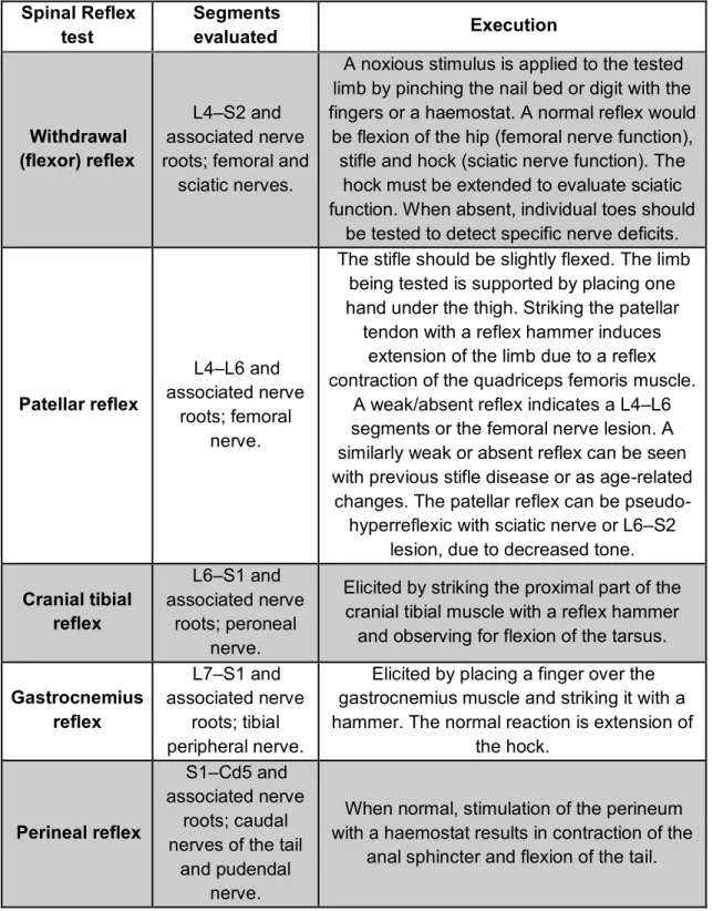
Table 5 – Important spinal reflex tests for the evaluation of a patient with CES ... 13
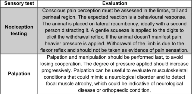
Table 6 – Sensory tests for evaluation of the sensory system of the neurologic patient with CES. ... 14
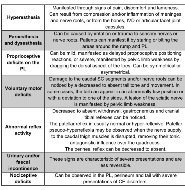
Table 7 – Clinical signs of CE disorders (summarized) ... 16
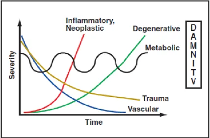
Table 8 – Disease categories based on onset and progression of signs. ... 17
Table 9 – Advantages and disadvantages of the myelography... 22
Table 10 – The appearance of fat, CSF and tissue oedema on commonly used imaging sequences. Intensity is described relative to normal CNS parenchyma ... 25
Table 11 – Therapy options according to the type of primary injuries... 43
Table 12 – Therapy for secondary injury due to SC trauma ... 44
Table 13 – Identification of the studied patients by breed, gender and neutering status, age and weight. ... 46
Table 14 – Mental status, posture, gait/movement and tail position evaluation of the patients in study ... 48
Table 15 – Evaluation of PL muscle tone, urinary/faecal function and sensory function of the patients in study. ... 49
Table 16 – Results of proprioceptive placing and flexor (PL) and patellar reflexes of the patients in study ... 50
Table 17 – Results of complementary laboratory tests requested for each case. ... 52
Abbreviations/Symbols/Acronyms List % – Percentage
AF – Anulus fibrosus disci intervertebralis
ALB – Albumin
ALT - Alanine Aminotransferase BCHM – Biochemistry
BID – Twice a day C – Cervical
Ca – Caudal vertebra
CBC – Complete blood count CE – Cauda equina
CES – Cauda Equina Syndrome CL – Compressive lesions CM – Contrast media
CNS – Central nervous system CSF – Cerebrospinal fluid CT – Computed Tomography
DH I – Disc herniation(s) Hansen type I DH II – Disc herniation(s) Hansen type II DLS – Degenerative lumbossacral stenosis
EOS – Eosinophils ERI – Erythrocytes
ESC - Extradural synovial cysts FCE - Fibrocartilaginous embolism GLOB – Globulins
HCT – Haematocrit HGB – Haemoglobin
HV-UTAD – Hospital Veterinário da Universidade de Alto Douro e Trás-Os-Montes
IM – Intramuscular IV – Intravenous
IVD – Intervertebral disc IVF – Intervertebral foramen
IVS – Intervertebral space L – Lumbar vertebra LEU – Leucocytes
LMN – Lower motor neuron LPL – Left pelvic limb LS – Lumbosacral LSTV - Lumbosacral transitional vertebra m – month(s) MC – meningocele Mg – Milligram MHCT – Microhematocrit Min - Minutes MMC – Meningomyelocele MONO – Monocytes MR – Magnetic ressonance
MRI – Magnetic ressonance imaging MVP - Mean plaquette volume N – Normal
NEU – Neutrophils NP – Nucleus pulposus
NSAIDs – Non-steroidal anti-inflammatories
PCR - Polymerase chain reaction test PL – Pelvic limb(s)
PO – Per oral
RETIC – Reticulocytes RPL – Right pelvic limb S – Sacral vertebra SC – Spinal cord SID – Once a day spp. – Species
T – Thoracic vertebrae T1W – T1 weighted image T2W – T2 weighted image
TID – Three times a day TL – Thoracolumbar
UMN – Upper motor neuron
VC – Vertebral canal WBC – White blood cells wk – Week(s)
CHAPTER I – Introduction
The cauda equina syndrome (CES) is a complex entity, with multiple possible aetiologies. Clinical signs may differ in each individual patient and vary according to the location and extent of the lesion. These are highly nonspecific and can be persistent or episodic and heterogenous. Neurological deficits are dependent of the nerve root involved and may differ between right and left sides. When present, they are LMN (lower motor neuron) in nature. CES is usually characterized by low-back pain, bilateral sciatica, saddle hypoesthesia or anaesthesia, motor weakness and neurological deficits of the pelvic limbs (PL), impairment of anal, rectal and bladder sphincter’s dysfunction as well as sexual impotence (Orendáčová et al. 2001; Platt, 2010).
For the diagnosis of this syndrome, the accuracy of the anamnesis and the physical, orthopaedic, and neurological exams play a very important role. A definitive diagnosis should be supported by imaging results. The therapy options usually include the conservative treatment, in cases considered less severe, and/or the surgical treatment for patients with more severe neurological deficits (Meij & Bergknut, 2010; Lanz & Rossmeisl, 2012). The prognosis should be established considering the neurological deficits, the aetiology of the disease and the chosen therapy (Griffin IV et
al., 2009b).
This study includes a literature review about the CES, as well as the clinical approach, diagnosis, treatment and prognosis for the different aetiologies that can be involved in the disease process. The practical component of the study includes the analysis of 11 clinical cases of dogs who were submitted to a CT scan, in the presence of clinical signs compatible with CES. The statistical analysis was based on collected data from Hospital Veterinário da UTAD (Vila Real, Portugal), between August 2017 and November 2018.
This study has the goals of: characterize the sample (breed size, age, gender, weight), characterize the severity and aetiology of the lesion analyse the clinical course of the disease and the recovery with and without decompressive surgery, and analyse and associate the treatment options with the recurrence of clinical signs.
CHAPTER II – Literature review
1. Anatomic description of the vertebral column 1.1. The vertebrae
The number of vertebrae and spinal cord (SC) segments is variable between species. Individual variations can occur, especially in transitional vertebrae. There are approximately 50 vertebrae separated into five distinctive regions: seven cervical (C1-C7), thirteen thoracic (T1-T13), seven lumbar (L1-L7), three fused sacral vertebrae (S1-S3) that constitute the sacrum and twenty caudal (Ca1-Ca20) vertebrae, approximately (Thomson and Hanh, 2012). When analysing the vertebral formula, the first letter of the word designates each group by means of abbreviation, followed by a digit, that designates the number of the vertebra in the specific group. For example, the canine vertebral formula is C7T13L7S3Ca20-23 (Sisson, 2002).
All vertebrae remain separate and articulate with adjacent vertebrae, creating movable joints, except for the sacral vertebrae. The vertebral column has considerable flexibility (Badoux, 2005). Vertebrae contribute to support the head, to provide attachment for the muscles that are responsible for body movements and to protect the SC and roots of the spinal nerves. There are two types of joints between vertebrae: one cartilaginous that involves the direct connection of the bodies of the vertebrae, and a synovial one on the vertebral arches, between articular processes (Sharp & Wheeler, 2005).
Between two adjacent vertebrae there is the interposed cartilaginous disc, the articulations between them and the connecting ligaments, which form a functional unit, complemented by the nerves and blood vessels that leave the vertebral canal (VC) through the intervertebral foramina (IVF), and the muscles, covering different regions (Liebich & König, 2004a).
The typical vertebra will present a cylindrical and dorsally flattened body, which faces into the VC. It may present a median crest ventrally and an arch that encloses the vertebral foramen, with right and left pedicles and laminae. The pedicles extend dorsally, on each side, from the dorsolateral surface of the vertebral bodies, with cranial and caudal vertebral notches. The notches of each side of adjacent vertebrae originate the IVF, through which the spinal nerves, arteries and veins pass. Occasionally, there can be an additional lateral vertebral foramen in the pedicle near to the IVF (Budras et al., 2007; Dyce et al., 2010b).
Each vertebra has processes for muscular or articular connections and insertions, classified by transverse, spinous, articular, accessory and mammillary processes, a convex cranial articular surface and a centrally depressed caudal articular surface. Dorsally, the vertebral arch has right and left laminae that unite in the dorsal midline to form a spinous process. The space between adjacent arches is the interarcuate space, where the yellow ligament is dorsally located. The union of the vertebral arch with the vertebral body originates the vertebral foramen, a set of tubes that come together to form the VC, which encloses the SC (De Lahunta et al., 2015).
In the lumbar region, the vertebrae are longer, with more uniform bodies than the thoracic vertebrae (Dyce et al., 2010b). The lumbar vertebrae are usually dorsoventrally flattened, increasing their width (until L7) and length (until L6) caudally. The body of the L7 has approximately the same length L1. The pedicles and laminae of the lumbar vertebrae are longer and larger. The spinous processes are higher and more massive in the mid-lumbar region, with their height decreasing caudally from L4. The accessory processes are well developed on the first three to four lumbar vertebrae and absent on the fifth or sixth (Sisson, 2002; Fletcher, 2013). Other regional features of the lumbar vertebrae are the prominent mammillary and, in some cases, accessory processes (Badoux, 2005).
The sacrum is short, wide and wedge-shaped. It lies between the ilium and articulates with it. The first sacral vertebra has a larger body than the other two combined. There’s a median sacral crest that originates from the fusion of the three spinous processes of the sacral vertebrae (Thomson & Hahn, 2002). In the dorsal surface of the sacrum, there are two pairs of dorsal foramina, through which the dorsal divisions of the sacral spinal nerves and spinal vessels will pass. Medially to these foramina, there are the fused mamilloarticular processes of adjacent segments that form the intermediate sacral crest (Budras et al., 2007). The pelvic surface bears two pairs of foramina, larger than the corresponding dorsal foramina, situated laterally to the sacral body. Blood vessels and the ventral branches of the first two sacral nerves pass through them. Laterally to the pelvic sacral foramina there are fused transverse processes that form the thin lateral sacral crest (Liebich & König, 2004b; Morales & Montoliu, 2012).
The wing of the sacrum is the enlarged, prismatic-shaped lateral portion, which has a rough auricular surface that articulates with the ilium. The base of the sacrum faces cranially, articulating with the last lumbar vertebra. Above its articular surface, begins the sacral canal, formed by the coalescence of the three vertebral foramina (Fletcher, 2013; De Lahunta et al., 2015). The sacral canal is compressed dorsoventrally (Sisson, 2002). The cranioventral part of the base of the sacrum has a transverse ridge, the promontory. The caudal extremity of the sacrum is the apex, which articulates with the first caudal
vertebra. Occasionally, the first caudal vertebra appears fused to the sacrum (Evans & De Lahunta, 2013).
1.2. The intervertebral discs
The intervertebral discs lie between adjacent vertebra, in the intervertebral spaces, except for the space between C1 and C2 and for the sacrum (Griffin IV et al., 2009b; Brisson, 2010). These are thick pads that form fibrocartilaginous joints and allow the movement of the vertebrae, keeping them together and contributing for the flexibility of the vertebral column, while protecting the SC from mechanical forces (Marinho et al., 2014; Smolders & Forterre, 2015).
The intervertebral disc (IVD) consists of an outer fibrous ring, the anulus fibrosus disci intervertebralis (AF), which surrounds an inner amorphous and gelatinous centre, the nucleus pulposus (NP), a semifluid remnant of the notochord. The AF is thicker ventrally, constituted by parallel fibres, creating oblique bands, between vertebral bodies. This fibrous ring is responsible for transmitting stresses and strains that are required by lateral and dorsoventral movements of the vertebral column. When approaching the NP, the AF becomes less fibrous and more cartilaginous. When the AF ruptures or degenerates, bulging can occur due to pressure caused by the vertebral body movements (Jeffery et al., 2013; Fingeroth & Thomas, 2015).
1.3. The ligaments of the vertebral column
The ligaments of the vertebral column can be organized into short ligaments, that connect successive vertebrae, and long ligaments, that go across several vertebrae (Liebich & König, 2004a).
The supraspinous, the ventral longitudinal and the dorsal longitudinal ligaments extend along considerable portions of the vertebral column. The supraspinous ligament is a thick band, particularly noticeable on the thoracic region, extending from the spinous process of the first thoracic vertebra (T1) to the third caudal vertebra (Ca3). During movements of flexion, the supraspinous ligament prevents abnormal separation of the spine, together with the thin interspinous ligaments (Griffin IV et al., 2009b; Evans & De Lahunta, 2013).
The ventral longitudinal ligament is located ventrally to the surfaces of the vertebral bodies, from the axis to the sacrum, attached to the intervertebral discs (Liebich & König, 2004b; Morales & Montoliu, 2012).
The dorsal longitudinal ligament is thicker than the ventral longitudinal ligament. It runs from the dens of the axis to the sacrum, along the dorsal surfaces of the bodies
of the vertebrae, constituting part of the floor of the VC. The dorsal longitudinal ligament narrows over the middle of each vertebral body and widens when crossing the intervertebral discs (Klopp, 2010a).
The intertransverse ligaments unite the transverse processes of the lumbar vertebrae and are only distinct in this region. These ligaments are tensed during lateral flexion and rotation movements (Liebich & König, 2004b).
The yellow ligaments are located between the arches of adjacent vertebrae, blended with the articular capsules that surround the articular processes. The epidural space is located ventrally to the yellow ligaments and draws apart the ligaments and arches of the vertebrae from the dura covering the SC (Evans & De Lahunta, 2013).
1.4. The spinal cord and the cauda equina
The SC is enclosed by the VC. In medium to large-breed dogs, the SC terminates at the L6–L7 vertebral level and in small-breed dogs, one-half to one vertebral body more caudally. Functionally, the SC can be divided into four regions: cranial-cervical (C1–C5); cervicothoracic (C6–T2); thoracolumbar (T3–L3); lumbosacral (L4–S3) (Dellman & McClure, 1986; Budras et al., 2007).
The innervation of the body is organized in a segmental pattern. Each cutaneous region of the body (dermatome) and group of muscle fibres (myotome) is innervated by one SC segment. A SC segment is defined as a portion of the SC that gives rise to one pair of spinal nerves. In the dog and cat, there are 8 cervical, 13 thoracic, 7 lumbar, 3 sacral and at least 2 caudal SC segments (Platt & Olby, 2012). Some SC segments lie within the vertebra of the same annotation, whilst others lie cranial to the corresponding vertebra. In the last two thoracic and the first two or three lumbar segments, the spinal segments are found within their corresponding vertebrae. All the other ones are located cranially to the vertebra of the same number (Dewey & Da Costa, 2016). Usually, the three sacral segments lie within the fifth lumbar vertebral foramen, and the caudal segments lie within the sixth lumbar vertebral foramen (De Lahunta et al., 2015).
There are two enlargements of the SC, caused by an increase in white matter and cell bodies, associated with the innervation of the thoracic (cervical intumescence) and PL (lumbar intumescence). The cervical intumescence can be found within the fifth to seventh cervical vertebral foramina (caudal cervical region) and the first thoracic segment. The lumbar intumescence is usually located between the fourth and sixth or seventh lumbar segments (Thomson and Hahn, 2012). Caudal to the lumbar intumescence, usually at the level of the L5 vertebra, the SC becomes conical, in what is called the conus medullaris. These segments appear successively smaller,
surrounded by caudally directed spinal roots. The conus medullaris is continued by the filum terminale, and, beyond it, by sacral and caudal spinal roots within the VC. The cauda equina (CE) is constituted by these nerve roots, which normally descend through the VC of vertebrae L6, L7 and sacrum. (Budras et al., 2007; Fletcher, 2013).
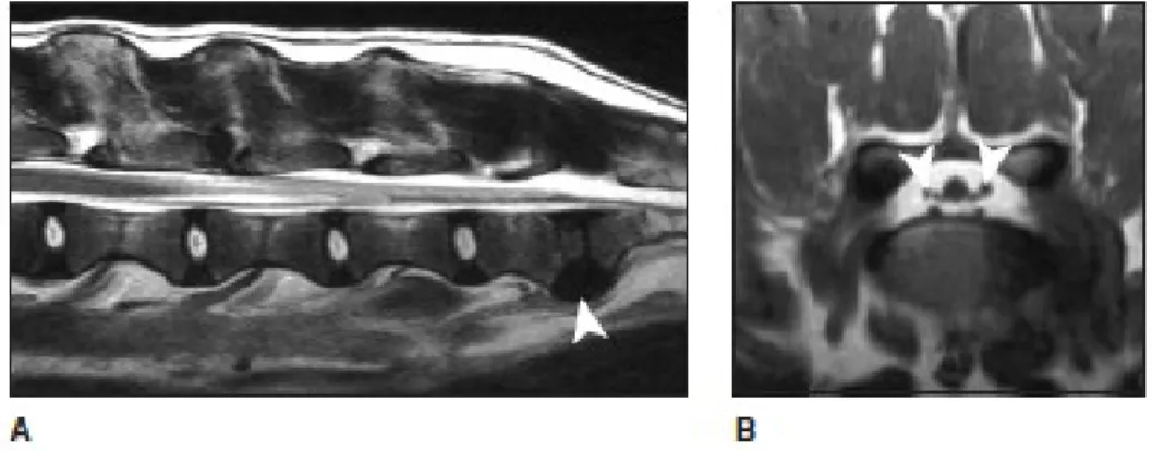
The CE nerve roots have the structure of a typical peripherical nerve and are partially unsheathed by the meninges. The CE is constituted by L6, L7, S1, S2, S3 and coccygeal 1 to 5 nerve roots. After leaving the foramina, L6, L7 and S1 nerve roots constitute the sciatic nerve. S2 and S3 nerve roots contribute to the formation of the pudendal and pelvic nerves. These nerve roots tolerate deformation better than SC. There is also a large epidural space in the region of the CE (figure 1). Thus, they are usually more resistant to injury than the SC tissue, but if severe damage occurs, recovery is unlikely (Sharp & Wheeler, 2005).
Figure 1 – Normal lumbosacral MRI. A) Sagittal, T2-weighted MRI of the lumbosacral spine in extension. There is loss of signal of the L7/S1 NP (arrowhead) but there is no dorsal displacement of the disc. B) Transverse, T1-weighted MRI through the L7/S1 disc space and foramen of the same dog as image A); the nerve roots (arrowheads) are surrounded by high-signal epidural fat (adapted from: Sharp & Wheeler, 2005).
1.5. Vascularization
A longitudinal ventral spinal artery and usually one or two dorsal spinal arteries course the full length of the SC, together with the spinal nerve (De Lahunta et al., 2015). On the surface of the SC, the longitudinal arteries are connected to a diffuse plexus, sending branches into the parenchyma, through the IVF. The origin of these branches is variable, according to the region of the vertebral column where they are situated. The first thoracic segments are supplied by the thoracic vertebral artery, a branch of the subclavian artery (Uemura, 2015). The remaining thoracic segments are supplied by branches of the intercostal and thoracic arteries. The lumbar area is supplied by lumbar segmentations of the abdominal and thoracic aorta (Morales & Montoliu, 2012).
Each spinal artery has one or two dorsal, dorsal spinal arteries, and a ventral root, the ventral spinal artery, that are responsible for the irrigation of the SC. The ventral spinal artery sends branches into the ventral median fissure, giving rise to the vertical artery, responsible for the irrigation of the grey matter (Budras et al.,2007; Uemura, 2015).
The SC veins have similar distribution to the arteries, presenting a ventral spinal vein in the ventral median fissure, a superficial venous plexus on the surface of the SC, with connections to the ventrolateral and dorsolateral veins, and a dorsal spinal vein. In the epidural space, on the floor of the VC, there are ventral internal venous plexuses. The venous sinuses of each side, on the floor of the VC, collect blood from vertebrae, meninges, and nerve roots. Dorsally, in the epidural space, interarcuate veins pass and connect the ventral internal vertebral venous plexus with the dorsal external vertebral venous plexus (Sjöström, 2003; De Lahunta et al., 2015).
2. Clinical assessment of the neurological patient
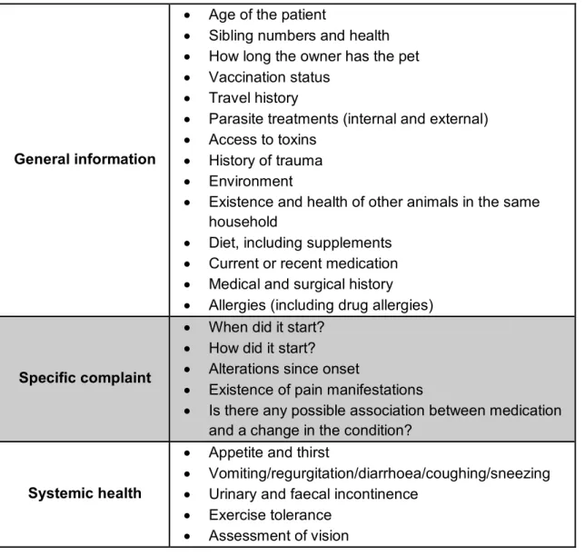
The first step of the clinical evaluation is to take a proper and complete history. The following table (table 1) exhibits the data that should be obtained from the owner (Lorenz et al., 2011). A clear and concise description of the main concern should be obtained from the owner. In the absence of clinical findings, the owner’s description can be the basis to institute an anatomical and differential diagnosis. Video footages could offer valuable information and clarify some doubts that the clinician may have. It is very important to get information about the onset, evolution and course of illness (see table 2), as it may provide insight into specific differential diagnoses (Schatzberg, 2017). The evolution of the condition should be characterized as progressive, static, improving or “waxing and waning”. It is important to identify factors that can trigger or improve the signs, previous therapy and its effect on disease course (Dewey & da Costa, 2016).
2.1. General physical examination
The neurological examination should be preceded by a general physical examination of all other body systems to detect abnormalities that can affect the nervous system, mimic a primary neurological disorder or influence the prognosis. An orthopaedic examination should be done in all animals with gait disturbances. Vital signs should be examined, and blood and urine collected for further laboratory analysis. Evaluation of the bladder function is very important, since urinary incontinence can be, sometimes, the only clinical sign of some CE disorders (Varejão et al., 2004; McKee, 2007).
General information
Age of the patient
Sibling numbers and health How long the owner has the pet Vaccination status
Travel history
Parasite treatments (internal and external) Access to toxins
History of trauma Environment
Existence and health of other animals in the same household
Diet, including supplements Current or recent medication Medical and surgical history Allergies (including drug allergies)
Specific complaint
When did it start? How did it start? Alterations since onset
Existence of pain manifestations
Is there any possible association between medication and a change in the condition?
Systemic health
Appetite and thirst
Vomiting/regurgitation/diarrhoea/coughing/sneezing Urinary and faecal incontinence
Exercise tolerance Assessment of vision
Table 1 – Information that should be obtained from the owner during the anamnesis (adapted from: Platt & Olby, 2012; Schatzberg, 2017).
Acute Onset over minutes to hours
Subacute Onset over days
Chronic Onset over several days, weeks or months
Episodic The animal returns to the normal status between episodes
2.2. Neurological examination
The neurologic examination intends to evaluate the integrity and function of the nervous system. It helps the clinician to elaborate a list of differential diagnosis, to identify the aetiology of the disorder, to determine its prognosis and to help choosing the most adequate treatment (Varejão et al., 2004). Having a list of all the steps can be a powerful and helpful tool in order to register the results and remember all parameters. Video recordings are important to ensure that the neurologic examination was fully done and to have a permanent record with the important data about the patient, allowing to evaluate the progression/regression of the clinical signs (LeCouteur & Grandy, 2010). The neurologic examination should aim to answer the following questions:
Do the clinical signs observed refer to a nervous system lesion? Within the nervous system, where is the lesion located?
What types of disease process can explain the clinical signs? What is the severity of the disease? (Platt & Olby, 2012).
It is very important to ensure that the animal is comfortable and cooperating during the examination. Ambulatory patients should be allowed to explore the examination room, in order to evaluate their mental status, behaviour, posture and gait. If the animal is anxious or nervous, the sensory evaluation should not be done right away. The clinician should be aware that sedation, analgesia or neurological conditions, such as epileptic seizures, can influence the results of this neurological evaluation (Platt, 2010; Thomas & De Risio, 2015).
Part I – hands-off examination
The first part of the neurological examination is constituted by evaluation of the mental status and behaviour, evaluation and characterization of posture and body positioning at rest, gait evaluation, and identification of abnormal involuntary movements (e.g. tremors) (Platt, 2010).
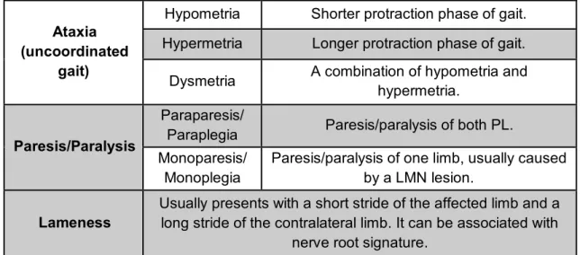
On CE disorders, one of the most common neurological presentations is gait disturbances (see table 3). The animal’s ability to generate and make coordinated movements should be assessed in a place where the patient can move freely, over a non-slip surface, preferably with the owner walking the animal. If the animal is not trying to walk, body support should be provided, with a sling or harness, so that any subtle voluntary movement can be noticed. Normal gait requires intact function of the brainstem, cerebellum, SC and sensory and motor peripheral nerves, neuromuscular junctions and muscles (Costes, 2012; Thomas & De Risio, 2015).
Ataxia (uncoordinated
gait)
Hypometria Shorter protraction phase of gait. Hypermetria Longer protraction phase of gait.
Dysmetria A combination of hypometria and hypermetria.
Paresis/Paralysis
Paraparesis/
Paraplegia Paresis/paralysis of both PL. Monoparesis/
Monoplegia
Paresis/paralysis of one limb, usually caused by a LMN lesion.
Lameness
Usually presents with a short stride of the affected limb and a long stride of the contralateral limb. It can be associated with
nerve root signature.
Table 3 – Important parameters to evaluate on a patient with CES (adapted from Platt & Olby, 2012).
Part II – hands-on examination
The hands-on examination should be constituted by an assessment of the cranial nerves; postural reaction tests; spinal reflexes, muscle tone and size examination and evaluation and sensory evaluation tests (Platt & Olby, 2012). Tables 4 to 6 describe the important tests to diagnose a CES.
The entire nervous system should be able to perform postural reactions (see table 4). The primary aim of postural reaction testing is to detect any subtle deficits that are not obvious on gait evaluation. Lesions affecting the anatomical sensory and/or motor components could result in abnormal postural reactions (Chrisman et al., 2003; Schatzberg, 2017).
The evaluation of spinal reflexes should be considered as a continuation of gait evaluation and postural reaction testing. It helps to classify the neurological disorder as UMN or LMN type, allowing to localize the lesion in specific SC segments or peripheral nerves (Lorenz et al., 2011). Spinal reflexes are segmental, which means that they only evaluate the spinal segment(s) within the intumescences corresponding to the stimulated nerve. In dogs, spinal reflexes and muscle tone must be evaluated with the patient in lateral recumbency. The animal can be unconscious. The most reliable spinal reflex tests for the PL’ evaluation are the patellar and withdrawal reflexes. Other spinal reflexes are more difficult to perform and interpret (see table 5) (Garosi, 2004; Parent, 2010).
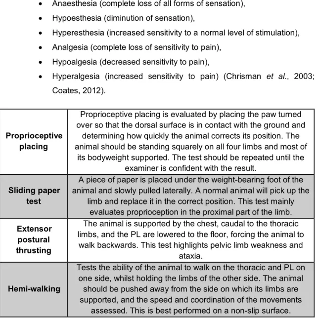
The evaluation of the sensory system depends on tests for pain perception (nociception). It requires a noxious stimulus and evaluation of the animal’s response. The purpose of testing pain perception is to detect and map out any areas of sensory loss (see table 6) (Platt, 2010). Pain perception can be classified as:
Anaesthesia (complete loss of all forms of sensation), Hypoesthesia (diminution of sensation),
Hyperesthesia (increased sensitivity to a normal level of stimulation), Analgesia (complete loss of sensitivity to pain),
Hypoalgesia (decreased sensitivity to pain),
Hyperalgesia (increased sensitivity to pain) (Chrisman et al., 2003; Coates, 2012).
Proprioceptive placing
Proprioceptive placing is evaluated by placing the paw turned over so that the dorsal surface is in contact with the ground and
determining how quickly the animal corrects its position. The animal should be standing squarely on all four limbs and most of
its bodyweight supported. The test should be repeated until the examiner is confident with the result.
Sliding paper test
A piece of paper is placed under the weight-bearing foot of the animal and slowly pulled laterally. A normal animal will pick up the
limb and replace it in the correct position. This test mainly evaluates proprioception in the proximal part of the limb. Extensor
postural thrusting
The animal is supported by the chest, caudal to the thoracic limbs, and the PL are lowered to the floor, forcing the animal to
walk backwards. This test highlights pelvic limb weakness and ataxia.
Hemi-walking
Tests the ability of the animal to walk on the thoracic and PL on one side, whilst holding the limbs of the other side. The animal
should be pushed away from the side on which its limbs are supported, and the speed and coordination of the movements
assessed. This is best performed on a non-slip surface.
Table 4 – Important postural reaction to evaluate in a patient with CES (adapted from Platt & Olby, 2012;De Thomas & De Risio, 2015).
Spinal Reflex test Segments evaluated Execution Withdrawal (flexor) reflex L4–S2 and associated nerve roots; femoral and
sciatic nerves.
A noxious stimulus is applied to the tested limb by pinching the nail bed or digit with the fingers or a haemostat. A normal reflex would
be flexion of the hip (femoral nerve function), stifle and hock (sciatic nerve function). The
hock must be extended to evaluate sciatic function. When absent, individual toes should
be tested to detect specific nerve deficits.
Patellar reflex
L4–L6 and associated nerve
roots; femoral nerve.
The stifle should be slightly flexed. The limb being tested is supported by placing one hand under the thigh. Striking the patellar
tendon with a reflex hammer induces extension of the limb due to a reflex contraction of the quadriceps femoris muscle.
A weak/absent reflex indicates a L4–L6 segments or the femoral nerve lesion. A similarly weak or absent reflex can be seen with previous stifle disease or as age-related
changes. The patellar reflex can be pseudo-hyperreflexic with sciatic nerve or L6–S2
lesion, due to decreased tone.
Cranial tibial reflex L6–S1 and associated nerve roots; peroneal nerve.
Elicited by striking the proximal part of the cranial tibial muscle with a reflex hammer
and observing for flexion of the tarsus.
Gastrocnemius reflex L7–S1 and associated nerve roots; tibial peripheral nerve.
Elicited by placing a finger over the gastrocnemius muscle and striking it with a hammer. The normal reaction is extension of
the hock.
Perineal reflex
S1–Cd5 and associated nerve
roots; caudal nerves of the tail
and pudendal nerve.
When normal, stimulation of the perineum with a haemostat results in contraction of the
anal sphincter and flexion of the tail.
Table 5 – Important spinal reflex tests for the evaluation of a patient with CES (adapted from: Dewey & da Costa, 2016).
Sensory test Evaluation
Nociception testing
Conscious pain perception must be assessed in the limbs, tail and perineal region. The expected reaction is a behavioural response. The animal is placed on lateral recumbency, ideally with a second person distracting it. A gentle squeeze is applied to the digits to
elicit the withdrawal reflex. If the animal doesn’t manifest pain, heavier pressure is applied. Withdrawal of the limb is due to the flexor reflex and should not be taken as evidence of pain sensation.
Palpation
Palpation and manipulation should be performed last, to avoid losing cooperation. The degree of pressure applied should increase
progressively. Palpation can be useful to evaluate musculoskeletal conditions that could mimic a neurological disorder and to detect
focal muscle atrophy, which could be indicative of neurological disease or orthopaedic condition.
Table 6 – Sensory tests for evaluation of the sensory system of the neurologic patient with CES (adapted from: Parent, 2010; Platt & Olby, 2012).
3. Cauda equina syndrome 3.1. Clinical signs
Problems affecting the caudal lumbar region are very common in large-breed dogs. Clinical signs associated with CES vary according to the location and extent of the lesion. These are highly nonspecific and can be persistent or episodic and heterogeneous. Clinical signs may be bilateral or unilateral (Parent, 2010).
Neurological deficits are dependent of the nerve root involved and are usually LMN in nature. Neurological dysfunction of the PL can range from paraplegia, paraparesis, monoplegia or monoparesis to mild proprioceptive deficits which do not affect gait (Danielsson & Sjöström, 1999). A shortened and stiff stride of the PL may be observed. Dragging the claws of the digits and ataxia are manifestations of decreased proprioception. The sciatic, cranial tibial, gastrocnemius, anal and flexor withdrawal reflexes may be normal, decreased or absent. If significant sciatic deficits are present, failure of hock flexion during withdrawal reflex test may be seen. Muscle atrophy may be observable in the sciatic distribution. The patellar reflex test may exhibit a false exaggerated response (patellar pseudo-hyperreflexia), which must be differentiated from the increased reflex that occurs with UMN deficits, observed in lesions cranial to the L4 segment (Ness, 1994; Dewey, 2013).
Patient history frequently includes lower lumbar or PL pain, which can be manifested in various ways. The tail can be flaccid or carried low due to pain, hypotonic, or paralytic. Lower lumbar pain can be triggered by the extension of the lumbosacral
joint, when the animal jumps, climbs stairs, stands from a prone position or crawls. Unwillingness to exercise or jump into a car, stiffness during physically demanding exercise, scuffing of the toenails of the PL, intermittent lameness, limb dysfunction exacerbated by activity, or LMN deficits may be indicative of a CES (Sjöström, 2003) (see table 7). The patient may show stiffness of the musculature surrounding the lumbosacral region (Orendácová et al., 2001; Mckee, 2007).
There are several techniques to isolate lumbosacral painful responses: traction or elevation of the tail; per rectum application of pressure to the L7-S1 disc space; performing the hyperextension or lordosis test, which consists in elevating the pelvic limbs off the ground, guaranteeing extension of the hips, and applying lumbosacral pressure; and rotation of the lumbosacral joint by swinging the PL bilaterally (Platt, 2010; Lanz & Rossmeissl, 2012).
In a general way, when analysing compressive lesions (CL), the first fibres to be compromised are the proprioceptive fibres and the last ones are the nociceptive fibres. Nociceptive dysfunction reflects a severe, extensive lesion (Meij & Bergknut, 2010; Lanz & Rossmeissl, 2012). In cases of CL of increasing severity, the clinical signs will start with spinal hyperpathia/sensitivity, due to meningeal irritation, proprioceptive dysfunction, loss of motor function manifested through paresis, loss of superficial pain, paralysis, and finally loss of all nociception. Loss of urinary and faecal continence can occur when the damage is severe enough to cause marked paresis or paralysis. Faecal incontinence is associated with poor anal tone and may be present even with a normal anal reflex. Urinary incontinence is usually a result of pelvic and pudendal nerve dysfunction (S1-S3), which causes urethral or anal sphincter hypotonia and possibly altered sensation in the perineal region (Platt, 2010; Thomson & Hanh, 2012). Occasionally, urinary incontinence, with dribbling of urine associated with an easy manual bladder emptying, and faecal incontinence may be the only clinical manifestations of lumbosacral disease. (Watt, 1991; Meij & Bergknut, 2010).
Hyperesthesia
Manifested through signs of pain, discomfort and lameness. Can result from compression and/or inflammation of meninges
and nerve roots, or from the bones, IVD or articular facet joint capsules.
Paraesthesia and dysesthesia
Can be caused by irritation or trauma to sensory nerves or nerve roots. Patients can manifest it by staring or biting the
areas around the rump and PL. Proprioceptive
deficits on the PL
Can be mild, manifested as delayed proprioceptive positioning reactions, or severe, manifested by pelvic limb weakness by dragging the dorsal aspect of the toes. Can be symmetrical or
asymmetrical.
Voluntary motor deficits
Damage to the caudal SC segments and/or nerve roots can be noticed by a decreased to absent tail tone and movement. In some cases, the tail can appear in an abnormally low position or
with a deviation to one of the sides. A lesion of the sciatic nerve is manifested by pelvic limb weakness.
Abnormal reflex activity
Decreased to absent withdrawal, gastrocnemius and cranial tibial reflexes can be noticed.
The patellar reflex in usually normal or hyper-reflexive. Patellar pseudo-hyperreflexia may be observed when the nerve supply
to the caudal thigh muscles is disrupted, removing their tonic antagonistic influence over the quadriceps.
The perineal reflex can be decreased to absent. Urinary and/or
faecal incontinence
These signs are characteristic of severe presentations and are less reversible.
Nociceptive deficits
Can be observed in the PL, perineum and tail with severe presentations of CE disorders.
Table 7 – Clinical signs of CE disorders (summarized) (adapted from Dewey & da Costa, 2016).
3.2. Differential diagnosis
A differential diagnosis list, according to signalment, history and neurological findings, is essential. Diagnostic tests should aim to confirm or rule out the differential diagnosis in the list, without replacing clinical evaluation. The primary differential diagnosis for the CE region are: degenerative lumbosacral stenosis, discospondylitis, neoplasia, and extradural synovial cysts (Platt & Olby, 2012).
After localizing the lesion, disease processes can be classified according to the cause, using the mnemonic DAMNITV. Each process has a typical signalment, onset, progression, and distribution within the nervous system (see figure 2 and table 8) (Varejão, 2004; da Costa & Moore, 2010). The list may be narrowed down through the neurological examination of the animal, depending on the presence, type, and severity of neurological deficits that arise from the CE region (L4-S3). Geriatric patients frequently
have more than one disease entity, which may interfere with the diagnosis. Orthopaedic conditions or other neurological diseases should be considered, since they may mimic or exaggerate signs of lumbosacral disease. Dogs with orthopaedic disease will have a normal neurological examination (Mckee, 1993; Meij & Bergknut, 2010).
Figure 2 – Onset and progression of neurological diseases of differing causes (adapted from: Platt & Olby, 2012).
Acute progressive with asymmetrical clinical signs
• Degenerative (e.g. IVD disease) • Neoplastic
• Inflammatory/infectious disease
Acute, progressive with symmetrical signs • Metabolic disorders • Nutritional disorders • Neoplastic • Inflammatory/infectious disease • Toxicity
Acute, non-progressive (often asymmetrical)
• Idiopathic • Trauma
• Vascular disorders
Chronic, progressive with symmetrical signs • Degenerative disorders • Anomalous disorders • Metabolic disorders • Neoplastic disorders • Nutritional disorders • Inflammatory/infectious disease • Toxicity
Chronic, progressive with asymmetrical signs
• Degenerative (e.g. IVD disease) • Neoplastic disease
• Inflammatory/infectious disease
Table 8 – Disease categories based on onset and progression of signs (adapted from: LeCouteur & Grandy, 2005; Da Costa & Moore, 2010; Dewey & da Costa, 2016).
3.2.1. The mnemonic DAMNITV a) D – Degenerative
Degenerative diseases involve morphological degeneration of the nervous tissue and can affect any part of the nervous system. Many are hereditary. The onset is typically insidious with slow progression of the clinical signs. The age of onset is variable, but it is more frequent in young animals. In young animals, shortly after birth, the clinical signs are often symmetrical. In adults, clinical signs can have an asymmetrical presentation (LeCouteur & Grandy, 2005; Da Costa & Moore, 2010).
b) A – Anomalous
Neurological signs can result from malformations that directly involve the nervous tissue or the surrounding tissue. These are usually recognized early in life and signs tend to be non-progressive or slowly progressive. In some cases, vertebral malformations only result in neurological signs when the animal reaches adulthood (Denoix, 2005; Coates, 2012).
c) M – Metabolic
Animals of any age can be affected by metabolic disorders. The clinical onset of neurological signs is variable, but usually acute. Clinical signs are often subacute to chronic. Neurological signs are typically bilateral and symmetrical. Most of these conditions tend to wax and wane with time (Lorenz & Kornegay, 2004).
d) N – Neoplastic, Nutritional
Neoplasia is more common in older animals (more than 5 years old) but can occur at any age. Neurological signs are usually chronic and progressive, although acute deterioration can be seen. Determining factors of the clinical expression are: lesion size, histological nature, growth rate, associated inflammatory response and location. Neurological deficits can be asymmetrical or symmetrical, often suggesting a focal lesion (Brehm et al., 1995; Lorenz & Kornegay, 2004).
As with metabolic diseases, neurological signs of nutritional diseases affecting the nervous system are typically bilaterally symmetrical. The onset of clinical signs is variable and often slowly progressive. The distribution of clinical signs can be diffuse or multifocal, as some nutritional diseases can affect selective areas of the CNS (LeCouteur & Grandy, 2005).
e) I – Inflammatory, Infectious, Idiopathic
The onset of sterile inflammatory (immune-mediated) or infectious diseases is cause-dependent and can be acute, subacute or a more insidious. Neurological signs usually progress without treatment, but, in some cases, may wax and wane early after onset. Neurological deficits can refer to a focal or multifocal lesion and can be asymmetrical or symmetrical.
Idiopathic disorders usually result in acute onset of non-progressive or regressive signs. Neurological deficits are variable, according to each syndrome. These disorders frequently affect the PNS (Greene, 1998; Tipold & Stein, 2010).
f) T – Traumatic, Toxic
The onset of traumatic disorders is usually peracute or acute. Signs usually remain static or improve over time. Neurological deficits can be symmetrical or asymmetrical and lesions can be focal or multifocal.
Toxicities often result in acute onset disease. Sings can be diffuse or bilaterally symmetrical and are non-progressive (McKee, 1990; Lorenz et al., 2011).
g) V – Vascular
Vascular disorders can result from loss of blood supply (ischaemia) or from haemorrhage into the nervous system. Their onset is typically peracute or acute, with non-progressive or regressive signs. Usually, deficits are initially focal and often asymmetrical. Haemorrhage may be an exception since it can be responsible for a more progressive onset over a very short period. Clinical signs usually regress after 24–72 hours, due to the diminution of the mass effect secondary to haemorrhage and reorganization or oedema resolution (Platt, 2010; Bartholomew et al., 2016).
3.3. Diagnosis
The diagnosis of a CE disorder should be based on a complete patient history and clinical signs and subsequent performance of careful physical, orthopaedic, and neurologic examinations. A definitive diagnosis should be supported by radiological and advanced imaging results (Meij & Bergknut, 2010; Lanz & Rossmeisl, 2012).
3.3.1. Laboratorial tests
Basic laboratorial tests should be done in all animals. These should integrate: an hemogram, a biochemical and type II urine analysis, in order to discharge some diagnosis in the differential diagnosis list and evaluate anaesthesia associated risks (Chrisman et al., 2003).
3.3.2. Neuroimaging a) Spinal radiography
Spinal radiographs can be used to identify fractures, luxations, discospondylitis, some vertebral tumours, congenital abnormalities and degenerative changes, and provide circumstantial evidence of disc herniation (Brisson, 2010).
Radiographs should be executed with a sedated animal in order to achieve adequate positioning, except if spinal instability is suspected. In this case, only lateral views should be acquired initially, to avoid iatrogenic neural injury. For an accurate interpretation, the spine should not be rotated, and padding ties and troughs are recommended (Varejão et al., 2004; Kinns et al., 2006).
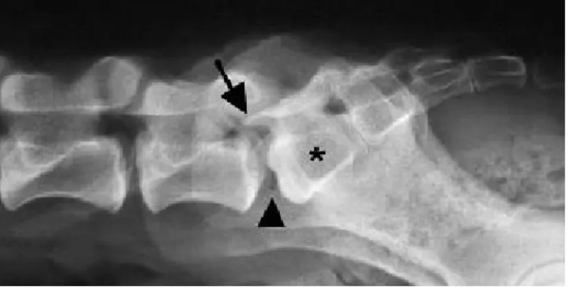
The following aspects should be evaluated on plain radiographs: basic anatomy (number of vertebrae, presence of processes and ribs); alignment of the vertebrae in two planes; width of the IVD space, comparing each space with the disc space immediately cranial and caudal; shape and opacity of the IVF; integrity of the vertebral endplates, which should be examined for lysis and sclerosis indicative of infection; evidence of vertebral neoplasia (lysis, sclerosis and distortion of the bone outline); degenerative changes of the vertebrae or articular processes (see figure 3) (Kinns et al., 2006; Gavin & Levine, 2015).
Radiographic studies are often the first diagnostic test performed in dogs with clinical signs of lumbosacral disease. These should extend from the L4 to Ca2 or Ca3, to allow examination of the entire lumbosacral region and CE nerve roots. Both lateral and ventrodorsal views should be obtained. Bone lesions can later be delineated more accurately using CT (Meij & Bergknut, 2010).
Dynamic radiographs have limited diagnostic value for CE disorders due to individual and breed variations in normal range of motion, VC diameter, and size of the L7-S1 IVD space. However, these can help to show instability and abnormal movement at the lumbosacral joint (Mattoon & Koblik, 1993; McKee, 1993; Schmid & Lang, 1993). Survey radiographs may help to exclude discospondylitis, neoplasia, fracture, luxation,
idiopathic lumbosacral stenosis or other causes of pelvic limb dysfunction which may mimic a CES (Morgan & Bailey, 1990; Steffen et al., 2007).
Figure 3 – Lateral radiograph of the lumbosacral region of a dog. Note the presence of a transitional vertebra (asterisk), elongation of the sacral lamina into the caudal aperture of L7 (arrow), and the vacuum phenomenon between L7 and S1 (arrowhead) (adapted from: Meij & Bergknut, 2010).
b) Myelography
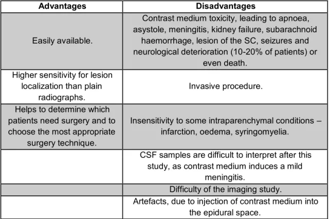
Myelography is a satisfactory imaging study, when CT and MRI are not available. This technique is performed by injecting a non-ionic contrast medium (CM) (iohexol or iopamidol) into the atlanto-occipital (cisterna magna) or lumbar (L5-L6) interarcuate space (Hecht et al., 2009). Advantages and disadvantages of this technique are expressed on table 9.
Injection of contrast media (CM) into the lumbar spine has a decreased risk of iatrogenic trauma and seizures, and improves delineation of CL, when compared to the cervical myelography. However, it is easier to introduce the needle into the atlanto-occipital subarachnoid space. Injection of contrast medium into the lumbar subarachnoid space may lead to epidural leakage, interfering with the assessment of the lumbosacral VC (Hecht et al., 2009; Newcomb et al., 2011).
Like survey radiography, myelography may be useful to exclude pathological changes that mimic the clinical signs of a CE disorder, such as neoplasia and IVD disease. Dogs have a variable and an unpredictable termination site of the dural sac. Therefore, myelography can only successfully be used to assess the CE if the patient’s dural sac extends caudally into the sacrum, which is an impossible feature to predict (Meij & Bergknut, 2010). If the patient has the dural sac elevated from the ventral vertebral floor or a dorsally located compressive lesion, myelography will not be successful. Laterally CLcan be missed in myelographic studies (Ramirez & Thrall, 1998). (Danielsson & Sjöström, 1999).
To interpret a myelogram, three basic pathological patterns must be recognized, according to the location of the lesion: intramedullary, intradural/extramedullary and extradural. For a precise interpretation, radiographs should be taken in ventrodorsal, lateral and right and left oblique views (Hecht et al., 2009; Newcomb et al., 2011).
This procedure is not indicated if the general anaesthesia can’t be performed safely, if there is an increase in the cerebrospinal fluid (CSF) pressure or an inflammatory lesion of the central nervous system. Since myelographic findings can raise doubts regarding the presence or absence of a lesion, CT should be used following myelography, since it allows a more precise anatomical lesion localization and allows to detect the CM in the subarachnoid space (Sharp & Wheeler, 2005; Gavin & Levine, 2015).
If needed, CSF analysis should be completed prior to performing a myelogram. CM induces a mild meningitis that complicates interpretation of CSF samples for up to one week after the study. CSF analysis is useful to dismiss inflammatory diseases. It can be collected either from the cisterna magna or the cisterna lumbalis, on the sedated animal, preferably caudally to the lesion (Griffin IV et al., 2009a; Brisson, 2010).
Advantages Disadvantages
Easily available.
Contrast medium toxicity, leading to apnoea, asystole, meningitis, kidney failure, subarachnoid
haemorrhage, lesion of the SC, seizures and neurological deterioration (10-20% of patients) or
even death. Higher sensitivity for lesion
localization than plain radiographs.
Invasive procedure.
Helps to determine which patients need surgery and to choose the most appropriate
surgery technique.
Insensitivity to some intraparenchymal conditions – infarction, oedema, syringomyelia.
CSF samples are difficult to interpret after this study, as contrast medium induces a mild
meningitis.
Difficulty of the imaging study.
Artefacts, due to injection of contrast medium into the epidural space.
Table 9 – Advantages and disadvantages of the myelography. (Adapted from: Hecht et al., 2009; Newcomb et al., 2011; Coates, 2012; Gavin & Levine, 2015).
c) Epidurography
For the evaluation of CE lesions, myelography is often inadequate, since the subarachnoid space ends at the conus medullaris (in the L6 region in most patients). Epidural contrast injection may help to delineate compressive CE lesions, especially at the L7/S1 IVD space. Epidurography has a low level of morbidity (Sisson et al., 1992; Ramirez & Thrall, 1998).
After aseptic preparation of the chosen site (L7-S1, sacrocaudal junction or between one of the caudal intervertebral spaces), the contrast material is injected (iohexol or iopamidol; 0.1–0.2 ml/kg body weight). L7-S1 contrast injection can produce an unsatisfactory epidurogram, in some cases (Garcia-Pereira et al., 2010).
The epidural space has an irregular contour, when compared to the subarachnoid space. Consequently, the contrast columns of an epidurogram will appear rough and uneven, in comparison to the contrast columns of a myelogram. Multiple radiographic views are helpful (lateral films taken with the coxofemoral joints in neutral, flexed and hyperextended positions). In a normal epidurogram, contrast fills the epidural space evenly, with the pelvis in any position. Potential epidurogram’s abnormalities include complete obstruction of cranial flow of CM past the L7-S1 space or dorsal deviation of the ventral contrast column over this space, which may be exacerbated on extended views and alleviated on flexed views. Occasionally, ventral deviation of the dorsal contrast column can occur (Roberts & Selcer, 1993; Dewey & da Costa, 2016).
d) Discography
Discography is also associated with low level of morbidity, but it is used less often than epidurography to evaluate L7-S1 disc lesions. This technique involves injection of iohexol or iopamidol (0.1-0.3 ml/kg body weight) directly into the NP of the disc. Then radiographs are taken (Kahanovitz et al., 1986; Ramirez & Thrall, 1998).
The injection site must be aseptic prepared. The needle is placed directly into the L7-S1 disc, preferably under fluoroscopic guidance. The injection of contrast is not easy in a normal disc (Weinstein et al., 1988).
It is possible to perform a combination discography/epidurography procedure, using a single needle puncture: after the discogram is performed, the needle is withdrawn from the disc into the epidural space and additional contrast is injected and radiographs are taken (Robertson & Thrall, 2011; Dewey & da Costa, 2016).