John Wiley & Sons. Printed in Singapore
J
ournal ofCutaneous Pathology
Borderline CD30
+ cutaneous
lymphoproliferative disorder: report
of a case with expression of cytotoxic
markers and response
to clarithromycin
CD30+ cutaneous lymphoproliferative disorders (CLPDs) are usually characterized by a benign clinical course. The prognostic value of cytotoxic markers in these lymphomas has not been evaluated in large series. We describe a case of borderline CD30+ CLPD with cytotoxic phenotype, presenting in a 22-year-old male patient as an ulcer on the forearm. He reported having had similar ulcers on the buttock and thigh that spontaneously regressed over the course of 1 year. The lesion resolved with a single course of clarithromycin; a subsequent lesion, too, responded to clarithromycin, and no recurrences or systemic
involvement have been documented in the 9-month follow-up. A conservative approach in the management of CD30+ CLPD is recommended. We believe that the anti-inflammatory and apoptotic effects of clarithromycin on T cells may have hastened the remission process.
Ponte P, Serr˜ao V, Viana I, Vale E, Jo˜ao A, Cerroni L. Borderline CD30+ cutaneous lymphoproliferative disorder: report of a case with expression of cytotoxic markers and response to clarithromycin. J Cutan Pathol 2011; 38: 301–305.© 2009 John Wiley & Sons A/S.
Pedro Ponte1, Vasco Serr˜ao1,
Isabel Viana2, Esmeralda
Vale2, Alexandre Jo˜ao1and
Lorenzo Cerroni3
1Department of Dermatology, Hospital dos
Capuchos, Centro Hospitalar de Lisboa Central, Lisbon, Portugal,
2Department of Dermatopathology, Centro de
Dermatologia M´edico-Cir´urgica de Lisboa, Lisbon, Portugal, and
3Department of Dermatology, Medical
University of Graz, Graz, Austria
Dr Pedro Fernandes da Ponte, Servic¸o de Dermatologia, Hospital de Santo Ant´onio dos Capuchos, Alameda Santo Ant´onio dos Capuchos, 1150-314 Lisboa, Portugal Tel: +351.21.3136300
Fax: +351.21.3136380 e-mail: [email protected] Accepted for publication October 16, 2009
The spectrum of CD30+ cutaneous lymphopro-liferative disorders (CLPDs) represents the second most common type of cutaneous T-cell lymphomas (CTCLs) after mycosis fungoides (MF), compris-ing approximately 30% of all CTCLs. This group includes lymphomatoid papulosis (LyP), cutaneous anaplastic large-cell lymphoma (cALCL) and border-line cases.1,2Both LyP and cALCL share a low-grade malignant behavior characterized by frequent spon-taneous regression of skin lesions and, in general, excellent prognosis.3 The term ‘borderline cases’ refers to those in which a clear distinction between LyP and cALCL cannot be made based on clinico-pathologic correlation.1 – 4
Case report
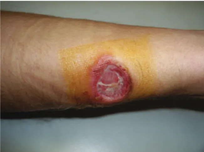
A 21-year-old white male patient in good general health presented with an ulcer of 4 months’ duration on his left forearm (Fig. 1). The lesion had started as an enlarging violaceous nodule that spontaneously ulcerated, leaving an asymptomatic, oval-shaped, 5-cm large ulcer, with undermined infiltrated bor-ders. He had similar but smaller nodules on his right upper eyebrow and on the back of his left thigh (Fig. 2).
On skin examination, there were two large atrophic scars on his left buttock and on his right thigh, with hypopigmented and erythematoviola-ceous areas, measuring 15 and 3 cm, respectively
Fig. 1. Asymptomatic, oval-shaped, 5-cm exsudative deep ulcer on the forearm, with undermined infiltrated borders.
Fig. 2. Violaceous nodule on the posterior aspect of the thigh.
(Fig. 3); according to the patient, these scars resulted from ulcers, similar to the one observed on the forearm, that had arose and healed spontaneously during the preceding year. There was neither lym-phadenopathy nor hepatosplenomegaly. His per-sonal and family histories were unremarkable.
Routine laboratory analysis (including autoim-mune markers and viral serologies), ultrasonography of the abdomen and radiologic examination of the chest were all normal. Mycologic and mycobacterial cultures from the lesions were negative.
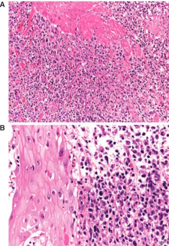
A skin biopsy showed dense lymphoid infiltrates occupying the whole dermis, with epidermal hyper-plasia and prominent edema of the papillary dermis (Fig. 4). There were some intraepidermal lympho-cytes, but prominent epidermotropism was lacking. In some areas, there was infiltration of the muscles. Cytomorphology showed predominance of medium-large atypical cells admixed with small lymphocytes and histiocytes (Fig. 5). Phenotypic analyses revealed strong positivity for CD30 (Fig. 6A); tumor cells were
Fig. 3. Large atrophic scar on the left buttock that resulted from the evolution of a previous noduloulcerative lesion.
also positive for the cytotoxic markers T-cell intra-cellular antigen-1 (TIA-1) and granzyme B (GrB) (Fig. 6B); they were positive for CD45RO and partly positive for CD2, MUM-1 and beta-F1. Other mark-ers (CD3, CD4, CD5, CD8, CD20, CD56) were negative. In situ hybridization for Epstein-Barr virus transcripts (EBER-1) was negative. The proliferation rate was over 90%.
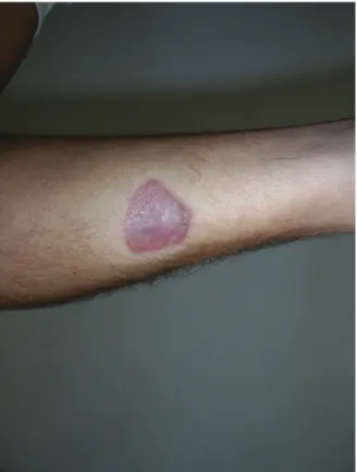
As there was some evidence of ulcer infection, a course of clarithromycin (CAM) was started at the initial observation. Within 5 days the ulcer of the forearm completely regressed, leaving an erythema-tous atrophic scar (Fig. 7); the papule and nodule of the eyebrow and thigh resolved with no residual lesions, too. After therapy, a single nodule developed on the posterior aspect of the leg, ulcerated and did not regress after treatment with topical antibiotics and corticosteroids; it resolved after another course of oral CAM.
The patient has been followed up closely with no recurrence of the disease to date (9 months after first observation). So far, full-body computerized tomog-raphy scan, bone marrow biopsy and peripheral T cells phenotype analysis excluded extracutaneous involvement.
Discussion
Clinicopathologic features of CD30+ CLPDs can pose difficult diagnostic challenges, and histologic criteria alone are often insufficient to differentiate clearly between LyP and cALCL, particularly concerning type C LyP.5 Primary lesions in LyP are typically multiple erythematous, dome-shaped papules or small nodules at different stages of development that spontaneously regress within weeks. In contrast, cALCL presents with solitary or localized nodules or tumors that often show
Fig. 4. Dense infiltrates of atypical lymphocytes within the entire dermis. Please note pseudoepitheliomatous epidermal hyperplasia and papillary dermal edema.
ulceration, but lack the spontaneous resolution observed in lesions of LyP.6 In unclear cases, the clinical course over time is used as decisive criteria for the definitive diagnosis and choice of treatment, and some authors suggest an observation period of 4–8 weeks to determine the natural evolution of the skin lesions.3
The recurrent, generalized, self-healing nature of the eruption observed in our patient clearly suggests a diagnosis of LyP. On the other hand, several lesions were of very large size, even exceeding 15 cm in diameter, and the ulcer on the forearm did not reveal a tendency to regression. In addition, the diffuse infiltrate of CD30+ large atypical lymphocytes with nuclear atypia suggests a histopathologic diagnosis of cALCL, or of type C LyP. In short, our patient presented with features of both entities, thus representing a ‘borderline’ case. Even close monitoring of the lesions during follow-up did not clearly clarify the exact classification. Owing to the spontaneous resolution of previous tumors, and to the presence of lesions at different sites of the body, we believe that our patient may have had a very
A
B
Fig. 5. A) Sheets of atypical lymphocytes with infiltration of a muscle; (B) anaplastic features of neoplastic cells.
unusual form of LyP with giant tumors that showed only partial spontaneous regression.
The time interval between the resolution of the lesions and the two courses of CAM suggests that the macrolide may have hastened the regression process. In vitro studies have clearly proven that macrolide antibiotics inhibit the proliferation of human lymphocytes stimulated with polyclonal T-cell mitogens;7 CAM was also found to suppress the production of proinflammatory cytokines via inhibition of nuclear factor-kB (NF-kB) activation, a mechanism similar to cyclosporine action.8 More recent studies showed that macrolide antibiotics, namely CAM, induce apoptosis of human peripheral lymphocytes through the Fas/Fas-ligand pathway and downregulation of Bcl-xL.9,10These effects could account for the striking response that occurred in our case. Nonetheless, due to the favorable course observed in this type of lymphoma and to the tendency for spontaneous regression, we cannot extrapolate these results, although the temporal relation is quite suggestive. Interestingly, CAM has been used in unresectable non-small-cell lung cancer patients, increasing the median survival of patients
A
B
Fig. 6. Positivity of neoplastic cells for (A) CD30 and (B) the cytotoxic marker TIA-1.
with advanced disease,11,12 and has shown efficacy in Helicobacter pylori-negative cases of low-grade malignancy MALTomas (lymphomas of mucosa-associated lymphoid tissue) of the rectum,13 by directly inducing apoptosis in lung carcinoma cells and B-cell lymphoma cells, respectively.
Another striking feature of this case was the cytotoxic phenotype of neoplastic cells, with loss of most T-cell markers. Expression of cytotoxic markers (TIA-1 and GrB) can be found in 70–90% of noncutaneous CD30+ ALCL of T-cell or null-cell phenotype,14 in addition to natural killer (NK)/T-cell lymphomas. As far as cutaneous lymphomas are concerned, some studies have shown that cALCL and LyP frequently express cytotoxic proteins.15,16 The prognostic value of cytotoxic markers is well established for noncutaneous CD30+ lymphomas,17
but no study has addressed this issue in cutaneous CD30+ lymphoproliferative disorders. Vermeer et al. and Massone et al. reported that cases of MF expressing GrB/TIA-1 do not have a more aggressive clinical behavior or a worse prognosis compared with cases of MF that do not express these cytotoxic molecules.18,19 An increase of expression of cytotoxic molecules can be observed in tumor
Fig. 7. Scar left from the resolution of the initial ulcer on the forearm.
stage MF, in particular in tumors showing blastic transformation.18
The correlation between clinical and histologic manifestations of CD30+ CLPD is not always straightforward, and particularly borderline cases present considerable difficulties in diagnosis and classification.20 Clinicians and pathologists need to be aware of the characteristic features of these entities to avoid misdiagnosis and inappropriate treatment. We believe that response to CAM in our case may warrant further studies on the anti-inflammatory and apoptotic effects on T cells of this molecule.
References
1. Willemze R, Jaffe ES, Burg G, et al. WHO-EORTC classifica-tion for cutaneous lymphomas. Blood 2005; 105: 3768. 2. Kempf W. CD30+ lymphoproliferative disorders:
histopathol-ogy, differential diagnosis, new variants, and simulators. J Cutan Pathol 2006; 33(Suppl. 1): 58.
3. Bekkenk MW, Geelen FA, van Voorst Vader PC, et al. Primary and secondary cutaneous CD30(+) lymphoproliferative disorders: a report from the Dutch Cutaneous Lymphoma Group on the long-term follow-up data of 219 patients and guidelines for diagnosis and treatment. Blood 2000; 95: 3653. 4. Kadin ME. Cutaneous Ki-1 lymphoma: pathology,
immunol-ogy and clinical characteristics. Princess Takamatsu Symp 1987; 18: 187.
5. El Shabrawi-Caelen L, Kerl H, Cerroni L. Lymphomatoid papulosis: reappraisal of clinicopathologic presentation and
classification into subtypes A, B, and C. Arch Dermatol 2004; 140: 441.
6. Querfeld C, Kuzel TM, Guitart J, Rosen ST. Primary cuta-neous CD30+ lymphoproliferative disorders: new insights into biology and therapy. Oncology 2007; 21: 689.
7. Morikawa K, Oseko F, Morikawa S, Iwamoto K. Immunomod-ulatory effects of three macrolides, midecamycin acetate, josamycin, and clarithromycin, on human T-lymphocyte func-tion in vitro. Antimicrob Agents Chemother 1994; 38: 2643. 8. Ichiyama T, Nishikawa M, Yoshitomi T, et al. Clarithromycin
inhibits NF-kappaB activation in human peripheral blood mononuclear cells and pulmonary epithelial cells. Antimicrob Agents Chemother 2001; 45: 44.
9. Ishimatsu Y, Kadota J, Iwashita T, et al. Macrolide antibiotics induce apoptosis of human peripheral lymphocytes in vitro. Int J Antimicrob Agents 2004; 24: 247.
10. Mizunoe S, Kadota J, Tokimatsu I, Kishi K, Nagai H, Nasu M.
Clarithromycin and azithromycin induce apoptosis of
activated lymphocytes via down-regulation of Bcl-xL. Int Immunopharmacol 2004; 4: 1201.
11. Mikasa K, Sawaki M, Kita E, et al. Significant survival benefit to patients with advanced non-small-cell lung cancer from treatment with clarithromycin. Chemotherapy 1997; 43: 288. 12. Hamada K, Mikasa K, Yunou Y, et al. Adjuvant effect of
clarithromycin on chemotherapy for murine lung cancer. Chemotherapy 2000; 46: 49.
13. Ohara T, Morishita T, Suzuki H, Masaoka T, Ishii H, Hibi T. Antibiotics directly induce apoptosis in B cell lymphoma cells derived from BALB/c mice. Anticancer Res 2004; 24: 3723. 14. Foss HD, Anagnostopoulos I, Araujo I, et al. Anaplastic
large-cell lymphomas of T-large-cell and null-large-cell phenotype express cytotoxic molecules. Blood 1996; 88: 4005.
15. Kummer JA, Vermeer MH, Dukers D, Meijer CJ, Willemze R. Most primary cutaneous CD30-positive lymphoproliferative disorders have a CD4-positive cytotoxic T-cell phenotype. J Invest Dermatol 1997; 109: 636.
16. Boulland ML, Wechsler J, Bagot M, Pulford K, Kanavaros P, Gaulard P. Primary CD30-positive cutaneous T-cell lym-phomas and lymphomatoid papulosis frequently express cyto-toxic proteins. Histopathology 2000; 36: 136.
17. Asano N, Suzuki R, Matsuo K, et al. Cytotoxic molecule expression is predictive of prognosis in Hodgkin’s-like anaplastic large cell lymphoma. Histopathology 2007; 50: 705.
18. Vermeer MH, Geelen FA, Kummer JA, Meijer CJ, Willemze R. Expression of cytotoxic proteins by neoplastic T cells in mycosis fungoides increases with progression from plaque stage to tumor stage disease. Am J Pathol 1999; 154: 1203.
19. Massone C, Crisman G, Kerl H, Cerroni L. The prognosis of early mycosis fungoides is not influenced by phenotype and T-cell clonality. Br J Dermatol 2008; 159: 881.
20. Cerroni L, Gatter K, Kerl H. Skin lymphoma – the illustrated guide, 3rd ed. Oxford: Wiley-Blackwell, 2009.