Andreia Sofia da Costa Vieira
Bachelor degree in Biochemistry
Rethinking Triazoles as Antifungals: Synthesis and
Evaluation of New Triazole Derivatives
Dissertation for the Master degree in Biochemistry for Health
Supervisor: Ana Petronilho, Auxiliary Investigator, ITQB-NOVA
Co-Supervisor: Catarina Pimentel, Auxiliary Investigator, ITQB-NOVA
Jury:
President: Doutor Pedro Manuel H. M. Matias, Principal Investigator ITQB-NOVA Arguer: Doutora Maria Rita Mendes B. Ventura, Auxiliary Investigator ITQB-NOVA Vowels: Doutora Margarida Archer Franco Frazão, Principal Investigator ITQB-NOVA
Doutora Ana Petronilho, Auxiliary Investigator ITQB-NOVA
Instituto de Tecnologia Química e Biológica António Xavier da Universidade Nova de Lisboa
iii Rethinking Triazoles as Antifungals: Synthesis and Evaluation of New Triazole Derivatives
Copyright – Andreia Sofia da Costa Vieira, ITQB/NOVA
O Instituto de Tecnologia Química e Biológica António Xavier e a Universidade Nova de Lisboa têm o direito, perpétuo e sem limites geográficos, de arquivar e publicar esta dissertação através de exemplares impressos reproduzidos em papel ou de forma digital, ou por qualquer outro meio conhecido ou que venha a ser inventado, e de a divulgar através de repositórios científicos e de admitir a sua cópia e distribuição com objetivos educacionais ou de investigação, não comerciais, desde que seja dado credito ao autor e editor.
v Acknowledgements
First of all, I would like to acknowledge my supervisors Dr. Ana Petronilho and Dr. Catarina Pimentel not only for the opportunity to allow me to work in a challenging project combining two distinctive areas, such as, but also for the continuous support and availability over the last year.
Also, a word of sincere gratitude to Maria Inês, Soraia Caetano and Catarina Amaral, mainly for their guidance and for always being present to assist me during the laboratory activities.
To my colleagues and friends, for all the companionship along these two fantastic years that we spent together, thank you.
Not less important, I cannot fail to mention my parents, for providing me with all the means, so that I could finish yet another stage of my academic career.
Finally, to Gustavo Marquês, thank you for the never-ending patience and encouragement throughout the years.
vii Resumo
Na última década, as infeções fúngicas tornaram-se um sério problema, principalmente devido ao excessivo uso de medicamentos antifúngicos, levando ao aumento das resistências adquiridas1. Assim sendo, é urgente desenvolver medicamentos antifúngicos mais eficazes.
O objetivo principal deste projeto de mestrado foi o desenvolvimento de novos agentes antifúngicos derivados de triazóis e pirimidinas, combinando numa só molécula dois grupos com atividade antifúngica. Na elaboração destes compostos foram utilizadas metodologias de cicloadição 1,3-dipolar de Huisgen, catalisadas por cobre2 ou ruténio3. Assim, foram sintetizados com sucesso 1,4- e 1,5-triazóis derivados de azidotimidina e correspondentes sais metilados. Foram sintetizados igualmente os 1.4-triazóis derivados de 5-fluorouracilo assim como um derivado metilado. Na sua maioria, os triazóis derivados da azidotimidina apresentaram rendimentos superiores aos do 5-fluorouracilo.
A atividade antifúngica destes compostos foi testada, utilizando como modelos os fungos Saccharomyces cerevisiae e Candida glabrata. Apenas um composto (8) apresentou capacidade de influenciar negativamente o crescimento do fungo Saccharomyces cerevisiae. Em relação ao fungo Candida glabrata nenhum dos compostos nas concentrações testadas apresentou capacidade de influenciar o crescimento deste fungo.
Adicionalmente, foram estudadas metodologias para a síntese de compostos de fluconazol estabilizados por ferro, bem como para a síntese de carbenos NHC de prata derivados de AZT. A coordenação do fluconazol a ferro foi bem-sucedida, obtendo-se um composto resultante da coordenação a Fe(II) de duas moléculas de fluconazol. Estudos de estabilidade em meio aquoso para este composto mostraram que a coordenação ao centro metálico nestas condições não se mantém, recuperando-se o fluconazol livre. Relativamente à formação de carbenos NHC de prata, o composto 8 reagiu com óxido de prata, com rendimento espectroscópico de 55 %. Tentativas de otimização do rendimento desta reação, e a separação de novo composto, não foram bem-sucedidas.
Palavras-chave: agentes antifúngicos, azóis, pirimidinas, metais de transição, carbenos N-heterociclicos
ix Abstract
In the last decades, fungal infections have become a serious problem, mainly due to the excessive use of this reduced number of drugs, leading to an increase of acquired resistances1. Therefore, it is urgent to develop new and more effective antifungal medicines.
The main objective of this master's thesis was the development of novel antifungal drugs, based on triazoles and pyrimidines, combining two groups with antifungal properties, known to be active in combinatorial therapies, in a single molecule. For the synthesis of these compounds, Huisgen's 1,3-dipolar cycloaddition methodologies, catalyzed by copper2 or ruthenium3 were employed. 1,4- and 1,5-triazoles derived from AZT (azidothymidine) were synthesized successfully as well as their methylated triazolium salts. Similarly, 1,4-triazoles derived from 5-fluorouracil were synthesized, as well as one methylated derivative. In general, the synthesis of the azidothymidine derivatives presented higher yields than those of 5-fluorouracil.
The antifungal activity of these compounds was evaluated, using as models the fungi Saccharomyces cerevisiae and Candida glabrata. Only one compound (8) displayed the ability to negatively influence the growth of the fungus Saccharomyces cerevisiae. Relatively, to Candida glabrata none of the compounds in the tested concentrations had the capacity to influence its growth.
Additionally, methodologies for the synthesis of fluconazole compounds stabilized by iron were studied, as well as synthetic methods for silver NHC carbenes derived from AZT. The coordination of fluconazole to iron was successful, obtaining a compound resulting from the coordination of Fe(II) to two molecules of fluconazole. Stability studies in aqueous solution showed that coordination to the metal center under these conditions is not stable, and free fluconazole can be recovered by crystallization. Concerning the formation of silver NHC carbenes, compound 8 react with silver oxide, but with a spectroscopic yield of 55%. Attempts to optimize the spectroscopic yield of this reaction were not successful.
Keywords: antifungal agents, azoles, pyrimidines, transition metals, N-heterocyclic carbenes
xi Index
1. Introduction ... 1
1.1. Currently used Antifungals ... 1
1.1.1. Polyenes ... 2
1.1.2. Echinocandins ... 3
1.1.3. Azoles ... 3
1.1.4. Pyrimidines... 5
1.2. Mechanisms of Resistance ... 6
1.3. Synthetic Methodologies utilized ... 8
1.3.1. Click Chemistry ... 8
1.3.1.1. Copper(I)-catalyzed azide-alkyne cycloaddition (CuAAC) ... 8
1.3.1.2. Ruthenium-catalyzed azide-alkyne cycloaddition (RuAAC) ... 10
1.4. Project Objectives ... 11
2. Results and Discussion ... 13
2.1. Synthesis of 1,4-triazoles derivatives from 3`-azido-3`-deoxythymidine ... 13
2.2. Synthesis of 1,5-triazoles derivatives from 3`-azido-3`-deoxythymidine ... 17
2.3. Methylation of 1,4- and 1,5-triazoles derivatives from 3`-azido-3`-deoxythymidine . 21 2.4. Synthesis of 1,4-triazoles derivatives from 5-Fluorouracil ... 24
2.4.1. Alkynylation of 5-Fluorouracil ... 24
2.4.2. Azide Synthesis ... 25
2.4.3. Synthesis of 1,4-triazoles derived from 5-fluorouracil and methylation ... 27
2.5. Synthesis of silver NHC compounds based on AZT ... 31
2.6. Synthesis of Iron fluconazole complexes: Studies on coordination and stability ... 33
2.7. Evaluation of the antifungal efficacy of the synthesized compounds against the yeasts Saccharomyces cerevisiae and Candida glabrata... 37
2.7.1. Derivatives of AZT in Saccharomyces cerevisiae ... 37
2.7.2. Derivatives of AZT in Candida glabrata ... 40
2.7.3. Derivatives of 5-Fluorouracil in Candida glabrata... 42
xii
3.1.2. Susceptibility tests of Saccharomyces cerevisiae... 45
3.1.3. Susceptibility tests of Candida glabrata ... 45
3.2. Synthetic procedures ... 47 4. Conclusions ... 67 5. Future work ... 69 6. Bibliography ... 71 7. Appendix ... 75 7.1. Fluconazole metalation ... 75
xiii Figure index
Figure 1.1-Most commonly used antifungal classes and corresponding targets. ... 2
Figure 1.2- Structure of Amphotericin B ... 2
Figure 1.3- Mechanism of action polyenes ... 3
Figure 1.4- Structure of Caspofungin ... 3
Figure 1.5-Types of azoles: imidazole, 1,2,4-triazole and 1,2,3-triazole ... 4
Figure 1.6-Mechanism of action of azoles ... 5
Figure 1.7-General structure of a pyrimidine ... 5
Figure 1.8-Mechanism of action 5-fluorocytosine ... 6
Figure 1.9-Mechanisms of antifungal resistance. (a) Overproduction of the target enzyme, (b) modification of the drug target, (c) increase of drug extrusion, (d) prevention of drug entry, (e) activation of bypass pathways, (f) impaired activation of the drug, (g) secretion of enzymes, which degrade the drug. ... 7
Figure 1.10-Model compounds developed in this project ... 11
Figure 1.11-Model compounds derivatives of AZT and 5-fluorouracil ... 12
Figure 2.1- Alkyne compounds used for the synthesis of 1,4-disubstiruted 1,2,3-triazoles from AZT ... 13
Figure 2.2-Fluconazole binding in the active site of Saccharomyces cerevisiae lanosterol 14-α demethylase. The Main chain is represented in gray, and fluconazole is represented in sticks (C atoms in cyan, N atoms blue, O atoms in red, and F atoms in pale blue). The heme is represented in sticks, with C atoms colored magenta. ... 14
Figure 2.3-1H NMR spectrum of AZT (top) vs phenylacetylene (middle) vs compound 1 (bottom) ... 16
Figure 2.4-13C NMR spectrum of AZT (top) vs phenylacetylene (middle) vs compound 1 (bottom) ... 17
Figure 2.5-1H NMR spectrum of AZT (top) vs phenylacetylene (middle) vs compound 5 (bottom) ... 19
Figure 2.6-13C NMR spectrum of AZT (top) vs phenylacetylene (middle) vs compound 5 (bottom) ... 20
Figure 2.7-1H NMR spectrum compound 1 (top) vs compound 5 (bottom) ... 21
Figure 2.8-1H NMR spectrums of compound 1 (top) vs 8 (bottom) ... 23
Figure 2.9-13C NMR spectrum of compound 1 (top) vs compound 8 (bottom) ... 24
Figure 2.10-5-fluorouracil and 5-fluorocytosine ... 25
Figure 2.11-1H NMR spectrum of aniline (top) vs phenyl azide (bottom) ... 27
Figure 2.12-1H NMR spectrum of compound 13 (top) vs compound 14 (middle) vs compound 16 (bottom) ... 28
xiv
Figure 2.13-13C NMR spectrum of compound 14 (top) vs compound 16 (bottom) ... 29
Figure 2.14- 1H NMR spectrum of compound 16 (top) vs compound 18 (bottom) ... 30
Figure 2.15-13C NMR spectrum of compound 16 (top) vs compound 18 (bottom) ... 31
Figure 2.16-1H NMR spectrum of compound 8 (top) vs compound with silver (bottom) ... 33
Figure 2.17-1H-NMR spectrums reaction (a) vs reaction (b) ... 34
Figure 2.18- Summary of the fluconazole metalations performed with FeCl2 ... 34
Figure 2.19-A: NMR spectrum of fluconazole and compound 19. B: Fluconazole representation ... 35
Figure 2.20-Mass spectroscopy spectrum of compound 19 (650-685 m/z) ... 36
Figure 2.21-Possible structures of compound 19 ... 37
Figure 2.22-Tested compounds in Saccharomyces cerevisiae ... 38
Figure 2.23- Antifungal activity of compounds 1, 2 and 8 against Saccharomyces cerevisiae. Yeast cells were spotted onto SC agar medium. Growth was recorded after 48 hours. The tested concentrations were 27 and 81 µM (compounds 1 and 2), 20 and 59 µM (compound 8) and 26 and 52 µM (Fluconazol - Fluc). ... 38
Figure 2.24- Antifungal activity of compounds 1, 2 and 8 against Saccharomyces cerevisiae growing in YPD medium. Yeast cells were spotted onto YPD agar medium. Growth was recorded after 48 hours. The tested concentrations were 27 and 81 µM (compounds 1 and 2), 20 and 59 µM (compound 8). ... 39
Figure 2.25- Antifungal activity of higher concentrations of compounds 1, 2 and 8 against Saccharomyces cerevisiae. Yeast cells were spotted onto SC agar medium supplemented with 1.35 mM of compounds 1 and 2 or 0.97 mM of compound 8. Growth was recorded after 24, 48 and 72h of incubation at 30ºC. DMSO was used as a mock control. ... 39
Figure 2.26- Antifungal activity of the AZT derivatives against Candida glabrata. The concentration range tested for each compound is listed in Table 3.1. Assays were performed as described in the Experimental procedures section. ... 42
Figure 2.27- Antifungal activity of the 5-FC derivatives against Candida glabrata. The concentration range tested for each compound is listed in Table 3.1. Assays were performed as described in the Experimental procedures section. ... 43
xv Scheme index
Scheme 1.1- Huisgen 1,3-dipolar cycloadditions ... 8
Scheme 1.2- CuAAC reaction for the formation of 1,4-disubstituted 1,2,3-triazoles ... 8
Scheme 1.3- CuAAC possible mechanism involving one copper(I) center ... 9
Scheme 1.4-CuAAC possible mechanism involving two copper(I) centers ... 10
Scheme 1.5- RuAAC reaction for the formation of 1,5-disubstituted 1,2,3-triazoles ... 10
Scheme 1.6- Catalytic Cycle of the RuAAC Reaction ... 11
Scheme 2.1-Scheme of the 1,4-disubstituted 1,2,3-triazoles synthesis ... 15
Scheme 2.2-Scheme of the 1,5-disubstituted 1,2,3-triazoles synthesis ... 18
Scheme 2.3-Methylation of the 1,4-disubstituted 1,2,3-triazoles ... 22
Scheme 2.4-Methylation of the 1,5-disubstituted 1,2,3-triazoles ... 22
Scheme 2.5- Scheme of the alkynylation of 5-Fluorouracil ... 25
Scheme 2.6- Mechanism of formation of azides by diazonium salts ... 26
Scheme 2.7- Scheme of the azides synthesis ... 26
Scheme 2.8-Scheme of the 1,4-disubstituted 1,2,3-triazoles from 5-Fluorouracil synthesis ... 27
Scheme 2.9- Scheme of the synthesis of compound 18 ... 29
Scheme 2.10- Silver reactions ... 32
xvii Table index
Table 2.1-Maximum concentration tested of AZT derivatives ... 41 Table 2.2-Maximum concentration tested of 5-Fluorouracil derivatives ... 42 Table 3.1- Range of concentrations tested for compounds 1-5, 7-12 and 16-18 ... 46
xix Acronyms list
13C-NMR Carbon-13 nuclear magnetic resonance
1H-NMR Proton nuclear magnetic resonance
5-FC 5-fluorocytosine
5-FU 5-fluorouracil
Ar Aromatic
AZT 3`-azido-3`-deoxythymidine
CuAAC Copper(I)-catalyzed azide-alkyne cycloaddition
d Doublet
DCM Dichloromethane
DMF Dimethylformamide
DMSO Dimethyl sulfoxide
DMSO-d6 Deuterated dimethyl sulfoxide
DNA Deoxyribonucleic acid
eq. Equivalent
et al. et alia (from Latin, meaning “and others”)
FdUMP 5-fluorodeoxyuridine monophosphate
Fluc Fluconazole
FUTP 5-fluorouridine triphosphate
HIV Human Immunodeficiency Virus
Hz Hertz
Ј Coupling constants
m Multiplet
MeOH Methanol
MIC Minimum inhibitory concentration
m/z Mass-to-charge ratio
Ph Phenyl
RNA Ribonucleic acid RNA
RPMI 1640 medium Roswell Park Memorial Institute 1640 medium RuAAC Ruthenium-catalyzed azide-alkyne cycloaddition
q Quartet
s Singlet
SCM Complete synthetic medium
t Triplet
Trz Triazole
xx
YPD Yeast extract peptone dextrose
1 1. Introduction
Fungi are a group of microorganisms spanning from unicellular forms (yeasts) to multicellular fungus, such as molds or mushrooms. Due to their metabolic abilities, many fungi play beneficial roles, for instance on the fermentation of bread, on the production of antibiotics, and on the decomposition of dead organisms. However, fungi can be opportunistic pathogens and cause diseases on plants, humans and other animals4.
During the past decades, fungal infections have become an alarming and serious topic, due to a great increase of fungal diseases and mortality caused by these infections on humans5. Pathogenic fungi are the cause of billions of infections annually, with ∼1.5 million attributable mortalities, a number comparable to that of tuberculosis and malaria6. Yet, the escalation of fungal infections on humans have been largely overlooked6. The rise of immunocompromised patients (such as those subject to haematopoietic stem cell transplantation, solid organs transplant, aggressive regimens of chemotherapy and HIV patients)5,7 led to an increase of fungal infections, while the relative low number of available antifungal agents forced an excessive utilization of these drugs. These factors combined potentiated the development of a large number of resistances against the existing antifungal agents. Most infections are caused by species belonging to four categories of fungi: Aspergillus, Candida, Cryptococcus, and Pneumocystis8, with invasive candidiasis being responsible for the majority of cases in what concerns hospital-acquire blood stem infections9.
In recent years, no considerable advances were achieved regarding antifungal therapies. Research on novel antifungals is particularly complex, since fungal pathogens are closely related to the host on a cellular level, decreasing the pathogen-specific targets9, and making difficult to develop broad spectrum compounds. In addition, reduced research funding in this area in recent years is also a major reason responsible for the limited drug development8.
This chapter will further describe existing antifungal drugs, their modus operandi and multiple antifungal resistances, as well as synthetic methods employed in this research project.
1.1. Currently used Antifungals
Fungal infections are treated by a variety of compounds. At present, circa 15 antifungal agents are approved for clinical use, but these drugs present issues regarding potency, safety and pharmacokinetic properties. Current drugs can be classified in four main classes: polyenes (e.g. amphotericin B); echinocandins (e.g. caspofungin); azole derivatives (e.g. fluconazole, ketoconazole, itraconazole, and voriconazole) and pyrimidines (e.g. 5-fluorocytosine)10. These antifungal drugs have a limited number of target structures, mostly affecting the fungal cell
2 membrane through disruption of ergosterol, β-1,3-d-glucan, or DNA/RNA synthesis11 (Figure 1.1).
Figure 1.1-Most commonly used antifungal classes and corresponding targetsI.
In the following sections, the modus operandi of these compounds will be briefly discussed, as well as their associated mechanisms of resistance.
1.1.1. Polyenes
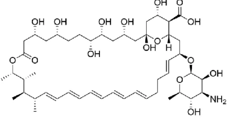
Amphotericin B (Figure 1.2) was the first polyene utilized for the treatment of fungal infections, since 1958, and was the most employed antifungal drug until fluconazole approval12.
Figure 1.2- Structure of Amphotericin B
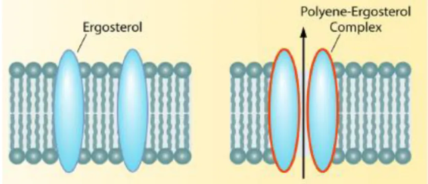
The mechanism of action of polyenes, such as Amphotericin B, lies on the stability of the fungal cell. Polyenes bind directly to ergosterol, a sterol found in fungi cell membranes with similar functions to those of cholesterol in animal cells, forming complexes that intercalate with the membrane12,13 (Figure 1.3), resulting in the formation of pores that lead to an increase in cellular permeability14, leakage of small molecules15 and death of the organism16. Ergosterol is absent on animals and as such it is a useful target for antifungal drugs17.
3
Figure 1.3- Mechanism of action polyenesII
1.1.2. Echinocandins
Echinocandins, such as caspofungin (Figure 1.4), are lipopeptide molecules, specifically cyclic hexapeptide with modified N-linked acyl lipid side chains18. They exert their antifungal activity by inhibiting the β-1,3-d-glucan synthesis, a fundamental component of the fungal cell wall.
Figure 1.4- Structure of Caspofungin
The disruption of β-1,3-d-glucan synthesis causes osmotic instability and eventually death14. Since β-1,3-d-glucan is not present in mammalian cell walls, echinocandins are selective to fungi and present lower toxicity and reduced side effects to the patient12.
1.1.3. Azoles
Azoles are the most commonly used antifungals. They are constituted by a five-member ring with at least one nitrogen atom. For the azoles currently used as antifungals, the main structures comprise exclusively imidazoles and triazoles, containing two and three nitrogen atoms respectively19.
4
Figure 1.5-Types of azoles: imidazole, 1,2,4-triazole and 1,2,3-triazole
Although the first report of antifungal activity of an azole compound (benzimidazole) was already described in 1944 by Woolley, it was not until the introduction of topical chlormidazole in 1958, that researchers became interested in the antifungal activity of this class of compounds19. Fluconazole, a broad-spectrum triazole antifungal developed by Pfizer was approved for use in 199011,20. Fluconazole presents high aqueous solubility, small molecular size, relatively low toxicity, its absorption is not influenced by gastric pH, and it can enter the cerebrospinal fluid extremely well. Fluconazole is generally utilized as first line antifungal therapy for the treatment of candida infections, disseminated candidiasis and cryptococcal meningitis17,21.
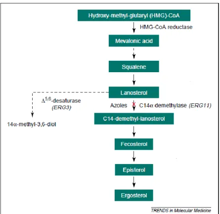
The mechanism of action of azoles targets the ergosterol (ergosta-5,7,22-trien-3β-ol) synthesis1 (Figure 1.6). Specifically, azoles will bind to the iron of the active site of the enzyme lanosterol 14-α-demethylase, a cytochrome P-450 dependent enzyme, through a nitrogen atom of the azole ring. This process precludes the ergosterol synthesis and promotes the synthesis and accumulation of 14α-methyl-3,6-diol, a toxic sterol. The latter causes increased permeability of the fungal cell membrane, disrupts the enzymes bound to the membrane, and inhibits fungal growth and replication13,20.
5
Figure 1.6-Mechanism of action of azolesIII
1.1.4. Pyrimidines
Pyrimidines are aromatic heterocycles containing two nitrogen atoms within a six-membered ring (Figure 1.7), well-known components of DNA and RNA and structural units of nucleobases, such as cytosine, thymine and uracil.
Figure 1.7-General structure of a pyrimidine
5-fluorocytosine (5-FC) was first synthesized as a potential anti-tumor agent in 195722. Later it proved to be active against candidiasis and cryptococcosis in mice, being utilized in such way to treat humans since 1960s22.
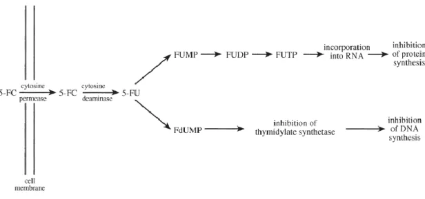
By itself 5-FC is inactive, but when inside a cell, it is deaminated to form 5-fluorouracil (5-FU) by the enzyme cytosine deaminase. For 5-fluorouracil two mechanisms of action have been suggested, one involving a conversion of 5-FU in 5-fluorouridine triphosphate (FUTP) and other involving the conversion of 5-FU into 5-fluorodeoxyuridine monophosphate (FdUMP). For the first, FUTP is incorporated into the fungal RNA replacing the uridylic acid, promoting
6 the inhibition of protein synthesis. In the second proposed mechanism, FdUMP inhibits the synthase of thymidylate, an important enzyme in the biosynthesis of DNA22 (Figure 1.8).
Figure 1.8-Mechanism of action 5-fluorocytosineIV
Due to the prevalence of intrinsically resistant strains and the frequent development of resistance during treatment, the 5-FC antifungal activity has used combination with other antifungal drugs, such as polyenes and azoles. This approach leads to an increase of the effectiveness of these drugs (synergetic effect), primarily against Cryptococcus and Candida infections15,23.
1.2. Mechanisms of Resistance
Fungi have acquired a large number of resistances to certain antifungals, during the past decades, (e.g. Candida krusei to fluconazole, Aspergillus terreus to amphotericin B, Cryptococcus spp. to the echinocandins, and Scedosporium spp. to all current antifungals)8. The development of fungal resistances is due to an excessive utilization of available drugs. Related to this is the rising number of immunocompromised patients requiring antifungal treatment1 and eventually with the excessive usage of fungicides such as azoles, in agriculture or other activities8.
As previously mentioned, several resistance mechanisms are known for 5-fluorocytosin, such as pathways that involve a mutation in cytosine permease decreasing the uptake of 5-FC or a mutation in cytosine deaminase and uracil phosphoribosyltransferase, blocking the formation of 5-FU and FUMP, respectively16,24 (Figure 1.9).
For azoles, their recurrent utilization has stimulated the development of resistance mechanisms in fungal pathogens, most relevantly for Candida species, but also for
7 Cryptococcus species25. Several molecular mechanisms of azole resistance have been described and are related to the following mechanisms (Figure 1.9).
• Decreased dug concentration: The development of active efflux pumps results in decreased drug concentrations at the site of action, this mechanism has been found in Candida species. Also, alterations in the plasma membrane composition affect membrane fluidity and asymmetry, disturbing the influx of drug into the cell, leading to a decreased uptake of drug24.
• Target site alteration: It has been demonstrated that mutations in ERG11, the gene encoding for the target enzyme lanosterol 14-α demethylase, prevent binding of azoles to the enzymatic site24.
• Up-regulation of the target enzyme: Overexpression of the target enzyme lanosterol 14-α demethylase, was found to be a mechanism of resistance in C. glabrata against fluconazole24.
• Development of bypass pathways: Mutation of the ERG3 gene prevents the formation of 14a-methyl-3,6-diol Without the formation of this toxic sterol azoles have no antifungal effect24.
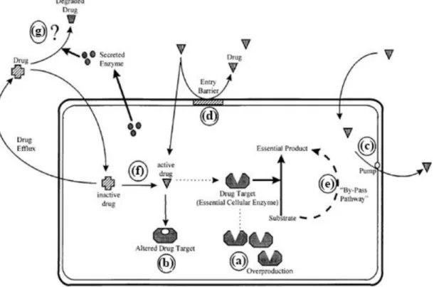
Figure 1.9-Mechanisms of antifungal resistance. (a) Overproduction of the target enzyme, (b) modification of the drug target, (c) increase of drug extrusion, (d) prevention of drug entry, (e) activation of bypass pathways, (f) impaired activation of the drug, (g) secretion of enzymes, which degrade the drugV.
8 1.3. Synthetic Methodologies utilized
1.3.1. Click Chemistry
Huisgen dipolar cycloadditions are reactions between a dipolarophile with a 1,3-dipolar compound that leads to the formation of 5-membered (hetero)cycles. For azides and alkynes this process usually generates a mixture of 1,4- and 1,5-disubstituted triazoles26 (Scheme 1.1). Since the discovery that the utilization of Cu(I) catalysts leads to the formation of 1,4-isomer26,27 and Ru catalysts produce to the 1.5-isomer28, the inconvenience with the selectivity for the Huisgen 1,3-dipolar cycloadditions between azides and alkynes was solved. This modification is a clear example of a click chemistry reaction. Click chemistry is a term that is commonly used to describe reactions that are high yielding, wide in scope and generate only byproducts, which can be removed without chromatography (e.g. through crystallization or distillation as purification methods). They are stereospecific (only one product), considered simple to perform (insensitive to oxygen and water), can be conducted in easily removable or benign solvents and the product is generally stable under physiological conditions29.
Scheme 1.1- Huisgen 1,3-dipolar cycloadditions
In this project, we employed copper and ruthenium-catalyzed azide-alkyne cycloadditions, to obtain specific structural differences and generate respectively 1,4-disubtituted triazoles and 1,5-1,4-disubtituted triazoles.
1.3.1.1. Copper(I)-catalyzed azide-alkyne cycloaddition (CuAAC)
For a CuAAC reaction the active catalyst is a Cu(I) center, which can be used directly or employing a copper(II) precursor alongside a reducing agent. The product of this reaction is an 1,4-disubstituted 1,2,3-triazole2 (Scheme 1.2).
Scheme 1.2- CuAAC reaction for the formation of 1,4-disubstituted 1,2,3-triazoles
Two different reaction mechanisms have been proposed in the literature for the CuAAC reaction. The first mechanism was suggested by Rostovtsev et al. in 200230 and is represented in Scheme 1.3. In this proposal, the formation of copper(I) acetylide (formation of a σ-bond) is followed by azide coordination to the metal (formation of a π -bond), forming the catalytically
9 active complex, which converts into a metallacycle with formation of a C–N bond. The next step is the copper oxidation, followed by a ring contraction that leads to the formation of the 5-member ring (triazolide). Finally, copper(I) triazolide is protonated via the acidic alkyne proton and copper is released, completing the catalytic cycle2,30.
Scheme 1.3- CuAAC possible mechanism involving one copper(I) centerVI
The second proposed mechanism, which is represented below, proposes an additional copper(I) participation (Scheme 1.4). In this proposal, the formation of a σ-bond and one π -bond between the alkyne and the two copper takes place, followed by the coordination of an azide, creating an azide/alkyne/copper(I) complex. In this step, to form the first covalent C-N bond, a nucleophilic attack by the carbon of the acetylide must occur at the N-3 of the azide. The second C-N bond closes the ring forming a triazolide. Finally, copper(I) triazolide is protonated, via the acidic alkyne proton and cooper is released, completing the catalytic cycle2,30.
10
Scheme 1.4-CuAAC possible mechanism involving two copper(I) centersVII
1.3.1.2. Ruthenium-catalyzed azide-alkyne cycloaddition (RuAAC)
The product of the RuAAC reaction is an 1,5-disubstituted 1,2,3-triazole. Scheme 1.5 shows a simplified scheme of this reaction. The ruthenium complexes generally utilized in RuAAC reactions are a Cp* complex, found to be especially active and selective catalysts for 1,5-disubstituted triazoles28.
Scheme 1.5- RuAAC reaction for the formation of 1,5-disubstituted 1,2,3-triazoles
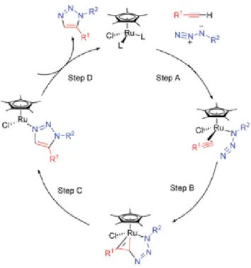
The cycle is initiated with the concerted coordination of the alkyne, azide and the ruthenium, forming a ruthenium complex, followed by the oxidative coupling of the alkyne and the azide originating a ruthenacycle. The first C-N bond will be formed between the terminal nitrogen of the azide and the carbon less sterically hindered of the alkyne. Subsequently, the complex undergoes reductive elimination, releasing the aromatic triazole and regenerating the catalyst3,28. A representation of this mechanism is depicted in Scheme 1.6.
11
Scheme 1.6- Catalytic Cycle of the RuAAC ReactionVIII
1.4. Project Objectives
The main goal of this project was the development of a library of molecules containing a triazole and a pyrimidine within the same core (Figure 1.10), aiming to develop more active antifungal agents based on the well-known synergetic effects for these drugs, when in combination therapies.
Figure 1.10-Model compounds developed in this project
The main building blocks for the development of the novel triazoles were 5-fluorouracil and 3`-azido-3`-deoxythymidine (AZT). Both are pyrimidines that can be further functionalized with different linkers and triazoles. The 5-flurouracil, when combined with the triazole provides as a linker a carbonated chain, and 3`-azido-3`-deoxythymidine a sugar (Figure 1.11).
12
Figure 1.11-Model compounds derivatives of AZT and 5-fluorouracil
The second main objective was the evaluation of the biological activity of the compounds, using as models the fungi Saccharomyces cerevisiae and Candida glabrata.
In addition, we targeted the development methodologies for the synthesis of fluconazole compounds stabilized by iron, as well as for the synthesis of silver NHC carbenes derived from AZT.
13 2. Results and Discussion
The main goal of this project was to develop of a library of compounds with high antifungal activity by gathering two commonly used antifungal agents in one single molecule, more specifically azoles and pyrimidines. To achieve this objective, it was performed the synthesis of triazoles derived from 3`-azido-3`-deoxythymidine (AZT) and 5-fluoruracil, employing the Huisgen azide-alkyne cycloaddition with copper and ruthenium catalysts, to achieve the desired connectivity. For the 5-fluoruracil derived triazoles, due to time constraints, only the CuAAC was used.
2.1. Synthesis of 1,4-triazoles derivatives from 3`-azido-3`-deoxythymidine
For our synthesis of 1,4-triazoles, a combination of copper (II) sulfate and sodium ascorbate was utilized. Sodium ascorbate was applied to reduce Cu(II) to Cu(I), since copper(I) is the active catalyst.
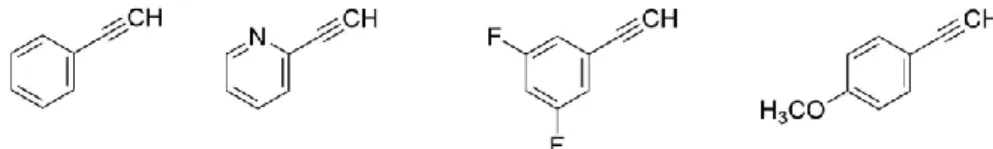
To synthesize the 1,4-triazoles derivative from AZT, four different alkynes were chosen. These were phenylacetylene, 2-ethynylpyridine, 1-ethynyl-3,5-difluorobenzene and ethynylanisole, and are represented in Figure 2.1.
Figure 2.1- Alkyne compounds used for the synthesis of 1,4-disubstiruted 1,2,3-triazoles from AZT
It is assumed that the modus operandi of azole compounds implies the coordination of one nitrogen from the azole ring to the iron of the heme group on the active site of the enzyme lanosterol 14-α demethylase. It has also been suggested that the backbone of the molecule interacts with the active site surroundings (Figure 2.2) and can influence the antifungal activity31. In agreement with this, some studies imply that the fluconazole difluorophenyl group induces an hydrophobic interaction with Met153 and the alkyl group of Lys157 in Cryptococcus neoformans lanosterol 14-α demethylase32, that fosters the antifungal activity of the compound. Thus, the above alkyne groups were selected to understand how the different features of the aromatic ring, in addition to the triazole core, would influence its antifungal activity and affect the fitting of the molecule to the catalytic pocket of the enzyme.
14
Figure 2.2-Fluconazole binding in the active site of Saccharomyces cerevisiae lanosterol 14-α demethylase. The Main chain is represented in gray, and fluconazole is represented in sticks (C atoms in cyan, N atoms blue, O atoms in red, and F atoms in pale blue). The heme is represented in sticks, with C atoms colored magentaIX.
In line with this, we chose a set of aromatic groups with different substituents, also, to be able to access different properties within the molecule. The substituents in the phenyl group were chosen to either increase polarity to the molecule (2-ethynylpyridine), therefore fostering the solubility of the molecule in water, or introduce variations in the basicity of the triazole (Ethynylanisole). In addition, one of the compounds used, bearing a 3,5-difluorophenyl group, provides access for a direct comparison with fluconazole. Our synthetic methodologies were initiated with phenyl acetylene, for which a synthetic procedure was already described. These methodologies were then extended to the remaining substrates.
The synthesis of compound 1, the phenylacetylene derivative, was performed according to Zhou et al.33. Phenylacetylene, 3'-azido-3'-deoxythymidine, CuSO4 and sodium ascorbate were mixed with H2O/tert-butanol and stirred at 100 ºC for 24 hours. At the end of the reaction, a yellow solid was formed. After filtration, this solid compound was initially extracted with dichloromethane, and the extract was washed with 10% ammonia, then water and brine. This purification, usually employed to ensure removal of Cu salts, did not work as expected, since the product did not dissolve completely in dichloromethane. Then, the remaining solid material was purified by silica gel chromatography (CHCl3: MeOH 10:1 v/v) and obtained as a white
powder with a yield of 73%. Compound 1 was again synthesized, after the optimization of the reaction conditions and work-up, to obtain 1 as a white powder with 83% yield.
With these first results, we determined the optimized reaction conditions for the synthesis of the novel AZT derived triazoles (Scheme 2.1). Thus, the syntheses of compounds 2, 3 and 4 were performed in ter-butanol, with CuSO4 and sodium ascorbate at 100ºC for 2 hours.
15 The purification of the compounds was performed using silica gel chromatography (CHCl3:
MeOH 10:1 v/v). While compound 2 was obtained as a brown solid with a yield of 50%, compounds 3 and 4 were obtained as white powders with yields of 95% and 33.3% respectively. Yields varied considerably depending on the alkyne substituent. Thus, for phenyl (1) and 3,5 difluorophenyl (3) yields were above 80%, with the pyridyl (2) performing somewhat lower and the methoxy group (4) with a very modest yield.
Scheme 2.1-Scheme of the 1,4-disubstituted 1,2,3-triazoles synthesis
According to Kolb et al.29 the compounds obtained from click reactions should be purified utilizing nonchromatographic techniques. However, in our case, all the compounds obtained through click reactions were purified by silica gel chromatography. It was the more effective methodology for the removal of copper salts.
To confirm the structure of the compounds, NMR spectroscopy measurements (1H and 13C) were performed. The formation of the triazole ring was confirmed through the appearance of a new signal, a singlet for all the 1,4-disubstituted compounds, corresponding to the proton of the C5 position of the newly formed triazole ring. This singlet resonates at around 8.94-8.67 ppm, and is concomitant with the disappearance of the signal corresponding to the terminal alkyne proton (Figure 2.3).
As a common feature, the thymine signals from the CH3 group resonate as a singlet at 1.83 ppm. The NH group, also a singlet, at 11.38-11.37 ppm, while the CH proton of the thymidine can be found between 7.85-7.84 ppm. As for the sugar ring, it gives rise to a set of six signals, between 6.46-2.67 ppm. For instance, the CH group, a quartet, resonates between 4.30-4.28 ppm, the triplet signal of the OH group appeared between 5.31-5.28 ppm and quartet of the CH-Ntrz group was located around 5.42-5.39 ppm.
16
Figure 2.3-1H NMR spectrum of AZT (top) vs phenylacetylene (middle) vs compound 1 (bottom)
In the 13C NMR spectrum, the thymine group signals are similar for all the compounds. For instance, the CH3 group resonates at 12.7 ppm, while the C=O groups can be found at 150.9 ppm and 164.2 ppm. As for the sugar ring carbons, the five signals can be found between 84.4-37.6 ppm. The formation of the triazole ring was confirmed, as was the case for 1H, due to the appearance of the new signals corresponding to C5 and C4 of the newly formed triazole ring. Both signals are located for all 1,4-disubstituted compounds around the same spectrum position (123.4-120.4 ppm and 147.9-145.0 ppm, respectively) (Figure 2.4).
17
Figure 2.4-13C NMR spectrum of AZT (top) vs phenylacetylene (middle) vs compound 1 (bottom)
2.2. Synthesis of 1,5-triazoles derivatives from 3`-azido-3`-deoxythymidine
Following the synthesis of 1,4-disubstituted triazoles AZT derivatives, the corresponding 1,5-disubstituted isomers were synthesized. Once again, Huisgen azide-alkyne cycloaddition was employed, but this time using a ruthenium catalyst. Ruthenium Cp* complexes are especially active and selective catalysts for 1,5-disubstituted triazoles, normally with excellent yields28. The ruthenium catalyst utilized was Cp*RuCl(PPh3)2, for which a number of procedures are available28. Phenylacetylene, 1-ethynyl-3,5-difluorobenzene and ethynylanisole (Figure 2.1) were selected as alkynes. The synthesis of this set of compounds was initiated with phenyl acetylene, for which a synthetic procedure was already described in the literature3 and this methodology was subsequently adapted to the remaining compounds.
To synthesize compounds 5, 6 and 7, AZT was mixed with Cp*RuCl(PPh3)2 and the correspondent alkyne. The reaction was conducted at 80ºC in dioxane under inert atmosphere for 48 hours (Scheme 2.2). All the new compounds were purified following similar procedures to those used for the 1,4-triazoles. Thus, we employed silica gel chromatography using a mixture of CHCl3: MeOH 10:1 v/v as eluent. Compounds 5 and 7 were obtained as brown
powders with yields of 85% and 33% respectively. We were unable to isolate compound 6 under similar conditions and did not explore other possibilities due to time constraints.
18
Scheme 2.2-Scheme of the 1,5-disubstituted 1,2,3-triazoles synthesis
To confirm the structure of all compounds, NMR spectroscopy measurements (1H and 13C) were performed. The formation of the triazole ring was confirmed due to the appearance of a new signal, a singlet for all 1,5-disubstituted compounds, corresponding to the proton of the C4 position of the triazole ring at 7.92 ppm in case of compound 5, 7.85 ppm for compound 7 and the absence of the peak corresponding to the terminal alkyne proton (Figure 2.5).
As a common feature, the thymine signals from the CH3 group resonate as a singlet at 1.79-1.78 ppm. The NH group, a broad singlet resonates at 11.36 ppm while the CH proton of the thymidine can be found at 7.79-7.78 ppm. The sugar ring protons appear as a set of six signals, between 6.57-2.57 ppm. For instance, the CH group, a quartet, is located at 4.40-4.38 ppm and the CH-Ntrz, a multiplet, around 5.19-5.14 ppm.
19
Figure 2.5-1H NMR spectrum of AZT (top) vs phenylacetylene (middle) vs compound 5 (bottom)
In the 13C NMR spectrum, the thymine group signals are similar for all compounds. The CH3 group resonates at 12.7 ppm, while the C=O groups can be found at 150.9 ppm and 164.1 ppm. As for the sugar, the carbon groups can be found between 85.3-38.1 ppm. The formation of the triazole ring was further confirmed through the observation of new signals corresponding to the carbons of the C5 and C4 position of the triazole ring, emerging for all the 1,5-disubstituted compounds around the same spectrum position (138.3-138.1 ppm and 133.3-133.1 ppm, respectively) (Figure 2.6).
20
Figure 2.6-13C NMR spectrum of AZT (top) vs phenylacetylene (middle) vs compound 5 (bottom)
Compound 5 was obtained as a brown powder with a yield of 85.55%, similarly to the corresponding 1,4-isomer. Following the tendency noted for the 1,4 triazoles, compound 7 was also obtained as brown powder, with a lower yield (33.3%).
Compounds 1 and 5 (1,4- and 1,5-triazoles) presented different spectroscopic features (Figure 2.7). The main difference between the two compounds relates to the singlet of the triazole ring proton. For the 1,4-triazole the singlet can be found around 8.67-8.94 ppm, while for 1,5-triazoles was detected at higher field around 7.85-7-92 ppm.
21
Figure 2.7-1H NMR spectrum compound 1 (top) vs compound 5 (bottom)
In addition, the phenyl protons also presented some differences. For the 1,4-triazole, the aromatic phenyl ring is shown as a doublet, triplet and triplet, in a 2:2:1 ratio, between 7.36-7.90 ppm, while for 1,5-triazoles, a large multiplet combines all the protons, around 7.60-7.53 ppm (Figure 2.7).
2.3. Methylation of 1,4- and 1,5-triazoles derivatives from 3`-azido-3`-deoxythymidine
Following the triazole synthesis, we developed methylation procedures for the triazoles synthesized previously. The main objective regarding compounds methylation was to form a triazolium salt and increase solubility. In addition, the methylation of the triazole ring can eventually influence the coordination to the iron on the active site of the enzyme lanosterol 14α-demethylase and affect the antifungal activity for these compounds.
The methylations were performed by reacting the triazoles with iodomethane or with methyl trifluoromethanesulfonate. Following several experiments, the optimized conditions were determined and are as follow: 1 eq. of the triazole for 10 eq. of the methyl compound. The reaction was heated at 100ºC in DMF for 24 hours. The reaction mixture of compound 8 had a yellow color upon completion and brown color for compound 10. After cooling to room
22 temperature, the compounds were precipitated by adding ether to the reaction mixture, followed by a filtration of the solid precipitate.
Scheme 2.3-Methylation of the 1,4-disubstituted 1,2,3-triazoles
To obtain compound 9, methylation with methyl trifluoromethanesulfonate was required, since the reaction with iodomethane was unsuccessful. Compound 9 precipitated as a brown liquid, and after several washes with ether, it was possible to isolate compound 9 as a brown solid with a yield of 8%. The low yield is due in part to the hydroscopic nature of the salt, requiring an extended work-up procedure (Scheme 2.3).
Scheme 2.4-Methylation of the 1,5-disubstituted 1,2,3-triazoles
Compound 8 was obtained as a yellow solid with a 72% yield and compound 10 as a brown solid, with a yield of 31.3%. The 1.5-disubstituted compounds 11 and 12 were obtained as brown solids with 88.4% and 77.2% yields respectively. Compound 8 had a slightly low yield compared to the 1,5-isomer compound 11, while compound 10 had a lower yield compared with compound 12.
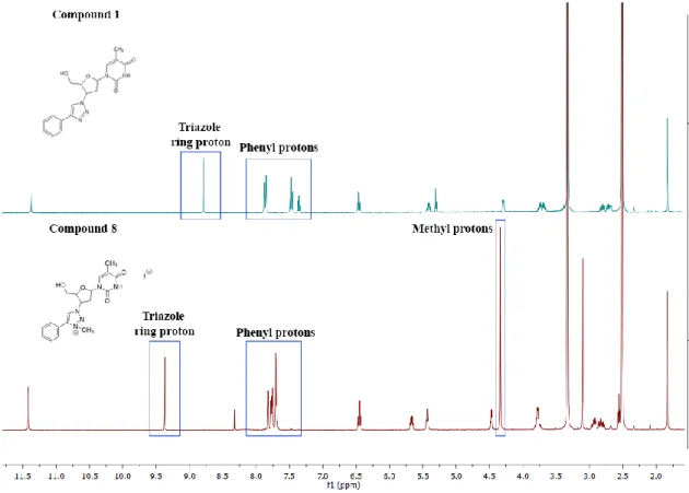
23 The structure of the compounds was confirmed by 1H and 13CNMR spectroscopy. The obtained spectra were examined and compared with those of the non-methylated compounds. The 1H NMR spectra from all methylated compounds showed an additional signal, corresponding to the new methyl group of the molecule (N-CH3). The new singlet can be detected at between 4.30-4.38 ppm for the 1,4-triazoles and between 4.44-4.42 ppm in the 1,5-triazoles. The thymine protons from the CH3 group was observed as a singlet that resonate at 1.83 ppm in the 1,4-triazoles and at 1.78 ppm for the 1.5-triazoles. The NH group, a broad singlet, is located between 11.42-11.39 ppm in the 1,4 and 15-triazoles. The sugar protons appeared as a series of six signals between 6.45-2.79 ppm. One important feature that led to the confirmation of the structure of the compounds was the significant shift to low field, observed for the singlet from the triazole ring (e.g. for 11 about 1.20 ppm). By contrast, the thymine protons did not undergo significant changes. Slight shifts were noted for the sugar protons, for instance, the CH-Ntrz group shifted around 0.26 ppm to low field and the CH shifted around 0.18 ppm, also to low field. Figure 2.8 represents the comparison between compound 1 and compound 8 1H NMR spectrums.
Figure 2.8-1H NMR spectrums of compound 1 (top) vs 8 (bottom)
For the methylated compounds, the 13C NMR spectrum also showed an additional signal, corresponding to the new methyl group of the molecule (N-CH3). The new signal appeared between 39.4-39.2 ppm for the 1,4-disubtituted and between 40.7-40.6 ppm in the 1,5-disubtituted. The pyrimidine CH3 group resonates at 12.7 ppm for, while the C=O groups can be
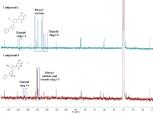
24 found at 150.9-150.8 ppm and 164.1 ppm. As for the sugar, the carbon groups can be found between 84.4-37.6 ppm. Figure 2.9 represents the comparison between compound 1 and compound 8 13C NMR spectrums. The structure of the compounds was confirmed by the rearrangement of the phenyl group signals positions and the shift of the triazole carbon C5 and C4 in the 1,4-triazoles (shifted around 8 ppm and 5 ppm, respectively) and in the 1,5-triazoles (shifted around 5 ppm and 3 ppm, respectively).
Figure 2.9-13C NMR spectrum of compound 1 (top) vs compound 8 (bottom)
The thymine and carbons do not undergo significant changes, however the sugar carbon from the CH-N- group near the triazole had a shift of 5 ppm in the 1,4-triazoles and 3 ppm in the 1,5-triazoles (Figure 2.9).
2.4. Synthesis of 1,4-triazoles derivatives from 5-Fluorouracil 2.4.1. Alkynylation of 5-Fluorouracil
fluorouracil is the active antifungal compound resultant of the deamination of 5-fluorocytosine (Figure 2.10). To synthesize the triazole from 5-fluorouracil, first it is necessary to introduce an alkyne group to form the precursor. In addition, syntheses of azides for the click reaction were also required.
25
Figure 2.10-5-fluorouracil and 5-fluorocytosine
To synthesize the 1,4-disubstituted triazoles from 5-fluorouracil, we chose to introduce an alkyne group at N1, making use of previously established procedures34.
In this approach, we adapted the procedure described by Weiss, J. T. et al.34. In this methodology, protection is not required and the alkyne group is bound directly to N1 in one sole step, employing propargyl bromide as reagent (Scheme 2.5).
Scheme 2.5- Scheme of the alkynylation of 5-Fluorouracil
This approach worked successfully, but purification via chromatography was required. Compound 13 was obtained as a white powder and the yield obtained was very low (4%).
To overcome this, the reaction was scaled-up from 1.5 mmol (200 mg) of 5-Fluorouracil to 4.6 mmol (600 mg). This resulted on a yield improvement to 13%. This outcome was slightly better, but could not be improved. As such, the repetition of the same reaction several times was required, in order to obtain the required amount of compound 13 to perform the remaining reactions. It must be stated that the published yield for this procedure is 40%.
The formation of compound 13 was confirmed by 1H NMR spectroscopy. In the 1H spectrum, the singlet corresponding to the NH proton can be detected at 11.92 ppm, while the 5-fluorouracil aromatic proton resonates at 8.14 ppm as a doublet. The doublet for the CH2 group resonates at 4.47 ppm and the alkyne terminal proton at 3.46 ppm as a triplet. The results matched the data reported by Weiss, J. T. et al 34 further confirming the structure of compound.
2.4.2. Azide Synthesis
With compound 13 already synthesized and to obtain our target molecule, our second step was to synthesize the azide. We synthesized two different azides, both aromatic, but with a varying substituent.
26 The synthesis of two azides, phenyl azide (14) and 4-bromo phenyl azide (15), was performed according to the procedure employed by Barral et al.35. This procedure employs anilines as azide precursors. The synthesis involves the formation of diazonium salts from the corresponding aniline, followed by the formation of the azide. The azide is formed by way of two competing reactions36 that can be observed in Scheme 2.6.
Scheme 2.6- Mechanism of formation of azides by diazonium saltsX
The residue was purified by silica gel chromatography using ether as eluent. Phenyl azide (14) was obtained as a yellow liquid with 76% yield and 4-bromo phenyl azide (15) as an orange liquid in 75% yield (Scheme 2.7).
Scheme 2.7- Scheme of the azides synthesis
To confirm the structure of the compounds, NMR measurements were performed. For the new azides, 1H NMR spectrum shows three signals for compound 14 and two signals for compound 15, these between 7.50-7.00 ppm. Important aspects that led to the confirmation of the compounds structure were the absence of the signal at 4.99 ppm corresponding to the amine
27 group (NH2) and the rearrangement of the aromatic signals that shifted from 7.02-6.47 ppm to 7.50-7.00 ppm (Figure 2.11).
Figure 2.11-1H NMR spectrum of aniline (top) vs phenyl azide (bottom)
2.4.3. Synthesis of 1,4-triazoles derived from 5-fluorouracil and methylation
The 1,4-disubstituted triazoles from 5-fluorouracil were synthesized in similar conditions used for the AZT derivatives described above, using CuSO4 and sodium ascorbate, at 80ºC (Scheme 2.8). Compounds 16 and 17 were obtained as white and light brown powders in relative low yields (36% and 15% respectively).
Scheme 2.8-Scheme of the 1,4-disubstituted 1,2,3-triazoles from 5-Fluorouracil synthesis
1H NMR spectroscopy measurements were also obtained, confirming the structure of compounds 16 and 17. The uracil signals from both compounds were observed as singlets
28 around 11.89-11.87 ppm for the NH group and at 8.23 ppm for the aromatic proton. The singlet from the CH2 protons appeared at 5.01 ppm in both cases. As was the case of the AZT derivatives, a new singlet corresponding to the CH group of the new triazole ring, at around 8.81-8.84 ppm, confirms the formation of the triazole (Figure 2.12).
Figure 2.12-1H NMR spectrum of compound 13 (top) vs compound 14 (middle) vs compound 16 (bottom)
In the 13C NMR spectra, for uracil signals, the C=O groups resonate at 158.1-157.9 ppm and 149.9 ppm, while the CH group at 130.5 ppm. The formation of the triazole ring confirmed through the observation of the new signals for C5 and C4 at 122.1 ppm and 144.0 ppm, respectively. Another important feature was the shift of the CH2 group signal from 37.0 ppm to 43.2 ppm (Figure 2.13).
29
Figure 2.13-13C NMR spectrum of compound 14 (top) vs compound 16 (bottom)
The methylation of compound 16 was performed following similar procedures as those used for the AZT derivatives. Compound 16 was reacted with 3 eq. of iodomethane in DMF, at 100ºC for 24 hours. After cooling to room temperature, the reaction mixture was precipitated with ether, obtaining compound 18 as a yellow powder in moderate yield of 46% (18) (Scheme 2.9).
Scheme 2.9- Scheme of the synthesis of compound 18
The confirmation of the structure of the compound 18 was performed by 1H and 13C NMR spectroscopy. The obtained spectra were evaluated and compared with the spectra of the non-methylated compounds. The 1H spectrums from the methylated compound showed an additional signal, corresponding to the new methyl group of the molecule (N-CH3), at 4.46 ppm. The uracil NH group, appeared as a broad singlet, at 12.08 ppm and the aromatic proton, as
30 duplet at 8.19 ppm. In addition, the triazole proton at C5 shifted from 8.81 ppm to 9.63 ppm. The uracil protons did not undergo significant changes, following the tendency observed for the AZT derivatives, Figure 2.14 represents the comparison between compound 16 and compound 18 1H NMR spectrums.
Figure 2.14- 1H NMR spectrum of compound 16 (top) vs compound 18 (bottom)
In the 13C NMR spectra of compound 18, the uracil signals were present at 158.1 ppm and 150.1 ppm for the C=O groups, while for CH group at 129.6 ppm. Following the tendency observed previously, a new signal at 40.5 ppm, corresponding to the N-CH3 is observed, as well as a shift at C5 and C4 of the triazole ring, by 6 ppm and 4 ppm respectively. (Figure 2.15).
31
Figure 2.15-13C NMR spectrum of compound 16 (top) vs compound 18 (bottom)
2.5. Synthesis of silver NHC compounds based on AZT
Metallic silver and silver compounds have numerous uses in medicine. Since the early days, silver with its notably antimicrobial activity, is not only utilized in medical devices (e.g. bone prostheses, surgical sutures and dentistry)37,38, but also particularly used against chronic ulcers, extensive burns, and difficult-to-heal wounds. Silver antimicrobial activity is related with its interference with electron transport system of the cell, interaction with cell membrane and the thiol groups of key enzymes in microorganisms39. Some studies report that silver NHC complexes have high antimicrobial activity, and perform better than their NHC precursors, such as imidazoles and triazoles39,40.
For the metallation of compound 8 we utilized Ag2O as silver source. The reaction was performed in DMF under inert atmosphere and in the absence of light, due to the sensitivity of silver salts. A variety of reaction conditions were tested, by changing the ratio of compound 8 versus Ag2O, temperature and reaction time (Scheme 2.10).
32
Scheme 2.10- Silver reactions
Analysis by 1H NMR spectroscopy shows the presence of a new compound, alongside with compound 8, in a 55:45 % ratio.
The new species is characterized by a signal at 4.21 ppm, corresponding to the protons of the methyl group (N-CH3) of the triazole ring, shifted by 0.12 ppm with respect to compound 8. In addition, a new set of signals in the aromatic region corresponding to the phenyl group could be observed (Figure 2.16).
It can be concluded that the metalation can be achieved, but only partially. The formation of the new silver compound is possible, but the conversion of compound 8 to the metallated derivative is not complete, at least under the tested conditions. Of note, independently of the reaction conditions, the result is similar in all cases, leading to 1:1 mixture approximately.
33
Figure 2.16-1H NMR spectrum of compound 8 (top) vs compound with silver (bottom)
2.6. Synthesis of Iron fluconazole complexes: Studies on coordination and stability
Some studies suggest that iron concentration can have an influence in the antifungal activity of fluconazole41. However, records on the coordination and stability of fluconazole and iron coordination compounds are extremely scarce. We decided to study the coordination of fluconazole to iron complexes and the evaluation of the stability of these complexes.
To synthesize these complexes, we tested a variety of conditions, using mainly FeCl2 as iron source. Different solvents were selected, such as DCM/MeOH, MeOH, D2O. Some reactions were performed under air while others under inert atmosphere.
The first reactions (a and b) were carried out with FeCl2 in (a) a mixture of DCM/MeOH (2/1) and (b) exclusively MeOH, and were monitored by 1H NMR for 24 hours. The 1H spectra showed a significant evolution with time. When comparing the two reactions, NMR analysis shows the formation of the same product in both cases, but the rate of its formation seems to be affected by the solvent (Figure 2.17).
34
Figure 2.17-1H-NMR spectrums reaction (a) vs reaction (b)
Reaction (b) was repeated, but using (d) 2 eq. of AgNO3 for each 1 eq. of FeCl2 under air and (e) under inert atmosphere. The utilization of silver salts to promote this reaction was based on the well-known insolubility of the AgCl2 salts, which leads to their precipitation in solution, leaving a vacant coordination site in Fe, thus favoring coordination of fluconazole. However, this approach was not fruitful.
Attempts to follow the reaction by NMR, using D2O as solvent, were not successful. Other iron precursor was tested (iron (II) acetate), but the results were not conclusive.
Figure 2.18- Summary of the fluconazole metalations performed with FeCl2
The reactions (f) and (g) were a repetition of the two first reactions (a and b), but this time under nitrogen. The results were, nevertheless, identical. Given these outcomes, we
35 decided to utilize the reaction conditions defined in (b). The optimized conditions are described in the Scheme 2.11.
Scheme 2.11-Optimized conditions for the novel compound
To characterize the newly formed compound, an analysis through NMR spectroscopy and mass spectroscopy was performed.
By analyzing the 1H NMR spectrum (Figure 2.19), an evident change of the signals in the left of the spectrum could be identified. These signals correspond to the hydrogen in the triazole ring. It can be concluded that the iron is coordinated with the nitrogen of the triazole ring.
Figure 2.19-A: NMR spectrum of fluconazole and compound 19. B: Fluconazole representation
The mass spectroscopy spectrum displays two peaks (Appendix Figure 7.1), one with 307 m/z corresponding to the fluconazole fragment that did not react or that underwent decoordination during measurement. The other peak has 667 m/z and it is most likely two
36 molecules of fluconazole and one iron (Figure 2.20), since the peak (667 m/z) presents the expected iron isotopic distribution.
Figure 2.20-Mass spectroscopy spectrum of compound 19 (650-685 m/z)
After the analysis of all the spectrums and mass spectrometry results, it can be concluded that the compound formed in reaction (b) contained one molecule of Fe bounded to two molecules of fluconazole. Some possible structures for this compound are displayed in Figure 2.21. The analytical methods utilized to characterize the compound 19 were not sufficient to elucidate the structure of the compound. Attempts to obtain suitable crystals for X-Ray analysis were not successful.
37
Figure 2.21-Possible structures of compound 19
To purify the newly obtained compound, we performed a recrystallization in water. Crystal were readily formed, but 1H NMR analysis of a sample indicated that it contained exclusively fluconazole. Thus, we conclude then the complex iron-fluconazole dissociates, when water.
2.7. Evaluation of the antifungal efficacy of the synthesized compounds against the yeasts
Saccharomyces cerevisiae and Candida glabrata
The first tests were performed using Saccharomyces cerevisiae to understand the potential of each of the compounds (compounds 1, 2 and 8), at a very early stage of the project. Following this, all the derivatives of AZT and 5-FU synthesized (14 different compounds) were tested against Candida glabrata, one of the most highly opportunistic fungal pathogens.
2.7.1. Derivatives of AZT in Saccharomyces cerevisiae
The tests started with a first screen in Saccharomyces cerevisiae of three derivatives of AZT (Figure 2.22). Because concentrations of fluconazole equal or above 26 M impair yeast growth42, concentrations around and greater than this value were assayed to evaluate the antifungal activity of compounds 1, 2 (27 and 81 µM) and 8 (20 and 59 µM).
38 The compounds were dissolved in DMSO (10 mg/ml) and the assay was performed in SC medium containing the drugs. Yeast cells were spotted onto the agar plates, incubated at 30ºC for 72 hours and growth was checked every 24 hours.
Figure 2.22-Tested compounds in Saccharomyces cerevisiae
The tests suggest that the antifungal activity of the novel compounds is very limited, when compared to that of fluconazole, since the yeast growth was not affected when the medium was supplemented with equimolar or higher concentrations of AZT derivatives (Figure 2.23).
Figure 2.23- Antifungal activity of compounds 1, 2 and 8 against Saccharomyces cerevisiae. Yeast cells were spotted onto SC agar medium. Growth was recorded after 48 hours. The tested concentrations were 27 and 81 µM (compounds 1 and 2), 20 and 59 µM (compound 8) and 26 and 52 µM (Fluconazol - Fluc).
39 To exclude any interference of the growth medium, we performed a similar assay, using YPD medium instead of SC. We observed no variations with respect to the SC tests (Figure 2.24).
Figure 2.24- Antifungal activity of compounds 1, 2 and 8 against Saccharomyces cerevisiae growing in YPD medium. Yeast cells were spotted onto YPD agar medium. Growth was recorded after 48 hours. The tested concentrations were 27 and 81 µM (compounds 1 and 2), 20 and 59 µM (compound 8).
We next assessed whether higher concentrations of the drugs would affect the yeast growth and found that only compound 8 exhibited fungistatic activity, when used at a concentration of 0.97 mM (Figure 2.25). Because DMSO per se, may be a stressor of yeast cells43, to exclude the influence of the solvent in the compounds activities, we have also monitored yeast growth in medium treated with similar volumes of DMSO, but without the compound (mock control). DMSO did not appear to contribute to the fungistatic activity of compound 8 (Figure 2.25).
Figure 2.25- Antifungal activity of higher concentrations of compounds 1, 2 and 8 against Saccharomyces
cerevisiae. Yeast cells were spotted onto SC agar medium supplemented with 1.35 mM of compounds 1
and 2 or 0.97 mM of compound 8. Growth was recorded after 24, 48 and 72h of incubation at 30ºC. DMSO was used as a mock control.