Sara Filipa Silva Pestana
Degree in Biomedical Science
Screening for asymmetrically expressed genes in the
left-right organizer of the zebrafish embryo
A thesis submitted in fulfillment of the requirements for the degree of the Masters in Molecular
Genetics and Biomedicine
Supervisor: Susana Santos Lopes, PhD, CEDOC-FCM
Jury:
President: Dr. Ilda Maria Barros dos Santos Gomes Sanches, PhD Arguer: Dr. Raquel de Amaro Lourenço, PhD
iii
Degree in Biomedical Science
Screening for asymmetrically expressed genes in the
left-right organizer of the zebrafish embryo
A thesis submitted in fulfillment of the requirements for the degree of the Masters in Molecular
Genetics and Biomedicine
v
Copyright © Sara Filipa Silva Pestana, Faculdade de Ciências e Tecnologia, Universidade Nova de Lisboa.vii
I would like to start by thanking to Susana Lopes, my supervisor, for the opportunity she gave me by accepting me in Cilia Regulation and Disease Lab, for all the support and mentor throughout this year, for her help when I needed.I also want to thank to Mónica, Bárbara and Pedro for all the things they taught me, all the advices and reviews given that helped me to build this project. I am honestly thankful for Raquel’s patience to teach me and for the great support showed to me since the beginning. This thesis was only possible due to your help. I am privileged for having worked with you all.
A special thanks to Inês and Marta, for being on my side throughout these last five years and for all good moments. I also want to thanks to Sara, Tânia and Neuza for their friendship over the years and for all the support.
I want to thanks to Gonçalo for his unconditional support and for his motivating sayings. A special thanks to Maria for help me and motivate me in these lasts weeks.
ix
The left-right axis is established during early development in four steps. First the asymmetry is broken through the cilia movement within the left-right organizer (LRO) generating a leftward fluid flow. Second, the flow, or what it transports, is sensed by some organizer cells that produce an asymmetric signal. Such signal triggers a genetic cascade that transmits the asymmetric information from the organizer to the lateral plate mesoderm. Ultimately, leading to an organ-specific morphogenesis with visceral organs being placed in the correct side of the body plan.Two hypotheses try to explain how the fluid flow is sensed. The Chemosensation model proposes that a morphogen accumulates on the left side of the LRO where it is perceived by ciliated cells. To test this model we studied several taste sensing-related genes. We focused on gnaia, a gene encoding a G protein alpha subunit, highly expressed in the zebrafish LRO. However, we could not draw definitive conclusions, as gnaia knockdown did not produce major left-right defects.
The second hypothesis, the ‘two cilia model’ is based on mechanosensation and predicts that two cilia populations have different functions in the LRO: the motile cilia generate the directional flow and the immotile cilia sense it through the Pkd1l1-Pkd2 complex. To test this hypothesis we looked for the expression patterns of possible downstream targets of Pkd2, screening for left-right asymmetries. However, we were not able to find new asymmetric genes, confirming that, so far, dand5 is still the only asymmetrically expressed gene in the LRO.
The two-cilia model also raised the question of what makes these two cilia populations different. In order to try to understand if the difference between motile and immotile cilia was structural, we looked for the localization of Dnal1, a crucial dynein component of outer dynein arms. Results showed mCherry-Dnal1 is expressed in both cilia types, suggesting that LRO cilia may be structural identical.
xi
O eixo esquerda-direita do plano corporal é estabelecido durante o desenvolvimento embrionário em quatro passos principais, coordenados pelo organizador esquerda-direita. O primeiro evento de quebra de Simetria consiste num fluxo direcional, sendo mais forte no lado esquerdo do organizador, gerado por cílios móveis. O fluido, ou o que nele é transportado, é posteriormente detetado pelas células do organizador, produzindo um sinal assimétrico que é transmitido ao longo da placa de mesoderme lateral através da ativação de uma cascada de genes assimétrica. Por último, esta informação assimétrica leva ao posicionamento correto dos órgãos viscerais ao longo do plano esquerda-direita do corpo.Existem dois modelos que tentam explicar como é que o fluido é detetado pelo organizador esquerda-direita. O modelo Quimiosensor propõem que um morfogénio seja acumulado no lado esquerdo do organizador e detetado pelos cílios do mesmo lado. Para testar este modelo, diferentes genes relacionados com a percepção de sabores químicos foram estudados, sendo que o gene gnaia, que codifica uma proteína G subunidade alfa, encontra-se ativo nas células do organizador. No entanto, a redução dos níveis desta proteína Gnaia por injeção de um morfolino bloqueador da tradução deste gene, não mostraram defeitos significativos no posicionamento dos órgãos internos.
O segundo modelo, ‘two cilia model’ também chamado de Mecanosensor, prevê que as duas populações de cílios existentes no organizador esquerda-direita desempenham funções distintas: os cílios móveis geram o fluxo direcional do fluido que, por sua vez é detetado pelos cílios imóveis. Neste projeto avaliámos os padrões de expressão de possíveis efetores do canal de cálcio, Pkd2, sendo que o objetivo seria encontrar novos genes assimétricos com um papel relevante do estabelecimento do eixo esquerda-direita. No entanto, nenhuma assimetria foi detetada, sugerindo que dand5 seja o único gene assimétrico no organizador.
O modelo dos dois cílios assume a existência de duas populações de cílios no organizador. Para tentar perceber a diferença entre cílios móveis e imóveis, usámos um componente da maquinaria ciliar necessária para a mobilidade do cílio, a dineina dnal1, fundido com um marcador de fluorescência para questionar se estava presente em ambas as populações ou só na móvel. A mCherry-Dnal1 foi observada nos dois tipos de cílios, o que indicia que ambos podem apresentar a mesma constituição estrutural.
xiii
ACKNOWLEDGMENTS vii
ABSTRACT ix
RESUMO xi
TABLE OF CONTENTS xiii
LIST OF FIGURES xvii
LIST OF TABLES xix
1. INTRODUCTION 1
1.1. Left-Right Axis 2
1.2. Cilia 3
1.2.1. Types of Cilia 4
1.2.2. Ciliogenesis 6
1.2.3. Dynein-mediated motility 8
1.2.4. Ciliopathies 10
1.3. Left-right organizer 12
1.4. Two left-right models 13
1.4.1. Morphogen model 13
1.4.1.1. Revisiting the Chemosensation 14
1.4.2. Two-cilia model 16
1.4.2.1. Evidence for Mechanosensation 17
1.5. Asymmetric Gene Cascade 18
1.6. Project goal 20
2. MATERIALS AND EXPERIMENTAL PROCEDURES 21
2.1. Microarray Analysis 22
xiv
2.4. High Resolution Whole Mount In Situ Hybridization (WISH) 27
2.4.1. In Situ Hybridization probes designing 27
2.4.2. In Situ Hybridization protocol 29
2.4.3. Mounting zebrafish embryos for photographic register 31
2.5. Morpholino Injection 32
2.5.1. Morpholino designing 32
2.5.2. Microinjection of morpholinos 32
2.6. Evaluation of organ position 33
2.7. Cloning dnal1 and mRNA injection 34
2.8. Live imaging 35
2.8.1. Mounting zebrafish live embryos for KV imaging 36
2.8.2. Confocal microscope setup 36
2.9. Statistical analysis 36
3. RESULTS 37
3.1. Testing the Chemosensation Hypothesis 38
3.1.1. Analysis of taste receptors and downstream effectors levels of expression 38
3.1.2. Expression pattern of gnaia 40
3.1.3. Molecular study of gnaia by knockdown 42
3.1.3.1. Evaluation of organ position 43
3.2. Screening for asymmetric gene expression in the KV 45
3.2.1. Expression pattern of target genes in wildtype embryos 48 3.2.2.. Expression pattern of target genes in pkd2 atgMO injected embryos 53
3.3. Kupffer’s Vesicle Cilia Motility 55
xv
4.1. Testing the Chemosensation Hypothesis 60
4.2. Screening for asymmetric gene expression in the KV 63
4.3. Kupffer’s Vesicle Cilia Motility 67
REFERENCES 69
ANNEXES 79
Annex I 80
Annex II 81
Annex III 83
xvii
Figure 1.1: Organ situs. 2
Figure 1.2: Cilia structures in vertebrates. 6
Figure 1.3: Cilia architecture. 7
Figure 1.4: Intraflagellar transport machinery. 8
Figure 1.5: Schematic diagram of the motile ciliary and flagellar axoneme. 10
Figure 1.6: Current models for establishing LR asymmetry. 18
Figure 1.7: Diagrammatic illustration of differences in GPCR signaling effectors in the different cell types. 16
Figure 2.1: Diagram of microarray setup. 23
Figure 3.1: Taste sense related genes are expressed in WT embryos. 39
Figure 3.2: tas2r201.2 and gnaia expression in the KV. 40
Figure 3.3: Whole-mount in situ hybridization showing expression pattern of gnaia in WT zebrafish larvae
at 4dpf. 41
Figure 3.4: Whole-mount in situ hybridization showing expression of gnaia in WT zebrafish embryos at
13hpf. 42
Figure 3.5: Heart laterality of gnaia atgMO injected embryos. 43
Figure 3.6: Toxicity of gnaia atgMO. 43
Figure 3.7: Gut laterality of gnaia atgMO injected embryos. 44
Figure 3.8: Scoring of heart and liver position in WT non-injected and gnaia morphants embryos. 44 Figure 3.9: Whole-mount in situ hybridization showing expression of wnt4a in WT zebrafish embryos at
13hpf. 49
Figure 3.10: Whole-mount in situ hybridization showing expression of fsta in WT zebrafish embryos at
48hpf. 50
Figure 3.11: Whole-mount in situ hybridization showing expression of fsta in WT zebrafish embryos at
13hpf. 50
Figure 3.12: Whole-mount in situ hybridization showing expression of fsta after a longer labelling
xviii
Figure 3.14: Whole-mount in situ hybridization showing expression of crb2a in WT zebrafish embryos at13hpf. 52
Figure 3.15: Whole-mount in situ hybridization showing expression of fsta in pkd2 morphants embryos.
54 Figure 3.16: Whole-mount in situ hybridization showing expression of crb2a in pkd2 morphants embryos.
54
Figure 3.17: PCS2+mCherry-dnal1 map. 56
Figure 3.18: Kupffer’s Vesicle cilia representation. 56
Figure 3.19: Dnal1 localization in transgenic Arl13b-GFP embryos. 57
xix
Table 2.1: Nucleotide sequence of forward and reverse primers for each gene used in qPCR assays andrespective probe length (bp). 25
Table 2.2: Nucleotide sequence of forward and reverse primers for each gene in study used in ISH assays
and respective probe length (bp). 28
Table 2.3: WISH experiment. 30
Table 3.1: Pkd2-independent target gene list and qPCR validation. 38
Table 3.2: Target gene list and their expression level ratio between pkd2 atgMO embryos and siblings for
2
1.1. Left-Right AxisThe formation of the Left–Right (LR) body axis throughout the development of vertebrate embryos has been an interesting and enigmatic subject of developmental biology for many years.
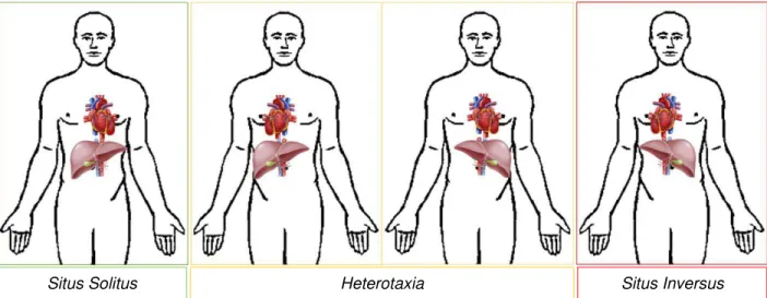
Although most vertebrate animals are bilaterally symmetric on the outside, they exhibit highly conserved LR asymmetries on the inside, mainly visceral organs regarding their position and morphology. Each thoracic and abdominal structure is located to one side or the other of the longitudinal midline, for instances the heart as well as stomach and spleen are positioned on the left side while the liver and gall bladder are on the right side. This arrangement, named situs solitus, corresponds to the normal anatomic left-right relation of the asymmetric viscera and such orientations are essential for their functions. According to epidemiological studies, situs anomalies are rare with an overall frequency estimated at 1 in 10 000 human births (Lin et al. 2014) which may result in significant morbidity and mortality. Reversal of visceral organ position can be either total or partial (Figure1.1). The complete reversal of all organs position, termed as situs inversus, can be isolated or may occur in combination with other abnormalities (Lamver et al. 2016). Whereas heterotaxia refers to the displacement of some organs but not all of them and represents the worst case scenario causing serious health problems due to the abnormal arrangement of the abdominal-thoracic organ-vessels in relation to each other (Shapiro et al. 2015).
Figure 1.1: Organ situs.
Schematic representation of the heart and liver position. In a situs solitus situation the heart is positioned on the left side of the chest, while the liver is located on the right side. Situs inversus is the complete mirror-imaged of situs solitus. Heterotaxia involves any laterality defects in the thoracic-abdominal internal organs with respect to the LR body axis.
During early embryogenesis, the temporally synchronized establishment of tissues that delineates the orientation and polarity along the three body axes – anteroposterior (AP), dorsoventral (DV) and left-right (LR) axes - coordinates the patterning of the body plan in vertebrates. The AP and DV axis are first patterned by Wnt and BMP (Bone Morphogenetic Protein family) gradients. And then the LR axis is established and oriented orthogonally to the pre-existing DV and AP axes (Niehrs 2004).
3
Although the mechanisms underlying AP and DV breaking symmetry have been studied exhaustively with the advent of molecular genetics, the initial steps of LR asymmetry patterning were, until a few decades, completely unknown. Through recent molecular and genetic studies, several discoveries have led to uncover mechanisms responsible for the generation of LR asymmetry patterning as well as genes with LR asymmetric expression, which propagate and translate the LR information into asymmetric organogenesis during development. These mechanisms are largely conserved among vertebrates, although some diversity has been identified.In the 1970s, Afzelius had identified cilia defects in Kartagener Syndrome patients, with respiratory difficulties, resulting from immotile cilia in the trachea, male infertility stemmed from sperm tail immotility, and frequently with situs inversus. This evidence suggested for the first time a link between visceral LR asymmetry establishment and cilia motility within an embryonic tissue (Afzelius 1976). Furthermore, Supp and colleagues later showed that inversus viscerum (iv) mouse, which result in LR complete inversion of fifty percent of homozygote embryos, carry a missense mutation on the ciliary axonemal dynein heavy-chain gene, left-right dynein (ldr) (Supp et al. 1997; Okada et al. 1999). A targeted mutation in left-right dynein gene showed similar phenotypes to iv mutants, confirming that lrd is required for normal LR development and also that lrd mutations leads to paralyzed cilia as a result of defective axonemal dynein motors (Supp et al. 1999). In addition, approximately half of live-born Kif3a or Kif3b null mutant mice showed LR laterality defects, resembling the phenotype of Kartagener Syndrome patients. These mutants were deficient in genes encoding two kinesin proteins, which normally form a motor protein complex ATP-dependent that track along microtubules, being involved in the trafficking of several cargoes within the cell and in ciliogenesis through intraflagellar transport. Thus in kif3 mutants, assembly of motile cilia is impaired (Nonaka et al. 1998; Takeda et al. 1999).
Such evidence that cilia-beating movement is involved in LR breaking asymmetry drew attention to this organelle, which has emerged as a key organelle in several physiological and developmental processes (Singla and Reiter 2006). Investigators have now identified a staggering correlation between human genetic diseases and dysfunctional ciliogenesis or cilia function.
1.2. Cilia
4
organelles have been associated with several cell functions as cellular motility, signal transduction, regulation of intracellular calcium levels, Wnt and Hedgehog signaling pathways (Fliegauf et al. 2007). Cilia have a complex ultrastructure with compartmentalization of molecular components that combine in functional modules, such as a basal body, transition zone, axoneme, ciliary membrane and the ciliary tip, comprising more than 650 proteins (Ishikawa and Marshall 2011).Depending on the cell type from which cilia protrude, ciliogenesis can lead to the formation of two different structures within the axoneme. The typical conformation is mentioned as “9+2”, since the nine doublet microtubules, organized in a circle, surround a central pair of singlet microtubules covered by the cell membrane. While another group of cilia have a “9+0” microtubule structure, meaning that they do not have a central pair of microtubules. At the ultrastructure level, each doublet microtubule itself consists of two conjoined tubulins, α and β tubulin heterodimers, making a total of 13 protofilaments of α tubulin and 10 protofilaments of β tubulin. The tubulin can undergo several post-translational modifications, as acetylation, glutamylation and glycylation, which have implications on ciliary assembly and motility (Ishikawa and Marshall 2011). And like the cytoplasmic microtubules, doublet microtubules polymerize with the plus end growing towards the ciliary tip.
1.2.1. Types of Cilia
Although all cilia types share the basic structural units, the specialization of cilia within a certain cell type to perform a specific function has resulted in significant variations of structure and regulation. Conventionally, depending on cilia axonemal structure, function and ability to move, cilia are categorized into two classes: primary and motile cilia.
Primary cilia are single and typically short organelles that protrude from almost vertebrate cell types between cell division, including stem, epithelial, endothelial connective-tissue, muscle cells and neurons. The majority of these type of cilia has a “9+0” microtubule conformation (Figure 1.2). Since primary cilia are immotile, the lack of ability to move was associated with the absence of the central pair microtubules. However, studies discovered non-motile with the typical conformation of “9+2” microtubules structure, in vestibular cilia of the auditory organ (Sobkowicz et al. 1995). Therefore, primary cilia are unable to move due to the lack of dyneins arms, which comprise the molecular motor proteins that power the cilia-beating movement, along with other interconnecting multiprotein complexes (Kobayashi and Takeda 2012)
5
Signaling in the cilium coordinates key processes during development and in tissue homeostasis, including cell migration, differentiation and/or re-entry into the cell cycle, specification of the plane of cell division, and apoptosis. Primary cilia are specialized morphologically and molecularly in order to participate in numerous biological process ranging from mechanosensation (by bending of the cilium) and chemosensation (detection of a specific ligands, growth factors, hormones or morphogens) to the transduction of essential signaling cascades, including Hedgehog and Wnt signaling and Planar Cell Polarity pathways. Furthermore, cilia also respond to light, sound waves, temperature, osmolarity or gravity (Singla and Reiter 2006). This impact of primary cilia in signaling has been highlighted by its defective function on specific organ diseases and developmental disorders, commonly referred to as ciliopathies.Motile cilia are usually longer and they can be found as a single cilium per cell (motile monocilia) or as many cilia at high density at the surface of the cells (multiple motile cilia) moving in a coordinated bending motion. To drive wave-like movement, motile cilia have a number of multiprotein complexes that interconnect the different components, within the microtubule core, including radial spokes, nexin links, central sheath and dynein arms. The two dynein arm classes, outer dyneins (ODA) and inner dyneins (IDA), encoded by different genes show distinct motor and ATPase properties that together generate the force necessary for rhythmic movement of the axonemes. Dynein arms are attached to the microtubules while the other components, mainly the central sheath apparatus and radial spokes, form a signal-transduction scaffold between the central pair microtubule and the dynein arms (Ibañez-Tallon et al. 2003).
6
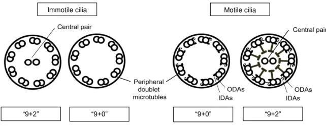
Thus, the simplistic classification of cilia as motile and immotile, according to their microtubule conformation, seems increasingly unsuitable as the number of exceptions rise. So far four cilia types have been identified and all of them have been associated with human disease, favoring the distinction of cilia into four subtypes (Figure 1.2): motile “9+2” cilia (such as respiratory and ependymal cilia); motile “9+0” cilia (embryonic nodal cilia); immotile “9+2” cilia (kinocilium of vestibular cells); and immotile “9+0” cilia (renal monocilia and photoreceptor-connecting cilia).Figure 1.2: Cilia structures in vertebrates.
A schematic representation of “9+2” and “9+0” axoneme cross-sections is shown. Both immotile and motile cilia can have either microtubule conformation, depending on their function within the cell type where they protrude. “9+0” structure comprise nine peripheral doublet microtubules whereas “9+2” conformation have an additional central pair of singlet microtubules. In addition, motility requires axoneme-associated dynein arms to generate the sliding of microtubules and thus motion. ODAs outer dynein arms. IDAs inner dynein arms. Adapted from Kobayashi and Takeda, 2012.
1.2.2. Ciliogenesis
Ciliogenesis is tightly coordinated with cell cycle progression and differentiation (Avasthi and Marshall 2012). During interphase, centrioles move to the plasma membrane and from a plasma membrane-associated foundation where the basal bodies are formed, docked onto actin-rich cortex and fused with the membrane (Ishikawa and Marshall 2011). Basal bodies position and their orientation dictates the alignment of the resulting cilia, creating an anchor and a template for the nucleation of the axoneme. After that, nine triplets of microtubules begin to form beneath an extension of membrane until the transition zone wherein nine axonemal doublets microtubules project radially around a central pair of singlet of microtubules, giving rise to the ciliary axoneme (Figure 1.3) (Mitchell 2007).
Motile cilia Immotile cilia
7
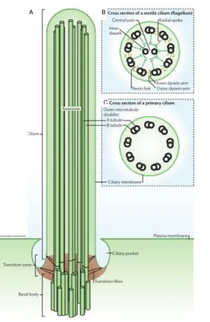
Figure 1.3: Cilia architecture.
Schematic diagram of a cilium: the cilium
(green) is produced once per cell and
extends from the basal body, forming the
transition zone, the axoneme and the ciliary
tip (A). Cross-section diagrams of a typical
motile cilium (which is identical to a
flagellum) (B) and a non-motile primary
cilium (C). Adapted from Ishikawa and
Marshall, 2011.
Since ribosomes are missing from the cilium, all proteins are synthesized in the cytoplasm and carried across the ciliary compartment and along the length of the axoneme by a bidirectional intraflagellar transport (IFT) (Vincensini et al. 2011). Proteins loaded onto the IFT particles are trafficked along the polarized microtubules, between the doubles and the membrane, from the basal body to the ciliary tip (anterograde movement) and in the opposite direction (retrograde movement). In the anterograde movement of cargo proteins, IFT is powered by heterotrimeric motor kinesin of the kinesin-2 family, whereas the retrograde movement is catalyzed by dyneins of the cytoplasmic dynein 2 family (Figure 1.4). It has been suggested that the transition zone acts as docking site for IFT particles and their motors, as well as it regulates the trafficking of proteins into and out of the cilium (Fisch and Dupuis-Williams 2011).
A B
8
Apart from the diverse functions of cilia, in response to cell cycle progression, cell differentiation or cellular stress, cilia can be shortened or reabsorbed, where the disassembled ciliary components return to the cell body for recycling or degradation.Figure 1.4:Intraflagellar transport machinery.
The canonical anterograde intraflagellar transport (IFT) motor, heterotrimeric Kinesin-2, transports IFT complexes A and B, axonemal proteins and cytoplasmic dynein 2 (previously known as cytoplasmic dynein 1b) to the tip of cilium. During this anterograde motion, Kinesin-2 is active and the retrograde motor, cytoplasmic dynein 2, is somehow kept inactive to allow smooth processive anterograde movement. At the tip of cilium, anterograde IFT trains release axonemal proteins and rearrange their conformation for retrograde IFT. Cytoplasmic dynein 2 is activated and transports retrograde IFT trains to the cell body. Figure and legend from Ishikawa and Marshall 2011.
1.2.3. Dynein-mediated motility
As previously mentioned, cilia have an intrinsic structure within their axoneme, that contains all the components necessary to generate and propagate waveforms. Although the path by which signals are propagated through the axonemal superstructure is complex and not yet well understood, ultimately cilia movement is powered by the molecular motors present in dynein arms.
Dyneins are highly complex macromolecular systems containing more than 20 different protein subunits, which are preassembled in the cytoplasm to generate fully assembly-competent dynein particles. An example of an assembly factor being ccdc151 which is a coiled coil domain containing 151 gene (Jerber et al. 2014). During the cilium growth, axonemal dyneins are transported from the cytoplasm into the
9
Thus dynein arms convert the chemical energy, released by the ATPase activity of heavy chain molecules, into mechanical work through the sliding movement of doublet microtubules, to produce the beating of the cilium. The intermediate and light chain components allow the motors to be regulated by external factors, such as calcium levels, phosphorylation status and the mechanical state or local curvature of the motors themselves (King 2016).Dynein arm function is dependent on the integrity of many dynein components. Studies using Chlamydomonas have reported so far 30-40 different axonemal dyneins. These dyneins combine to form
two significantly different dynein arms, in subunit structure content and arrangement within the axoneme (reviewed in Kamiya 2002). Nevertheless, both rows of dynein arms are capable of generating motive force. The outer dynein arms (ODA) are positioned proximally to the cilia membrane, being responsible for increase cilia-generated propulsive force output (cilia beat frequency), while the inner dynein arms (IDA) are proximal to the central pair and they are required the proper control necessary to generate and propagate specific ciliary waveforms (Brokaw and Kamiya 1987, Kamiya et al. 1991). In Chlamydomonas, the IDA and ODA are spaced as specific liner repeats of 96 nm and 24 nm, respectively, along the axis of each doublet microtubule (Goodenough and Heuser 1985).
One of the most highly conserved component of axonemal ODA is DNAL1, which stands for axonemal dynein light chain 1, known as LC1 in Chlamydomonas. New insights on this protein came from analysis of Chlamydomonas oda2-t mutants, that express a truncated form of γ-HC, one of the three heavy chain motors. In this mutant, LC1 is not present, unlike the other components (Liu et al. 2008). Like LC1, DNAL1 physically interacts with DNAH5, the human orthologue of the Chlamydomonas ODA γ-HC (Horváth et al. 2005). And when dnal1 is mutated leads to the absence of ODAs within the cilia causing PCD (Mazor et al. 2011). PCD patients with dnal1 mutations present situs inversus, showing that DNAL1 is also important
10
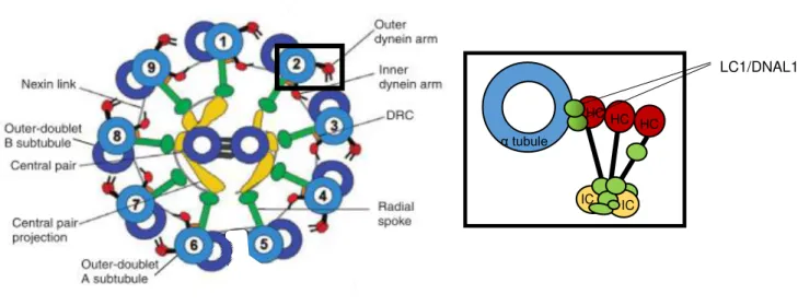
central pair to determine bend direction, shape (motion pattern) and speed in response to specific chemical inputs (Smith and Yang 2004)(Figure 1.5).Figure 1.5: Schematic diagram of the motile ciliary and flagellar axoneme.
The cross-section illustrates the “9+2” conformation have nine doublet microtubules linked to two outer and inner dynein arms, the dynein regulatory complex (DRC) and the radial spokes, that connect it to the central pair apparatus (central pair projection plus central pair microtubule). Nexin link connect the adjacent doublet microtubules. The expanded view of the ODA schematically depicts several light (in green), intermediate (IC, in yellow) and heavy (HC, in red) chains, where two LC1/DNAL1 proteins link the heavy chain to the α tubule (in blue). Adapted from Kobayashi and Takeda 2012.
The sliding of the doublet microtubules relative to one another constitutes the basic movement-inducing interaction in the cilium structure. Dynein-generated forces cause this sliding, which is consequently converted to axonemal bending by the restraining influence of the basal bodies that anchor the axonemal microtubules to the cell.
Although much has been learned about dyneins in terms of their composition and location, there remain many outstanding questions concerning how they are assembled, transported and located at precise sites within the cilium. In addition, the dynamics of the dynein motor itself need to be better understood.
1.2.4. Ciliopathies
Ciliated cells are present in almost all organs throughout the human body, with a nearly ubiquitous appearance among the vertebrates. Adding to the fact that cilia exert innumerous functions during development, tissue morphogenesis and homeostasis, it is not surprising that cilia defects lead to an inadequate function of these tissues. Consistently, cilia dysgenesis and dysfunction have been associated to many different human disorders, generally named as ciliopathies. These cilia-related diseases can either involve single organs or can occur as multisystemic disorders with phenotypically variable and
LC1/DNAL1
IC IC
HC HC HC
11
overlapping disease manifestations. Consequently, these diseases have been associated with developmental defects affecting the central nervous system and the skeleton, reproductive system dysfunction, chronic airway diseases, cystic disorders of the kidney, liver and pancreas, defects in vision, smell and hearing and even in oncogenesis (Fliegauf et al. 2007).Regarding the immotile cilia, defects appear to underlie a broad range of phenotypes, probably due to their nearly ubiquitous presence and their emerging role in signal transduction. Some major ciliopathies include Polycystic Kidney Disease (PKD), Bardet−Biedl Syndrome (BBS), Joubert Syndrome, Oral-Facial-Digital Syndrome, Alström Syndrome or Meckel Gruber Syndrome (Fliegauf et al. 2007, Hildebrandt et al. 2011) .
PKD or Autosomal Dominant PKD (ADPKD) is the most common potentially lethal disease in Europe, affecting 1 person in 1000. The disease hallmark is the development of hundreds of microscopic fluid-filled cysts in the kidney, which grow exponentially and continuously causing loss of normal renal tissue. ADPKD is caused by loss of function mutations in either Pkd1 or Pkd2, which encode the proteins polycystic kidney disease-1 and polycystic kidney disease-2, respectively. These two proteins interact with each other forming a complex localized in the cilia of the renal epithelial cells (Qian et al. 1997, Nauli et al. 2003) which is thought to be required to sense the fluid flow inside renal tubules. The mechanical stimulation of Pkd2, a calcium channel, leads to the influx of extracellular calcium, probably to regulate cell growth and differentiation. Recently, Kim and colleagues (2016) suggested that Pkd1/Pkd2 complex actively respond to non-canonical wnt ligands during kidney development, where Pkd1 is likely to function as a co-receptor. Consequently in ADPKD, the defective wnt-induced Pkd1/Pkd2-mediated calcium signaling would adversely affect multiple processes, contributing to cyst initiation/formation (S. Kim et al. 2016). Since polycystins are widely expressed, wnt/Ca²⁺ signaling is thought to mediate responses in other tissues rather than only in the kidney. Such assumption is supported by the wider phenotypic spectrum caused by Pkd1 and Pkd2 mutations compared with the phenotypic spectrum of ADPKD patients.
12
phenotypic movement of cilia can vary: when both ODA and IDA are affected, the majority of cilia are immotile; while IDA defects cause abnormal cilia beating pattern with reduced beating amplitude. Also anomalies in the radial spokes can lead to abnormal cilia beating (Chilvers et al. 2003). PCD is a group of heterogeneous disorders and so far more than 30 genes have been implicated as causative of these disease, which is predicted to explain roughly 70% of PCD cases (Horani et al., 2016).1.3. Left-right organizer
In 1976, after the movement of cilia in a, yet to discover, embryonic epithelial tissue was linked to LR asymmetry (Afzelius 1976), several studies suggested that the node in mouse embryos was indeed the structure responsible for the LR axis formation, being designated as left-right organizer (LRO). Motile monocilia were observed on the ventral surface of the node in early developed embryonic mice (Sulik et al. 1994, Bellomo et al. 1996) and the gene encoding the left-right dynein, lrd, was found to be specifically
expressed at the node, providing randomized situs when mutated (Supp et al. 1997). Ultimately, node ablation disrupted LR positioning at early organogenesis of the mouse embryos (Davidson et al. 1999). The node is a quiescent structure located at the anterior tip of the primitive streak in mouse embryos, flanked by endoderm and the lateral plate mesoderm (LPM). The cup-shaped ventral node contributes to the formation of the notochordal plate and the floor plate (Sulik et al. 1994). Ultrastructural studies have shown that monocilia present on the ventral side of the node rotate in a counterclockwise direction, suggesting that a fluid flow is necessary for establishing LR asymmetry (Nonaka et al. 1998, Okada et al. 1999). Nonaka and co-workers demonstrated a leftward flow of embryonic fluid, which they called nodal flow, as the earliest LR asymmetric event in mouse development. Furthermore, they provided strong elegant arguments for the pivotal role of mechanical nodal flow in the patterning of LR asymmetries, through a culture system in which embryos grow under artificial constant flow (Nonaka et al. 2002). Applying a rapid rightward flow in WT embryos, which should develop a normal organ situs, resulted in reverse direction of observed nodal flow and consequently most of the embryos exhibited complete reversal of nodal and pitx2 (LR markers of the LPM that precede organ situs). In opposition, when an externally leftward flow was applied in mutant embryos, which would normally lead to inverted LR orientation, it was sufficient to completely rescue the phenotype. For instances, the randomized LR patterning exhibited by the impairment of motile cilia in iv mutants was restored when the embryos were cultured under an external artificial leftward flow (Nonaka et al. 2002).
13
In zebrafish, the left-right organizer homologue of the mouse node is the Kupffer’s Vesicle (KV). Thus, the KV is a ciliated organ of asymmetry in the zebrafish embryo that initiates LR development of the brain, heart and gut (Essner et al. 2005), and it is conserved among teleost fishes.Cilia within the zebrafish KV arise from a group of approximately two-dozen of dorsal surface epithelial cells, known as dorsal forerunner cells (DFCs). These cells migrate at the leading edge of the embryonic shield throughout gastrulation and, ultimately DFCs cluster and undergo a mesenchymal-to-epithelial transition to differentiate into KV cells. Epithelial KV cells form a monolayer ellipsoid vesicle with fluid filled lumen at the tail region. During early somite stages, the KV rapidly expands as each cell forms and elongates a single cilium from their apical surface facing the lumen (Matsui and Bessho 2012).
Consistently with being analogous to node cells, KV cells express the left-right dynein-related 1 gene (lrdr1), the mouse homologue of the lrd gene. When lrdr1 is knocked down, the motile cilia-driven fluid flow is impaired, which consequently alters LR development (Essner et al. 2005, Kramer-Zucker et al. 2005). Although the mouse and zebrafish LRO architectures are different, the net fluid flow in zebrafish KV is analogous to the nodal flow in the mouse, producing a leftward counterclockwise movement essential for LR patterning (Okabe et al. 2008). In addition, two populations of cilia, one motile and other immotile, were observed recently within the KV-lining cells (Sampaio et al. 2014) in agreement to McGrath and colleagues previous report in the mouse node (McGrath et al. 2003). Nevertheless, cilia within these two organizers have different conformations. Mouse node motile cilia are widely distributed through the ventral node and have a “9+0” structure, which generate an almost perfect circular motion by stiff cilia. As for the motile cilia within KV cells, they show both a bending and rotational motion, and present a ”9+2” microtubule conformation. The fish cilia make a cluster on the anterior dorsal roof, in order to produce the directional flow. The presence of immotile cilia in the zebrafish KV supports the hypothesis that they may function as sensors of flow, as described for crown cell cilia of the mouse node (Yoshiba et al. 2012).
1.4.Two left-right models
The leftward fluid flow is the first LR asymmetric event and once it is settled leads to the second step which is sensing the flow. Two models have been proposed to explain how cells recognize the flow and translate the physical left-sided information into gene expression. Each model is based on different cilia properties, such as chemosensation and mechanosensation.
1.4.1. Morphogen model
14
be the determinant for both sides, where the left side determinant activated the expression of the left side-specific genetic pathway, while the right sided determinant inhibits its expression. Thus the nodal flow would maintain a concentration gradient of the morphogen along the LR axis in the node (Okada et al. 1999, Okada et al. 2005). Tanaka and colleagues’ work added important evidence to this model with regard to the identity of the putative morphogen. The authors observed flowing material composed by a lipid core and an outer membrane, which they called nodal vesicular parcels (NVPs). NVPs were described as being released from dynamic protruding cilia into the flow and then broken by crashing into cilia or cells, thereby releasing their contents in proximity to the left wall of the node. Tanaka and co-workers have shown that NPVs carry Sonic Hedgehoc (Shh) and Retinoic acid (RA), which are known to act synergistically. NVPs release was dependent on fibroblast growth factor (FGF) signaling (Tanaka et al. 2005). However, Cartwright and colleagues took these observations into account, modeled the movement of NVPs and showed that the flow could indeed cause them to accumulate on the left side of the node, for proper symmetry breaking. However, they argued that due to properties of the nodal flow there could not be any impact process involved in the NVPs rupture by cilia nor by the node wall. Instead, they predicted that a yet undiscovered biochemical mechanism that actively ruptures NVPs must exist, presumably chemical in nature (Cartwright et al. 2007).Despite these interesting insights, several observations argue against this morphogen hypothesis. Although critical diffusivity indicates only proteins with mass higher than 20 kDa (kilo Dalton) can be directional transported, supporting the Shh and RA as morphogens, genetic analysis of mutants does not provide convincing support of their roles as nodal flow morphogens (Vermot et al. 2005, Zhang et al. 2001).
1.4.1.1. Revisiting the Chemosensation
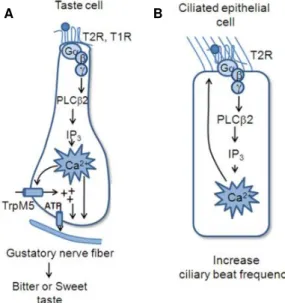
In airways epithelial cells, Shah and colleagues (2009) discovered that not only taste receptors were expressed but also the main downstream effectors of the taste sensing pathway. Different Tas2 receptors were expressed throughout the ciliary membrane, while the G protein α-gustducin and the channel TRPM5 were localized within the cilia. As for phospholipase Cβ2, its expression was detected below cilia in the apical portion of the cell (Figure 1.7). The authors showed that bitter compounds induced both a transient, dose-dependent increase in intracellular calcium in ciliated cells as well as an increase in ciliary beat frequency. The response is thought to provide protection for the airway epithelium from noxious compounds and products of bacterial infection by speeding up the process of mucociliary clearance (Shah et al. 2009).
15
Such evidence reinforce the idea that motile cilia, highly enriched with receptors and channels, are capable to sense, interpret and transmit external environment information through chemical signals, supporting the chemosensory hypothesis of the morphogen model.The taste 1 and 2 receptors are part of the G protein-mediated system which is relatively complex comprising a receptor, a heterotrimeric G protein and an effector, which can be regulate independently by accessory proteins, soluble mediators or on the transcriptional level (Wettschureck and Offermanns 2005). G protein-coupled receptors (GPCRs) are seven transmembrane receptors that actively respond to several signals, including light, odor, chemokines, hormones, growth factors and neurotransmitters, thus affecting numerous cellular processes. Heterotrimeric G proteins function as molecular switches that activate intracellular signaling cascades in response to the stimulation of GPCRs, having a central role in cell biology. Heterotrimeric G proteins are divided in three subunits: alpha (α), beta (β) and gamma (γ), where the α-subunit controls the switching function through its own ability to cycle between an inactive GDP-bound conformation and an active GTP-bound conformation that modulates the activity of downstream effectors. Additionally, α-subunits can be distinguished into four families according to their structural and functional properties: Gs,Gi/Go, Gq/G11, and G12/G13, and βγ subunits form a functional unit to regulate ion channels, particular isoforms of adenylyl cyclase (AC), phospholipase C (PLC) and phophoinositide-3-kinase (PI3K). Therefore, heterotrimeric G proteins modulate cyclic AMP (adenosine monophosphate) through the enzymes adenylyl cyclase and phosphodiesterase (PDE) and lead to an increased calcium release from intracellular stores, via phosphoinositide-3-kinase enzyme (Oldham and Hamm 2008). Several components of the G protein mediated signaling are present in primary cilia, which have been associated with many sensory functions and also with ciliary defects (Hilgendorf et al. 2016).
16
Tas1 and Tas2 receptors and phospholipase Cβ2 orthologues have been found also in fish taste bud cells sharing a significant homology to each mammalian cognate taste receptor families (Ishimaru et al. 2005). Although the diversity of taste cells varies among species, the presence of the main components of taste sensing suggests a common mechanism of taste reception and an intracellular signal transduction pathway, at least at the level of effector molecules, among vertebrates (Matsumoto et al. 2013). Consistently, fish species respond to water-soluble chemical compounds and avoid a diet highly enriched with bitter components, where Tas2r are involved in avoidance feeding behaviors. However, phylogeny studies revealed a major exception concerning the G protein protein α-gustducin specialized gene for taste signaling in mammalians. No orthologue was found in several teleost and amphibian genomes, which could be due to the acquisition of gustducin gene by the land-living vertebrates or to the loss of it during evolution of teleost lineage (Oka and Korsching 2011). Nonetheless, two other G protein α subunits, gnaia and gna14, were discovered to be expressed in zebrafish taste-related tissues (Ohmoto et al. 2011). gnaia is a member of Gi α subunit class, which function as inhibitors of different adenylylcyclases, and gna14 is a member of Gq/G11 class, orthologue of the mammalian equivalent G14 α -subunit, that is thought to act as activator of phospholipase C isoforms. The outcome of these two G proteins is reduced cAMP levels, similar to the effect of G protein α-gustducin in mammalians, suggesting similar roles within taste sensing pathway.
Shah and Meyer’s findings (Shah et al. 2009, Meyer et al. 2012) suggest an unexplored possibility for taste sensing-related signaling in LR patterning, through the Chemosensation Model.
Figure 1.7: Diagrammatic illustration of differences in GPCR signaling effectors in the different cell types.
In both cases taste GPCRs activate the downstream PLC signal effectors, but the effects of increased calcium differ among the taste cells (A) and the ciliated epithelial cells (B). Adapted from Kinnamon 2012.
1.4.2. Two-cilia model
The second model was proposed by McGrath and colleagues (2003) upon the observation of two different populations of cilia in the mouse node, therefore they named it as the two-cilia model (Figure 1.6). The
17
authors reported that nodal cells in the center of the node had cilia expressing the left-right dynein (Lrd), the mouse orthologue of human DNAH11 (dynein axonemal heavy chain 11), whereas cilia of the surrounding horseshoe-shape ring cells were negative for the motor protein. Since left-right dynein is important for cilia movement, its presence or absence was interpreted as cilia capable or not to rotate. Hence, this model predicts that motile cilia in the center of the node generate the leftward fluid flow and the immotile cilia located at the node periphery are able to sensing it (McGrath et al. 2003, Tabin and Vogan 2003). This implies that a mechanosensor must be present in the ciliary membrane and that it undergoes a distortion in response to the external fluid shear stress, which can lead to the opening or closing of the conductive pathway through the channel. The force was envisaged to be applied either through cytoskeletal and extracellular matrix elements attached to the channel, or through membrane tension by the lipid bilayer itself (Sukharev and Corey 2004).1.4.2.1. Evidence for Mechanosensation
Pennekamp and colleagues (2002) reported distinct LR laterality defects in Pkd2 null mouse embryos (Pennekamp et al. 2002). These homozygous mutants fail to produce the polycystic kidney disease 2 (Pkd2), a calcium channel that if mutated in humans causes autosomal dominant polycystic kidney disease (ADPKD). Moreover, McGrath and colleagues also reported that all nodal cilia expressed Pkd2, suggesting that it could have a sensory role in the negative lrd cilia population (McGrath et al. 2003). An important cue to the mechanosensory model came from the finding that polycystic kidney disease 1 (Pkd1) and Pkd2 were expressed in primary cilia in kidney tubule cells, forming a complex that detects fluid flow and transduces it into an increase of intracellular calcium (Nauli et al. 2003). Nauli and colleagues proposed that Pkd1 functions as a mechanosensor that detects the bending of cilia induced by fluid flow, leading to conformational changes that in turn would open the Pkd2 channel. Pkd2, as a calcium channel, would then give rise to an influx of calcium that ultimately would alter gene expression, growth, differentiation and apoptosis.
18
In zebrafish, left-right patterning defects were also observed using Pkd2 morphants and mutants (Bisgrove et al. 2005, Schottenfeld et al. 2007). Motile and immotile cilia have also been found in the zebrafish KV (Sampaio et al. 2014) and Pkd2 is present in all zebrafish KV cilia along the axoneme and the basal body (Roxo-Rosa et al. 2015) reinforcing the idea that Pkd2 plays an important and conserved role in LR patterning among species. So far, no Pkd2 partner has been identified in the zebrafish KV.Figure 1.6: Current models for establishing LR asymmetry.
(A) The nodal vesicular parcel (NVP) model
predicts that vesicles filled with morphogens
are secreted into the fluid and are
transported to the left side by nodal flow,
where they are smashed releasing content.
(B) In the two-cilia model argues that motile
cilia in the node create a leftward nodal flow
that is mechanically sensed through passive
bending of non-motile sensory cilia at the
periphery of the node. Bending of the cilia on
the left side leads to a left-sided release of
Ca2+ that initiates the establishment of body
asymmetry (Fliegauf et al. 2007).
Both models can explain certain aspects of LR establishment, but a lot of questions remain unanswered, it is thus unclear which is the correct view of how nodal flow is interpreted by the embryo. It is plausible that both chemosensation and mechanosensation can be correct, acting together to form a robust process from which results the proper LR development.
1.5. Asymmetric Gene Cascade
Once the nodal flow is transduced by the node cells, it then triggers a left-sided genetic signaling cascade in the node, (that is not well explored) which is later transmitted to the lateral plate mesoderm (LPM). The requirement of Pkd2 calcium channel in LR axis formation strongly supports that calcium signaling plays a crucial role between sensing the flow and the activation of asymmetric gene pathway on the left side of the embryo.
19
In mouse, Pkd2 was found to be required only in the immotile ciliated crown cells, at the node periphery, regulating calcium signaling, which was increased in both sides of the node (Yoshiba et al. 2012). Dynamic calcium signals in the node were observed to become gradually biased to the left side, during development (Takao et al. 2013). Similar Pkd2-dependent calcium oscillations were observed within zebrafish cilia that were transduced to the cytosol and surrounding cells on the left side of the KV (Yuan et al. 2015). These data suggest that both calcium signal amplitude and frequency may regulate asymmetricpatterning.
In mouse, two secreted proteins are expressed bilaterally in the node: the Nodal gene, which encodes a member of the transforming growth factor beta (TGFβ) (Collignon et al. 1996), and the Cerberus-like 2 (Cerl2) gene, Nodal antagonist. Upon the activation of the Nodal cascade signaling, Cerl2 levels become higher on the right side of the node and Nodal becomes restricted to the opposite side (Marques et al. 2004). Expression of Cerl2 appears to be the most direct outcome of the flow signal, hence it initiates Cerl2 mRNA degradation on the left side (Nakamura et al. 2012). Being a secreted protein, Nodal spreads out to the left side of the LPM, where exerts a positive feedback to induce its own expression and to activate its own negative regulators. Among these, the Lefty genes are crucial to prevent ectopic Nodal induction in the right side of LPM and to control the Nodal domain after initiation, where Lefty1 is expressed in the midline barrier and Lefty2 is expressed in the left lateral plate mesoderm. In addition, Nodal in the LPM regulates the expression of the downstream effector Pitx2, a key transcription factor responsible for the situs-specific organogenesis.
In zebrafish, the left-sided calcium increase through Pkd2 activates transiently CaMK-II, a Ca²⁺ /CaM-dependent protein kinase (Francescatto et al. 2010). CamK-II activation was observed only in cells lining the left sided functional KV, suggesting that CamK-II could enhance left sided Nodal-related protein, Southpaw (Spaw) in the node, or could influence the secretion of it into the LPM. Spaw and Dand5, a member of the Cerberus family, are the orthologues of Nodal and Cerl2 present in mouse node. Similarly to the genetic signaling cascade in the mouse, dand5 is first expressed bilaterally in KV. At 8-10 somite stage, in response to the fluid flow the dand5 expression pattern becomes stronger on the right side of KV (Lopes et al. 2010), where Dand5 antagonizes Spaw function and prevents spaw expression in the right LPM (Hashimoto et al. 2004). Despite in zebrafish spaw remains always bilateral in the KV, only on the left side of KV, Spaw can activate its own expression via an auto-regulation mechanism throughout the left LPM.
20
1.6. Project goalsThe main goal of this Master project was to investigate the molecular basis of biochemical and mechanical pathways activated by motile cilia in the zebrafish KV.
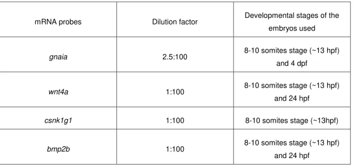
To test the chemosensory pathway, we studied several G protein couple receptors as well as the downstream effectors. This model is based on the presence of a morphogen flowing in the LR organizer, however there is yet no available technology sensitive enough to analyze the content of such small volume as is the fluid inside the KV. Therefore, we reasoned that the putative morphogen would trigger the chemosensory signaling through binding to a receptor expressed in motile KV cilia. We investigated the sweet taste receptors, tas1r1, tas1r2.2 and tas1r3, because they were found to have chemosensory functions in spermatozoa (Meyer et al. 2012), the bitter taste receptors, tas2r200.2 and tas2r201.2, as well as the downstream effector plcb2, and two other putative downstream effectors gnaia and gna14 based on their capacity of sensing chemical signals within motile cilia, as reported by Shah and colleagues (Shah et al. 2009). These genes were expressed in an unpublished microarray from Lopes lab that analyzed the genes specifically expressed in the KV cells in WT embryos.
Regarding the mechanosensory model, we tested ten different genes potentially involved in LR signaling pathway, including wnt signaling (wnt4a, csnk1g1), bmp signaling (fsta, bmp2b, smad4), calcium binding signaling (cacng2a) and others (foxi1, crb2a, azin1a, rragca, dlk1). We wanted to find more asymmetric genes that could be intermediate players between flow, pkd2 pathway and dand5 expression, a link not fully characterized. For this propose we used the previously mentioned microarray dataset for genes specifically expressed in the KV cells of WT embryos and Pkd2 morphants. The candidate genes were all expressed in both analyses and in order to test for asymmetries we did an in situ hybridization screen for their expression pattern in the KV. The two-cilia model gave rise to another interesting question: what determines the difference between the two cilia populations involved in LR axis formation. In the mouse node, the two types of cilia are proposed to be distinguished by the presence or absence of left-right dynein - Lrd. Where motile cilia are positive for Lrd and immotile cilia are negative for the Lrd (McGrath et al. 2003). However, in zebrafish KV cells we noticed that the percentage of motile and immotile cilia varied
during the LR development. As the total number of cilia remains the same, we can observe that 80% of immotile cilia become motile. We hypothesized two possible explanations for this: (1) all cilia undergo the same ciliogenesis process, being structural equal to each other, and over time a specific molecule or signal switches the motility on in 80% of cilia. (2) 20% of cilia, the true immotile cilia, may have different ultra-structure, where plausibly the dynein motor proteins are absent. To test this, we evaluated the localization of Dnal1, an outer dynein arm component that links the motor proteins to the microtubules, within the KV cilia.
22
In the best interest to answer our defined objectives, we performed different experimental procedures as described below.2.1. Microarray Analysis
In order to identify genes which expression is regulated by Pkd2 function, before I joined Lopes’ lab they decided to do a very strict tissue specific mRNA profiling analysis by microarray. They used non-injected embryos and pkd2 knockdown morphants at 14 hpf (10 somites stage) to extract KV-specific RNA. For that purpose they used one Foxj1a: GFP zebrafish reporter line, which expresses the minimum promoter region of a forkhead domain-containing transcription factor (foxj1a) driving the green fluorescent protein (GFP) tag. This line labels the KV cells and slighty the neural tube cells in green and it was used to purify this cell population by Fluorescence Activated Cell Sorter (FACS). Therefore, they ended up with two collection tubes, one with the KV cell population marked by GFP and another with the remaining cells without GFP fluorescence. After that, they extracted RNA from control embryos and pkd2 morphants KV cells and the transcripts were accurately detected as their expression were measured.
The aim of this transcriptomic study was to catalog and quantify the RNA content of KV cells, regardless of their structure or function, like a snapshot of all activated genes. By the time I came to the lab, a long gene list had already been compiled, thus we selected a shorter list based on literature of target genes that could have a role within left-right patterning.
We looked at genes whose expression was altered by the knockdown of pkd2 as possible targets of Pkd2 function. The rational was that by changing pkd2 expression levels, the calcium signaling through cilia would be impaired and the corresponding downstream pathway would be affected. From this point of view, we selected the following genes: antizyme inhibitor 1a (azin1a, NCBI Gene ID: 550230); casein kinase 1, gamma 1 (csnk1g1, NCBI Gene ID: 494092); forkhead box i1 (foxi1, NCBI Gene ID: 353313); follistatin a (fsta, NCBI Gene ID: 100004116), bone morphogenetic protein 2b (bmp2b, NCBI Gene ID: 30632); crumbs homolog 2a (crb2a, NCBI Gene ID: 723994); wingless-type MMTV integration site family, member 4a (wnt4a, NCBI Gene ID: 30123), ras-related GTP binding Ca (rragca, NCBI Gene ID: 573492), SMAD family member 4 (smad4, NCBI Gene ID: 559111), calcium channel, voltage-dependent, gamma subunit 2a (cacng2a, NCBI Gene ID: 393614) and protein delta homolog 1 (dlk1, NCBI Gene ID: 101883341). We named them as Pkd2-dependent target genes.
23
type 1, member 1 (tas1r1, NCBI Gene ID: 654781); taste receptor, type 1, member 2, tandem duplicate 2 (tas1r2.2, NCBI Gene ID: 664686); taste receptor, type 1, member 3 (tas1r3, NCBI Gene ID: 562318); taste receptor, type 2, member 200, tandem duplicate 2 (tas2r200.2, NCBI Gene ID: 562318); taste receptor, type 2, member 201, tandem duplicate 2 (tas2r201.2, NCBI Gene ID: 100333330); guanine nucleotide binding (G protein), alpha inhibiting activity polypeptide a (gnaia, NCBI Gene ID: 323509); guanine nucleotide binding protein (G protein), alpha 14 (gna14, NCBI Gene ID: 445297) and phospholipase C, beta 2 (plcb2, NCBI Gene ID: 569376). These genes were considered as Pkd2-independent target genes.A diagram with the main steps involved in this microarray dataset is presented below.
Figure 2.1: Diagram of microarray setup.
KV cells labeled with foxj1a:GFP from pkd2 atgMO injected embryos, 5-mismatch MO injected embryos and their non-injected siblings were sorted by FACS. A 5-mismatch MO was used as a control of pkd2 atgMO injection, specificity and toxicity. The expression levels of activated genes in KV cells were detected by the microarray analysis, and then we compared both transcriptomes. Genes that were found significant and differentially misregulated in Pkd2 injected embryos were grouped in a Pkd2-dependent targets list, while genes that were not differentially expressed were listed as Pkd2-independent targets.
2.2. Zebrafish breeding
Zebrafish general maintenance and breeding, and egg collection were performed at the CEDOC fish facility (Lisbon, Portugal), according to standard procedures described in the zebrafish book by Westerfield (1995).
foxj1a:GFP
Inject pkd2 - MO
Non-inject
FACS sorting Microarrays analysis
KV
24
For zebrafish breeding, we used mating tanks that have a removable insert with holes that allow eggs to fall through. This feature protects the eggs from being eaten by adult fish. Zebrafish adults are collected from the main tank system and are placed in a mating box during the afternoon or evening. Usually, in each mating tank we put one male and two females separated with a divider. This transparent partition avoids fishes from breeding before the desired time and allows the visual contact initiating the courtship behavior between the male and females. In the following morning, we remove the divider and reduce the water level, which favors the mating ritual behavior to continue. As the males chase the females around the tank, they stimulate spawning of eggs while releasing sperm into the water for external fertilization. After a few minutes, adults are removed from the tank by using a net and placed again into the main tank system. The eggs are collected and placed in one or more Petri dishes, in order to achieve an optimal density of 50 – 100 embryos per dish, with embryonic medium (5 mM NaCl, 0.2 mM KCl, 0.3 mM CaCl2, 0.3 mM MgSO4, ddH2O – pH 6.5). Embryos stayed in temperature-controlled incubators either at 25ºC or 28ºC until the desired developmental stage for our experiments.Wild type AB line and transgenic line Arl13b: GFP zebrafish were used for this thesis purpose and embryos were staged according to Kimmel et al (Kimmel et al., 1995). The period of embryogenesis is given in hours post-fertilization (hpf) and days post-fertilization (dpf) according to morphological features. For somite-staged embryos, we also took into account the number of somites to infer their developmental time.
2.3. Quantitative Real-Time Polymerase Chain Reaction (qPCR)
The qPCR analysis associates PCR amplification and detection into a single step, which allows to quantify gene expression even when we have minute amounts of nucleic acids. Amplicon recognition is achieved by monitoring the accumulation of fluorescent signal during each cycle.
For Pkd2-independent target genes, we wanted to evaluate the low transcription levels that we obtained with the microarray dataset. In order to do that and to confirm taste receptors and their downstream effectors expression, we used the qPCR technique in WT embryos. Regarding Pkd2-dependent target genes, the qPCR was also done, in order to compare and validate the gene expression levels from the microarray data between wt and Pkd2 knockdown embryos.
25
To design the primers for cDNA amplification, we used the Primer-BLAST bioinformatics tool from NCBI, which allow us to choose complementary sequences with exon-exon boundaries. This is an important feature because primers will bind specifically to messenger RNA and not to DNA sequence that may have been left in the sample. Primer sequences are described in Table 2.1. Regarding the reference genes, rpl13a (ribosomal protein L13a, NCBI Gene ID: 560828,) and eef1a1a (eukaryotic translation elongationfactor 1 alpha 1a, NCBI Gene ID: 336334), the primers used had already been optimized in our lab.
Table 2.1: Nucleotide sequence of forward and reverse primers for each gene used in qPCR assays and respective probe length (bp).
Code Primer Fw Primer Rv Probe
length
rpl13a TGACAAGAGAAAGCGCATGGTT GCCTGGTACTTCCAGCCAACTT 117
eef1a1a CCTTCAAGTACGCCTGGGTGTT CACAGCACAGTCAGCCTGAGAA 182
tas1r1 GTGCGCTCTTTTCAGGTTGTTTGT AGCATGCGGAGAACAGAGATGAA 146
tas1r2 CACTCTCTTGCCCTGGCATAAC GGGGTCCCGAGTATAATCCACATA 154
tas1r3 TCTGTCCAGTTCCCTTCAGATTGT TAGGCCATACCAGCCGCATAGT 148
tas2r200.2 GGAGTTGGGGTTGTCGGTGTTT TGCCAACCACCATGCTGAGAATA 163
tas2r201.2 CCCACCTGTCTGGCTGTACATT CAAGGCCATCGGACTGGAACAA 126
gnaia GGCCGCCTGAGGATTGACTTT CCCTCCAGAGCCGCTTGATTAC 133
gna14 CGCTGAGTGCCATGCAGTCTAT GAATCCCGCTGTCGCTCCATAA 177
pbcβ2 AGCTCATCGGTTTGCCCAAAGA GGGATGATGCGGTGACCAATGA 190
wnt4a GACGCAAGTTTTGCGCTGAGAA GCTTGGCCAGGTATAGCCAGTT 177
csnk1g1 CTCAGCACCAACCCTTGAGGAA AAACAGCAGCACTTGGCCTCAT 145
bmp2b AATCGGCAGTGGTCCCTCAGTA GCCTCGAAAGCCTCTTCGTGAT 166
fsta ACGCCTACTGTGTGACATGCAA TCATCGCATGACTTGGCCTTGAT 184
rragca GTCATCGACGCTCAGGATGACTA AGCTTGCATCTGCCAGGTCAT 196
crb2a GTTCCTGTTGGCCAGGTTTTGAA TCCTGGTTGGCACTGACATACAT 179
foxi1 AGTTGCACGGGATGAGGATGAT TAAGCGCGTCCTCGGATTTCA 152
26
The qPCR reactions were performed in a LightCycler ® 96 Real-Time PCR system (Roche), using FastStart Essential DNA Green Master (Roche).We performed a qPCR validation assay for each primer pair with a serial dilution of cDNA (1:10, 1:1, 1:0.1) with three replicates for each concentration. A negative control (water) reaction was also done for each primer in order to evaluate primer-dimers and possible contaminations. We used the following cycling parameters: pre incubation at 95ºC for 10min, three step amplification at 95ºC for 10s, 60ºC for 10s and 72ºC for 20s in 45 cycles, melting at 95ºC for 10s, 65ºC for 60s and 97ºC for 1s, and cooling at 37ºC for 30s.
Then we analyze the obtained melting peaks and standard curves to access primer annealing specificity and reaction efficiency, respectively. The melting curves represent melting peaks by plotting the first negative derivative of the fluorescence as a function of temperature (-dF/dT). Therefore, products of different lengths and sequences will melt at different temperatures and are observed as distinct peaks. If the chosen primers are specific, a single sharp peak appears. If we obtained more than one sharp peak that probably means that more than one amplicon was transcribed and means the designed primers are not suitable. Regarding the standard curve, it is constructed by plotting the log of the starting quantity of the template against the Cq values obtained (Taylor et al. 2010), to access qPCR efficiency. A 100%
efficient reaction represents the maximum increase of amplicon which is 2-fold per cycle. Efficiencies lower than 90% or higher than 110% are indicative of reaction problems that can cause artifact results.
All data obtained were normalized with the reference genes. In these experiments, we used the rpl13a and eef1a1a genes that were validated as reference genes for developmental time course studies of zebrafish. They have not only a high transcript abundance but also a high expression stability during embryonic development (Tang et al., 2007). Reference genes are important for proper normalization, in which they are responsible to compensate for the intra and inter-kinetic qPCR variations that result from basic difficulties during the method itself.
dlk1 TGTGAATCTCCCGGCGAGTGTA GGCAGTGTGTAGCAGTGCAGAT 163
cacng2a CCGTGCGAGCGTCTAGTATCTT GTTACTGAGGCCTGCAGACACGAA 149