José Guilherme Pereira de Almeida Santos
Licenciado em Ciências Biomédicas
Molecular tools to dissect the role of
Dmrt2a and Dmrt2b
in the left-right axis formation
in zebrafish
Dissertação para obtenção do Grau de Mestre em Genética Molecular
e Biomedicina
Orientadora: Maria Leonor Tavares Saúde, Professora Doutora,
Instituto de Medicina Molecular
Faculdade de Medicina da Universidade de Lisboa
Júri:
Presidente: Prof. Doutor José Paulo Nunes de Sousa Sampaio
Arguente: Prof. Doutora Solveig Thorsteidóttir
Vogal: Prof. Doutora Maria Leonor Tavares Saúde
iii
Molecular tools to dissect the role of
Dmrt2a and Dmrt2b
in the left-right axis formation in zebrafish
Submitted by
José Guilherme Pereira de Almeida Santos
Thesis to obtain the degree of
Masters in Molecular Genetics and Biomedicine
v
Molecular tools to dissect the role of Dmrt2a and Dmrt2b in the left-right axis formation in zebrafish
Copyright José Guilherme Pereira de Almeida Santos, FCT/UNL, UNL
vii
Acknowledgements
First of all I would like to thank my supervisor Leonor Saúde for taking the risk of giving me the chance to do this work. I would like to thank all your help and for making us believe that everything is going to work fine. I really enjoyed this year in UDEV.
I also would like to thank all my colleagues, Margarida Figueira, “buddy”, João I miss you so much :p, Rita Pinto this project is haunted haha, Ana Ribeiro espectacular, Rita Serrano and Susana Pascoal the forbidden bench, Lara and Sara Matos I promise I will free some space. A lot! Thank you all for helping me during this year.
And now the winners are… HAHA In no particular order, Margarida Pereira, who taught me so much and helped me put this work on the right track, Aida who helped me a lot with fish maintenance, crosses, and sexual dimorphism prediction LOL, and Sara Fernandes, for the wisdom and patience.
I also want to thank Rita Fior and Raquel Mendes for showing me last year how cool developmental biology could be.
Although their effort is not presented in this work, Andreia and Ana Farinho also helped me during this year. Napkins for science!!
ix
Abstract
We tend to view the vertebrate body as bilaterally symmetric, but in fact, this only happens from the outside. Internally, most of the organs from heart to liver are asymmetrically positioned. Skeleton and its associated muscles, symmetric structures of the vertebrate body, have its origins in the transient symmetric blocks of mesoderm called somites whereas the asymmetric morphogenesis of the internal organs is due to asymmetric gene expression in the lateral plate mesoderm (LPM).
Previous studies using Morpholino (MO) technology have shown that dmrt2a is involved in these two processes in zebrafish. When Dmrt2a levels are reduced, asymmetric gene expression in the LPM becomes randomized and symmetric gene expression in the presomitic mesoderm (PSM) is disrupted. The paralogous of dmrt2a, the fish specific dmrt2b has been shown to be involved in regulating asymmetric gene expression in the LPM as well.
Here we used the recent Transcription activator-like effector nucleases (TALENs) technology to generate dmrt2a and dmrt2b mutant alleles that will allow us in the future to uncover the downstream effectors of these transcription factors using high-throughput experiments. In addition, we overexpressed
dmrt2a at the one-cell stage to characterize asymmetry versus symmetry phenotypes.
The results show clearly the ability of TALEN technology to generate mutant alleles in zebrafish. Nevertheless, dmrt2a and dmrt2b homozygous mutants developed so far fail to recapitulate their previously described MO phenotypes which raise the question on what molecular mechanism(s) allow(s) zebrafish to cope with frameshift mutations.
The overexpression of dmrt2a shows that a time window of opportunity during which symmetric embryonic territories are able to respond to asymmetric signals does exist during embryonic development.
xi
Resumo
Olhamos geralmente para os vertebrados como sendo simétricos. No entanto, isto apenas é correcto de um ponto de vista exterior, visto que no interior dos vertebrados, a maioria dos órgãos desde o coração ao fígado estão posicionados de forma assimétrica. O esqueleto e os seus músculos associados, estruturas simétricas dos vertebrados, têm a sua origem em estruturas transientes chamadas de sómitos. Já a morfogênese assimétrica dos órgãos internos, deve-se à expressão genética assimétrica na mesoderme da placa lateral.
Estudos anteriores mostraram através do uso de Morpholinos que o gene dmrt2a está envolvido nestes dois processos em peixe-zebra. Quando os níveis de Dmrt2a são reduzidos, a expressão genética assimétrica na mesoderme da placa lateral apresenta-se randomizada, enquanto a expressão genética na mesoderme pré-somítica deixa de ser simétrica. O gene dmrt2a tem um parálogo no peixe, o dmrt2b.
Este último também está envolvido na manutenção da expressão genética assimétrica na mesoderme da placa lateral.
Neste trabalho usamos a recente tecnologia TALEN para criar alelos mutantes para os genes
dmrt2a e dmrt2b que nos vão possibilitar no futuro identificar os genes alvo destes factores de transcrição usando metodologias de larga escala.
Além disto, sobre-expressamos o gene dmrt2a no estádio de uma célula para caracterizar o fenótipo assimetria vs. simetria.
Os resultados claramente demonstram a capacidade da tecnologia TALEN para criar alelos mutantes em peixe-zebra. No entanto os mutantes homozigóticos desenvolvidos até agora, não revelaram um fenótipo semelhante ao que já havia sido descrito utilizando Morpholinos, o que lança a pergunta sobre qual será, ou quais serão o(s) mecanismo(s) que o peixe-zebra usa para lidar com mutações que alterem a grelha de leitura de um gene.
A sobre-expressão de dmrt2a mostrou que a janela de oportunidade durante a qual territórios embrionários simétricos podem responder a sinais assimétricos efectivamente existe durante o desenvolvimento embrionário.
xiii
Contents
Acknowledgements ... vii
Abstract... ix
Resumo ... xi
Contents ... xiii
List of Figures ... xv
List of Tables ... xxi
Abbreviations ... xxiii
Chapter 1 Introduction ... 1
1.1 Background ... 1
1.2 Left-right asymmetry within the vertebrate body ... 1
1.3 Bilateral symmetry of the vertebrate body ... 5
1.4 Symmetry versus asymmetry during development ... 7
1.5 The role of dmrt2a ... 8
1.6 Transcription activator-like effectors (TALEs) ... 9
1.7 Genetic engineering with TALE nucleases (TALENs) ... 10
1.8 TALENs assembly ... 11
1.9 Focus of this work ... 11
Chapter 2 Materials and Methods ... 13
2.1 List of Primers ... 13
2.3 Transformation of competent cells ... 14
2.4 Colony PCR ... 14
2.5 Cloning of dmrt2a and dmrt2b ... 14
2.6 TALENs design and assembly ... 15
2.6.1 dmrt2a and dmrt2b TALENs design ... 16
2.6.2 dmrt2a and dmrt2b TALENs assembly using the Golden Gate cloning system ... 17
2.7 Genotyping ... 18
xiv
2.7.2 NaOH genomic DNA extraction ... 19
2.8 Total RNA extraction ... 19
2.9 mRNA synthesis ... 19
2.10 Anti-sense mRNA probes synthesis... 20
2.11 Whole mount in situ hybridization ... 20
2.12 High Resolution Melting ... 21
2.13 Mutant zebrafish line ... 22
Chapter 3 Results ... 25
3.1 Generating zebrafish dmrt2a and dmrt2b mutant alleles with TALENs ... 25
3.1.1 Deciding on the TALEN target sequence ... 25
3.1.2 TALENs design and assembly ... 28
3.1.3 TALENs mRNA injection (Mosaic G0 generation) ... 30
3.1.3.1 Single TALEN pair mRNA injection ... 30
3.2.3.2 Multiple TALEN pair mRNA injection ... 35
3.2.4 Germline transmission of acquired mutations (Heterozygous F1 generation) ... 39
3.2.4 Homozygous mutants (F2 generation) ... 46
3.2.4.1 Failure of homozygous dmrt2a and dmrt2b mutants to recapitulate its respective MO phenotype ... 46
3.2 Overexpression study of the gene dmrt2a ... 48
Chapter 4 Discussion and Future Work ... 53
4.1 TALENs, as a new tool to generate mutant alleles in zebrafish ... 53
4.2 Zebrafish TALEN mutants phenotype versus zebrafish morphants phenotype ... 54
4.3 An overexpression analysis reveals a time window of action of dmrt2a ... 56
Chapter 5 Conclusion ... 59
xv
List of Figures
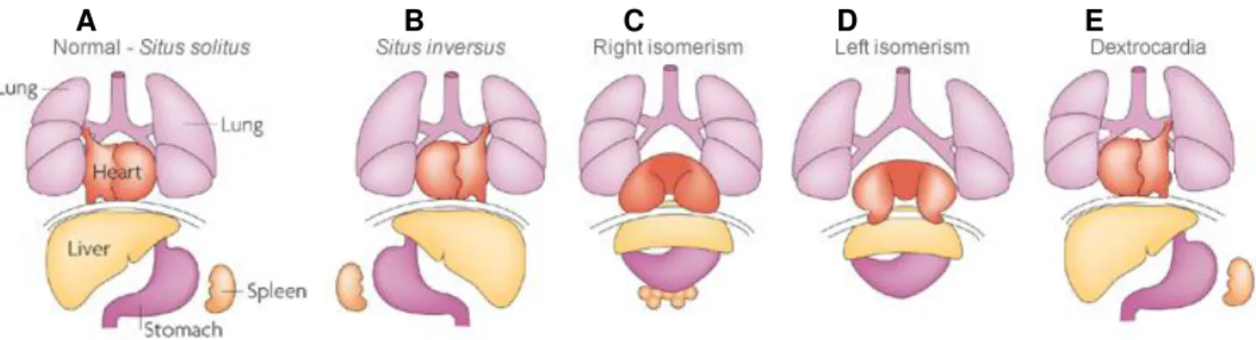
Figure 1.1 Human laterality problems. (A) Normal organization of the internal organs referred to as situs solitus. (B) Situs inversus. The position of the internal organs is a complete mirror-image of the normal situation. (C) Right isomerism. Two right sides are formed. (D) Left isomerism. Two left sides are formed. (E) Dextrocardia. The heart is the only organ that is reverted from the normal situation. Adapted from (Fliegauf et al. 2007). ... 2 Figure 1.2 Clock and Wave front model of somite formation. The synchronized mRNA oscillations of cyclic genes along the presomitic mesoderm (PSM) describe a wave of expression that is initiated in the posterior region of the PSM (phase I) and moves towards the anterior region of the PSM (phase III) where it slows down culminating with somite formation. At the same time, the determination front marks the position where each new pair of somites is formed. The determination front is defined by opposing gradients of FGF/Wnt and Retinoic acid and moves posteriorly until all the somites are formed. Adapted from (Dequeant and Pourquie 2008). ... 5 Figure 1.3 Simple representation of cell-cell communication via the Notch signaling pathway. A signal sending cell, expresses the Notch ligand Delta. Once Delta binds the Notch receptor, the Notch intracellular domain (NICD) is released upon cleavage of Notch, and translocates into the nucleus where it acts as a transcriptional regulator. The detached fragment of Notch (NECD) is endocytosed along with Delta into the signal sending cell. Adapted from (Lewis et al. 2009). ... 6 Figure 1.4 Retinoic acid (RA) protects the presomitic mesoderm (PSM) from asymmetric signals. At the same time asymmetrical signals (red arrow) are being transferred from the node to the left lateral plate mesoderm (Left LPM), RA signaling (red) protects the PSM allowing symmetric somite formation. Both Notch and FGF signaling are transiently lateralized (blue), but by the action of RA these regain its symmetric activity in PSM. Adapted from (Kawakami et al. 2005). ... 8 Figure 1.5 Simple representation of Transcription activator-like effectors (TALEs). (A) The N-terminus is required for type III secretion and the C-N-terminus contains nuclear localization signals (NLS). The effector domain acts as a transcription activator. Binding specificity comes from a region of typically 33-34 amino acid repeats (letters in black). Concentrated at residues 12 and 13 the RVDs (red) specify the nucleotide that is targeted by each repeat. (B) The most common RVDs and theirs target nucleotides. ... 10
xvi
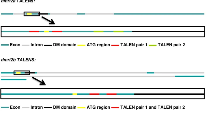
Figure 3.1 Representation of dmrt2a and dmrt2b genomic sequences. dmrt2a is composed of 3155 bp, with 4 exons (blue) and 3 introns (grey). The ATG and the DM domain of dmrt2a are situated in the second exon distancing 171 bp from each other (upper black rectangle). dmrt2b is composed of 6671 bp, with 3 exons (blue) and 3 introns (grey). The ATG and the DM domain of dmrt2b are situated in the first exon and distance 138 bp from each other (lower black rectangle). ... 25 Figure 3.2 Wild type adult zebrafish genotyping. (A, B) 10 wild type adult zebrafish were genotyped for dmrt2a (panel A) and dmrt2b (panel B). For both genes, the DM domain extends further than the sequenced region. No polymorphisms were found for both genes in between the ATG and the DM domain. (A) The genotyped region of dmrt2a only revealed one polymorphism (A/T) upstream of the ATG. (B) The genotyped region of dmrt2b, revealed several polymorphisms upstream of the ATG. ... 27 Figure 3.3 Schematic representation of each TALEN pair target region within the dmrt2a and
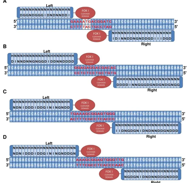
dmrt2b genomic sequences. For both genes a magnification of the genomic region that compromises the ATG and the start of the DM domain is shown. For dmrt2a two TALEN pairs were designed: one pair (upper red stripes) to target the ATG region (yellow stripes) and one pair (upper green stripes) to target a region prior to the start of the DM domain (black stripes). For dmrt2b also two TALEN pairs were designed but these target the same region in between the ATG and the DM domain, and so are represented together in the lower red stripes. ... 28 Figure 3.4 RVD sequence representation for each TALEN monomer assembled during this work. (A) Left and Right monomers of dmrt2a-pair1 with the ATG highlighted in white. (B) Left and right monomers of dmrt2a-pair2. (C) Left and right monomers of dmrt2b-pair1. (D) Left and right monomers of
xvii
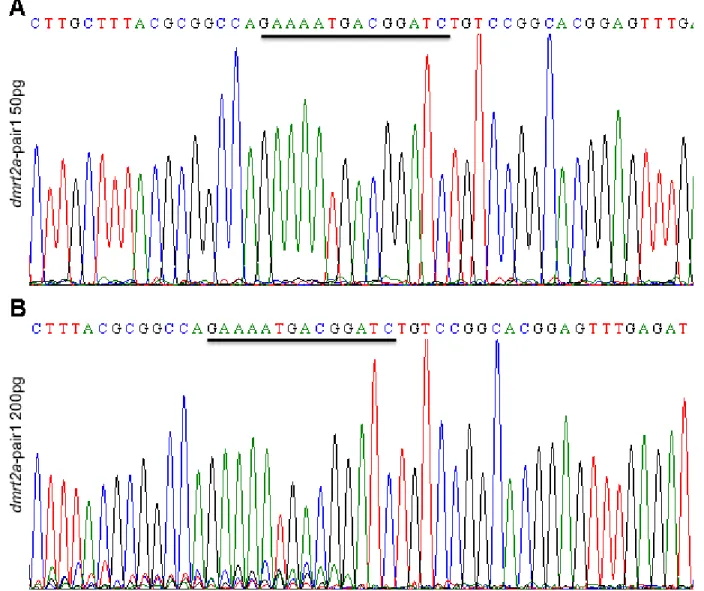
at the ATG region. (B) The sequencing reaction is performed with a reverse primer to assess the presence of mosaic peaks at the target site of dmrt2a-pair2. Each pair has its target site underlined... 36 Figure 3.10 Electrophoresis run of dmrt2a-pair1+pair2 samples reveal the presence of faint bands below 200 bp. Genomic DNA samples from mosaic fish that were injected with dmrt2a-pair1+pair2 were used to amplify a fragment of 200 bp spanning the two target sites (distancing 100 bp from each other). As a negative control, a genomic DNA sample from an embryo that had not been exposed to TALENs was used (lane 2). Faint bands of approximately 100 bp are visible only in lanes 3 to 9. These correspond to the genomic DNA samples from fish injected with the two TALEN pairs simultaneously, revealing the possibility that some fragments are lacking 100 bp. ... 37 Figure 3.11 Extraction of portions of agarose gel below 200 bp. The full amplification content of 4 of the previously amplified products was run on an agarose gel. Portions of agarose gel were extracted from below the 200 bp fragments to assess if they consisted on fragments where a big genomic deletion had occurred. These were purified and then reamplified. ... 38 Figure 3.12 PCR reactions run on a 4% low melting agarose gel reveal the possible existence of big genomic DNA lesions. The 4 samples that were previously reamplified were run on a 4% low melting agarose gel so that any existing small fragments could be separated. 100 bp fragments were clearly visible on all 4 amplification reactions (red rectangle). These could possibly belong to fragments lacking the 100 bp that separate each dmrt2a TALEN target site. Apart from these 100 bp fragments, smaller fragments are clearly visible, as in lane 2 (yellow rectangle). These may belong to genomic DNA fragments where TALENs introduced a genomic deletion bigger than 100 bp. ... 38 Figure 3.13 Sequencing result from a small DNA fragment amplified from genomic DNA samples of
dmrt2a-pair1+pair2 exposed fish. A small fragment from the yellow rectangle in Figure 3.12, was extracted from the gel, purified and sequenced. The sequencing result reveals that this fragment belongs to dmrt2a.
The sequencing reaction was performed with HRM_dmrt2a_pair1_FW. The nucleotides on the left of the
xviii
xix
shown in red and mutant samples are shown in green. (A) HRMA of dmrt2a heterozygous incross progeny. (B) HRMA of dmrt2b heterozygous progeny. (C) Overall percentages of formed alleles. ... 47 Figure 3.23 The frequency of asymmetric expression of the cyclic genes decreases dramatically after the 12-somite stage. Asymmetric gene expression of the cyclic genes from 8 to 14-somite stage assessed by in situ hybridization (A) More than 50% of the analyzed embryos show asymmetric expression of the cyclic genes between 8 to 12-somite stages. This asymmetric gene expression then decreases dramatically with only a small number of embryos showing asymmetric gene expression further on. Although in a lower percentage, some wild type sibling embryos also show asymmetric gene expression before the 12-somite stage. (B) Representative images of deltaC, her7 and her1 expression patterns at the 8-somite stage. Upper panels show the wild type sibling controls whereas lower panels show the embryos where dmrt2a was overexpressed. Around 450 embryos were used during this experiment. ... 49 Figure 3.24 Failure of the leftward displacement of the heart cone in a dmrt2a overexpression context. (A, B) Representative images of cmlc2 expression between 28 and 32 hours post fertilization in embryos overexpressing dmrt2a. Left “normal” jog was observed in 79% of the studied embryos (A), whereas Right jog could be observed in 21% of the embryos (B). ... 50 Figure 3.25 Sporadic appearance of a kink in the notochord at the level of the 12th somite. Embryos overexpressing dmrt2a sporadically develop a kink in the notochord around the 12th somite. ... 50 Figure 3.26 Comparison between the expression of dmrt2a at the onset of the time window, in a wild type embryo and in an embryo where dmrt2a was overexpressed. (A, B, C D) In situ hybridization for
dmrt2a at the 8-somite stage. The expression of dmrt2a does not seem to change much between sibling wild type controls (A, C) and the dmrt2a overexpression embryos (B, D). (A, B) Flat mount view at the level of the somites. (C, D) Whole mount dorsal view. ... 51 Figure 3.27 Complete randomization of the heart cone displacement. (A, B) Representative images of cmlc2 expression between 28 and 32 hours post fertilization in embryos where dmrt2a was overexpressed by the injection of 200pg dmrt2a mRNA at the one cell stage. Left “normal” jog was
xxi
List of Tables
xxiii
Abbreviations
AAD Acidic activation domain
DSB Double-stranded breaks
HR Homologous recombination
HRM High resolution melting
HRMA High resolution melting analysis
LPM Lateral plate mesoderm
MO Morpholino
NECD Notch extracellular domain
NHEJ Non-homologous end joining
NICD Notch intracellular domain
NLS Nuclear localization signals
PSM Presomitic mesoderm
RA Retinoic acid
RVD Repeat-variable di-residues
TALE Transcription activator-like effector
TALEN Transcription activator-like effector nuclease
1
Chapter 1
Introduction
1.1 Background
It is amazing to realize that the human body is not all about symmetry. We tend to view all the vertebrates as bilaterally symmetric, but that only happens from the outside. Internally, most of the organs from heart to liver are asymmetrically positioned. It is a feature that is conserved throughout chordate evolution, and although varying among different species, the normal individuals within a given species show the same kind of asymmetries. Among higher mammals, left-right asymmetry extends even higher to the brain and nervous system (Levin 2004).
The external bilateral symmetry of the vertebrate body, resides on the skeleton and its associated muscles. These, have their origins in the somites which are transient embryonic structures, formed in pairs along the anterior-posterior axis and in a cyclic and symmetric way. Upon formation, somites then differentiate, giving rise to axial skeleton and skeletal muscles.
The origins of the internal asymmetry can be traced back to the gastrulation stage, before any morphological asymmetries can be observed. During this stage, asymmetric gene expression in the node is initiated, and a conserved cascade of asymmetrically expressed genes referred to as the nodal-lefty-pitx2 cassette will lead to the morphological asymmetric organization of the organs.
1.2 Left-right asymmetry within the vertebrate body
2
Figure 1.1 Human laterality problems. (A) Normal organization of the internal organs referred to as situs solitus. (B)
Situs inversus. The position of the internal organs is a complete mirror-image of the normal situation. (C) Right isomerism. Two right sides are formed. (D) Left isomerism. Two left sides are formed. (E) Dextrocardia. The heart is the only organ that is reverted from the normal situation. Adapted from (Fliegauf et al. 2007).
It is therefore a challenge to modern scientists to answer a fundamental question: what is the driving force that triggers left-right patterning among so many different species and to what extent it is evolutionary conserved. Also hidden behind this question is the mechanism that ensures that at the same time an internal asymmetric body is being formed, external bilateral symmetry is not compromised.
Being the asymmetrical organization of the internal organs the most visual event, it can only be explained by earlier mechanisms of symmetry breaking. As so, the development of an asymmetric body plan is divided into 3 distinct steps. First, bilateral symmetry has to be broken, forming a left-right axis that is oriented relative to the dorsal-ventral and anterior-posterior axes. Then, differential gene expression between the two sides has to be triggered. And finally, in response to this differential gene expression, changes in cell behavior, such has migration rates, will contribute to a morphological difference between the left and right sides of the body (Vandenberg and Levin 2013).
Differential gene expression between the two sides has one common and conserved feature among different phyla which is the Nodal-Lefty-Pitx2 cassette. At the onset of gastrulation, both around the
mouse node, the chicken Hensen’s node, the Xenopus gastrocoel roof plate and the zebrafish Kupfer’s vesicle, Nodal activity becomes restricted to the left side. The expression of nodal, a gene encoding a member of the transforming growth factor beta (TGFβ) family, spreads out to the lateral plate mesoderm (LPM), and activates not only the expression of the genes lefty1, lefty2 and pitx2 but also his own. Lefty2 encodes another member of the TGFβ family, competing for the same Nodal receptors, but unlike Nodal that functions has a dimer, Lefty2 functions has a monomer which allows much more diffusion than Nodal, thus limiting Nodal’s activity to the left side. The genetic program leading to subsequent asymmetries is thought to be triggered by Pitx2, a paired-like homeodomain transcription factor that is the effector of Nodal signaling (Nakamura and Hamada 2012, Babu and Roy 2013).
Despite the evolutionary conservation of this Nodal activity on laterality, it remains to be explained the first step of forming an asymmetric body plan: Which event triggers the formation of the left-right axis and how is Nodal localization on the left side of the node initiated?
3
It was thought that the answer to this question resided in the action of cilia (Nonaka et al. 1998). These are microtubule organelles that extend from the surface of many cells and that are present at the node of mouse, as well as in Kupfer’s vesicle of zebrafish. In mouse, a leftward flow is created by the rotation of cilia, at the ventral pole of the embryo. This is called nodal flow and when an artificially nodal flow was generated independently from ciliary motility, laterality was also determined (Nonaka et al. 2002). Two models attempt to explain this mechanism. With cilia being divided into motile and immotile, the “two cilia model” defends that motile cilia generate the leftward flow and that this is sensed by nonmotile mechanosensory cilia. These are present at the periphery of the node and it has been observed that only this type of cilia exhibit polycystin-2, a protein thought to be involved in mechanosensation (McGrath et al. 2003). Another perspective comes from the morphogen gradient model which predicts that a hypothetical morphogen is carried out through the leftward flow. This transport has been described in mouse. Called Nodal Vesicular Parcels, these are membrane-sheathed vesicles budding from node cells. Upon release of their content Sonic Hedgehog and retinoic acid could be asymmetrically concentrated. In agreement, both models report an asymmetrical Ca2+ release that would be defining for the following establishment of the Nodal-Lefty-Pitx2 cassette (Fliegauf et al. 2007, Speder et al. 2007).
Cilia as the answer to laterality could also be supported from clinical data. Individuals with ciliopathies such as Kartagener’s syndrome, apart from having situs inversus, usually suffer from respiratory dysfunctions like chronic rhinosinusitis and bronchiectasis. Cilia are also present in the respiratory epithelium being part of a mechanism called Mucociliary clearance that enables the airways to protect the lungs from harmful substances in the surrounding environment, (Morillas et al. 2007, Babu and Roy 2013).
Nevertheless, cilia flow cannot be the only crucial mechanism defining laterality. Many phyla establish a left-right axis without the help of cilia, including vertebrates like chick and vertebrate mammals like pig. Chick and pig have morphologically asymmetric nodes, consequence of leftward cell movements, with pig not even showing to have cilia in the notochordal plate as well as space for the flow to be generated. Moreover, both in chick and in pig, asymmetrical gene expression domains form hours before the asymmetric gene expression of nodal on the left side of the node.(Vandenberg and Levin 2010) Other examples come from zebrafish, one of the animal models where cilia flow in the Kupfer’s vesicle is thought to possibly play a key role in left-right patterning. Two distinct studies in zebrafish with mutants of the gene seahorse, a gene that is involved in multiple cilia-mediated processes (Kishimoto et al. 2008), report incoherent results. In one case a seahorse zebrafish mutant shows very little laterality defects even though its ciliary flow is almost absent. On the other hand another study reveals a seahorse zebrafish mutant with normal cilia, yet, half of the studied fish develop laterality defects (Vandenberg and Levin 2010). Overall there are many studies today that show little support on a causal link between cilia and laterality in many different phyla, and even if a question of conservation is raised, hypothesizing that mouse has a particular role for cilia in establishing laterality, how to explain that mutations for the gene
4
ciliopathies (Valente et al. 2010) but do not seem to affect laterality, both in zebrafish and mice (Vandenberg and Levin 2010).
Results from chick, pig, and other model organisms, raise another interesting question. Could laterality be a case of convergent evolution? It is difficult to accept that a characteristic spanning so many different phyla might have had different origins. Indeed, the quest for the earliest possible event that could break bilateral symmetry, and that could be a common ancestor of laterality, has retrieved new models on its origins.
The ion flux model resides on the idea that an asymmetric distribution of K+ channels and H+ pumps could be the driven by cell chirality during the first embryonic cleavages. This would lead to an accumulation of serotonin on the right side of the embryo where it represses the expression of nodal or its homologues, considering the species. Cilia model supporters considered that the studies involved in this model were inconsistent because they were altering pathways that could consequently affect cilia parameters. Nevertheless it was already confirmed a role for serotonin in the early cell cleavages without affecting node precursor cells (Vandenberg and Levin 2013).
Another model focusing on the earliest possible definition of a left-right axis is the chromatid segregation model. During the first cell cleavage the chromatids would be differentially imprinted and segregated. It has been shown in yeast that differentially segregated chromatin, mRNAs and proteins are sufficient to maintain asymmetry. Some similar mechanisms have been found in eukaryotic cells and embryos (Vandenberg and Levin 2013).
Finally the PCP model, based on a highly conserved mechanism used to correctly orient cell division, like in the Drosophila eyes and wings, mammalian kidney and vertebrate limbs, has also been shown to be associated with left-right patterning. First adopted by the cilia model supporters, since the PCP pathway has the ability to properly position cilia in the node, it has been shown that disrupting the PCP pathway in chick causes laterality defects even though chick do not use cilia to define their left-right axis. The same was shown in frog embryos, with the disruption of the PCP pathway on cells that do not contribute to the node causing laterality defects (Segalen et al. 2010, Vandenberg and Levin 2013).
Non-mutually exclusive, these models confirm that the definition of a left-right axis might happen very early in development. Also they leave open the possibility that not just one mechanism is the ultimate responsible for the correct placement of the asymmetrical body components. Might be that different amplification steps of an initial left-right axis definition occur throughout development, not only in a sequential order but also in a redundant manner. This would explain why most of the experiments trying to address laterality, only achieve randomization phenotypes, with only one small percentage of the studied individuals showing complete reversal of its left-right axis (Vandenberg and Levin 2013).
5
1.3 Bilateral symmetry of the vertebrate bodyThe external symmetrical appearance of the vertebrate body plan is mostly due to the symmetric organization of the skeleton and its muscles. These have its origins in the somites, transient embryonic structures, composed of blocks of epithelial mesoderm, that form in pairs on both sides of the axial structures, neural tube and notochord, and at a time rate and number that is species specific. In zebrafish each new pair of somites is formed in a period of 30 minutes, whereas in chicken this takes 90 minutes, in mouse 120 minutes and in humans this period lasts approximately 4 to 5 hours. (Dequeant and Pourquie 2008, Lourenço and Saúde 2010).
Extensively reviewed, the process behind the formation of the somites is explained by the clock and wavefront model. A set of genes, called the cyclic genes, are continuously oscillating their expression along the presomitic mesoderm (PSM). This was first discovered in chicken with the gene hairy1 showing a dynamic and cyclic expression pattern along the PSM with the same periodicity of somite formation (Palmeirim et al. 1997). Other cyclic genes were then discovered. These are called hes genes in the mouse and her in zebrafish and show a similar oscillating pattern of expression along the PSM. The expression of the cyclic genes starts in the most posterior part of the PSM and through what can be observed as 3 distinct phases of expression, it reaches the most anterior part of the PSM, setting the time for the formation of a new pair of somites. At the same time the cyclic genes are oscillating, opposing gradients of Fgf8/Wnt and Retinoic Acid (RA) mark the position of what is called the determination front. Immature cells supplied from the tail bud, posteriorly, are under the influence of Fgf8 and Wnt signaling and they will only start to differentiate when they reach the most anterior part of the PSM. The process is continuously repeated with the determination front moving posteriorly, until all the somites are formed (Figure 1.2) (Dequeant and Pourquie 2008, Lourenço and Saúde 2010).
6
The synchronized oscillations of the cyclic genes reside mainly on the Notch signaling pathway. The function of this pathway is to coordinate gene expression in contiguous cells. A signal-sending cell expresses a Notch ligand, as it is Delta, and the binding of this ligand with the receptor, Notch, leads to the cleavage of Notch, releasing the Notch intracellular domain (NICD), that translocates to the nucleus where it acts as a transcriptional regulator. The detached extracellular fragment of Notch (NECD) along with Delta is endocytosed into the Delta-expressing cell (Lewis et al. 2009).
Figure 1.3 Simple representation of cell-cell communication via the Notch signaling pathway. A signal sending cell, expresses the Notch ligand Delta. Once Delta binds the Notch receptor, the Notch intracellular domain (NICD) is released upon cleavage of Notch, and translocates into the nucleus where it acts as a transcriptional regulator. The detached fragment of Notch (NECD) is endocytosed along with Delta into the signal sending cell. Adapted from (Lewis et al. 2009).
The Notch signaling pathway was first associated with the synchronized oscillations of the cyclic genes based on the fact that the her genes are targets of the Notch pathway and also by its salt-and-pepper expression pattern in the PSM of notch mutants which could be due to a failure of synchrony in its oscillations. Her1 and Her7, transcription factors, establish a negative feedback loop that leads to a periodic repression of DeltaC. This way, it is possible for neighbouring cells to be synchronized, at the same time they oscillate along the PSM (Holley et al. 2002, Dequeant and Pourquie 2008, Lewis et al. 2009). The cell-cell communication that is the center of this Notch signaling based synchronization was further confirmed with the implantation of cells overexpressing deltaC in a zebrafish embryo resulting in the desynchronization of the cyclic gene expression (Ishimatsu et al. 2007).
7
Upon formation, the somites undergo a differentiation process. It begins with the formation of different cellular compartments, each one with its unique gene expression, which leads to the development of the different tissue progenitors. Dorsally, the dermomyotome is an intermediary structure that gives rise to the progenitors of the skeletal muscles (myotome), limb muscle progenitors and dermis of the back, whereas ventrally the somite gives rise to the sclerotome, progenitors of the axial skeleton (Hollway et al. 2007).
Zebrafish, supported by its swim bladder, has no use for a robust skeleton. Consequently the zebrafish somite is predominantly composed of myotome, with the sclerotome restricted to a minor fraction of the somite. Although no significant structure resembles the dermomyotome, anterior somitic cells constitute its functional equivalent (Stickney et al. 2000, Hollway et al. 2007).
1.4 Symmetry versus asymmetry during development
At the same time symmetric somites are being formed in the PSM, asymmetric signals are being transferred to the lateral plate mesoderm (LPM). The mechanism that prevents these signals from reaching the PSM and thus disrupting symmetric somite formation resides on RA signaling.
Apart from its role in positioning the determination front during somitogenesis, RA is also involved in this process, buffering these asymmetric signals. Mouse raldh2 mutant embryos, which lack the enzyme that produces RA, exhibit fewer somites on one side of the PSM (Vermot et al. 2005). This is caused by a desynchronization of the waves of expression along the PSM which leads inevitably to an asymmetric somite formation between both left and right sides. This has also been shown in chick (Vermot and Pourquie 2005) and zebrafish (Kawakami et al. 2005).
In zebrafish, studies using a translation blocking Morpholino (MO), observed that the initiation of somitogenesis is bilaterally symmetric even in the raldh2 morphants. Nevertheless, as somite formation proceeds, raldh2 morphants start developing an uneven number of somites between both sides of the PSM. Interestingly, after the 13 somite stage, symmetric somite formation is recovered in the raldh2
8
Figure 1.4 Retinoic acid (RA) protects the presomitic mesoderm (PSM) from asymmetric signals. At the same time asymmetrical signals (red arrow) are being transferred from the node to the left lateral plate mesoderm (Left LPM), RA signaling (red) protects the PSM allowing symmetric somite formation. Both Notch and FGF signaling are transiently lateralized (blue), but by the action of RA these regain its symmetric activity in PSM. Adapted from (Kawakami et al. 2005).
To buffer the asymmetric signals, RA also presents a transient asymmetrical signaling. rere, encoding a chromatin-remodeling protein, positively regulates RA signaling by forming a complex with the nuclear receptor NR2F2 (COUP-TFII), p300 (EP300) and RARs. nr2f2, was found to be asymmetrically expressed in the right PSM. Combining to the fact that a mutation in the mouse rere leads to a similar phenotype as the one observed in raldh2 mutants, a revised model was proposed where the action of RA signaling as a buffer, may itself be transiently asymmetric (Vilhais-Neto et al. 2010).
1.5 The role of dmrt2a
The focus of this work, the gene dmrt2a, belongs to a family of transcription factors called DMRT (DM related transcription factors). These are genes encoding a zinc finger like DNA binding motif, the DM domain, and although initially associated with sex determination, some of these genes have since been linked with other developmental processes (Hong et al. 2007).
9
In respect to the LPM, dmrt2a knock down or overexpression lead to the randomized expression pattern of genes like pitx2a and spaw, which normally are restricted to the left side of the LPM. Consequently, the correct positioning of the organs is compromised. Concerning the PSM, the cyclic genes and several other genes like myoD, fgf8, raldh2 or cyp26a, become bilaterally asymmetric. Nevertheless this asymmetric gene expression is only observed until the 12 somite stage with no distinctive phenotype in later stages of development being observed.
In the mouse, where dmrt2a has its homologous gene dmrt2, considerably different observations were made. Here, Dmrt2 is not involved in left-right patterning. In the null dmrt2 mouse nodal is restricted to the left side of the node and LPM and pitx is restricted to the left side of the LPM. Also, this mutant does not express dmrt2 in the node which correlates with its normal organization of internal organs (Lourenco et al. 2010). On the other hand axial skeleton and rib patterning defects can be observed in a null dmrt2
mouse but bilaterally symmetric gene expression of hes7 is not affected (Seo et al. 2006, Lourenco et al. 2010). The null dmrt2 mouse has problems in somite differentiation. Here dmrt2 is specifically expressed in the dermomyotome, and although no significant alterations were detected in terms of muscle development, it is clear that the normal arrangement of the myoblasts in the myotome is affected with the myocytes failing to elongate and occupy the entire rostral-caudal domain of the myotome. These changes may arise from an essential role of dmrt2 in providing extracellular matrix components within the mouse dermomyotome which are essential for correct myocyte differentiation (Seo et al. 2006).
The gene dmrt2a has also a paralogous, the fish specific dmrt2b. Similarly to dmrt2a, knock down of dmrt2b randomizes the asymmetrical gene expression of genes in the LPM leading to an incorrect organ positioning. However this similarity is restricted to the LPM with no involvement of dmrt2b in the synchronization of the cyclic genes during somitogenesis. Nevertheless dmrt2b is also expressed in the somites and has its role during somite differentiation, since its knock down inhibits the sonic hedgehog pathway leading to slow muscle defects (Liu et al. 2009).
1.6 Transcription activator-like effectors (TALEs)
Transcription activator-like effectors (TALEs) are a class of DNA binding proteins that can be found in some species of plant pathogenic bacteria of the genus Xanthomonas. Consisting on these bacteria key virulence factors, TALEs are translocated into the plant cell cytoplasm via type III secretion system, and have the ability to reprogram host cells by mimicking eukaryotic transcription factors. They enter the nucleus, bind to specific sequences in the host gene promoters and activate transcription of downstream genes (Boch et al. 2009, Cermak et al. 2011).
10
concentrated at residues 12 and 13, referred to as repeat-variable di-residues (RVD). These are the residues that specify the target, one RVD to one nucleotide, and although many different RVDs can occur across TALEs, four of them, HD, NG, NI, and NN, account for 75% of the total and respectively associate with one of the four DNA bases. Also, and common across TALEs, is the requirement for the binding site
to be preceded by a 5’T (Boch et al. 2009, Bogdanove et al. 2010, Cermak et al. 2011).
Figure 1.5 Simple representation of Transcription activator-like effectors (TALEs). (A) The N-terminus is required for type III secretion and the C-terminus contains nuclear localization signals (NLS). The effector domain acts as a transcription activator. Binding specificity comes from a region of typically 33-34 amino acid repeats (letters in black). Concentrated at residues 12 and 13 the RVDs (red) specify the nucleotide that is targeted by each repeat. (B) The most common RVDs and theirs target nucleotides.
1.7 Genetic engineering with TALE nucleases (TALENs)
The simple code that governs TALE activity made it possible to customize these proteins in order to achieve modifications in DNA sequences of interest.
Transcription activator-like effector nucleases (TALENs) are TALE-nuclease chimeras, where the TALE region required for high-affinity DNA binding is fused into the catalytic domain of FokI. The result is a gene modification tool capable of inducing double-stranded breaks (DSB) in vivo. It is important to point out that FokI cleaves as a dimer, so two opposite TALENs are needed to create this DSB leaving a spacer in between them so that the two FokI domains can act. (Cermak et al. 2011, Miller et al. 2011) Two major processes can be involved in repairing these DSB: non-homologous end joining (NHEJ), resulting in small insertions or deletions (indels), and homologous recombination (HR), used for sequence modifications, provided by a donor template (Cade et al. 2012).
11
1.8 TALENs assemblyTALENs assembly is the process by which all the RVDs that confer binding specificity to a TALEN are put together in a previously constructed vector that includes, among others, the catalytic FokI domain. Considering that each TALEN monomer has around 16 RVDs and that at least one TALEN pair is needed to produce a cut, it would be a very time consuming, expensive and error-prone protocol to make if traditional molecular cloning techniques were used. This was made easier following the work of (Cermak et al. 2011) where the recent method of Golden Gate cloning was applied to TALENs assembly. Through Golden Gate cloning several separate plasmids can be efficiently cloned into an acceptor vector in one single reaction mixture in one tube. Using type IIS restriction endonucleases which cleave outside their recognition sites (sticky ends), and a ligation enzyme, the reaction mixture is subjected to multiple steps of digestion/ligation, according to the optimal temperatures for each of the enzymes used. Since the correct assembly eliminates the restriction enzyme recognition site, the efficiency of this procedure is high with almost all of the colonies after transformation possessing the desired construct (Engler et al. 2009, Cermak et al. 2011).
1.9 Focus of this work
Previous studies that tried to unveil the function of dmrt2a and dmrt2b in zebrafish were done using MO (Saude et al. 2005, Liu et al. 2009). Although broadly used in genetic studies MOs have a transient effect and are not transmitted through the germline. Also, MOs may have off-target effects which can lead to phenotype misinterpretation (Huang et al. 2012).
During this work recent TALEN technology (Bogdanove et al. 2010, Cermak et al. 2011, Miller et al. 2011) will be used to try to generate homozygous mutants for dmrt2a and dmrt2b. With homozygous mutants for these two genes high-throughput analysis using Microarray technology (Sobek et al. 2006) can be made without having the risk of variability which can be introduced by the MO.
Also the possibility of these mutants being viable will allow the generation of a double mutant for
dmrt2a and dmrt2b. This would help us understand better the roles of these two paralogous genes, and possibly unveil to what extent they are related to the homologous mouse dmrt2.
13
Chapter 2
Materials and Methods
2.1 Zebrafish lines and maintenance
The zebrafish strain TU was used. Adult fish and embryos were maintained and bred according to standard procedures (Westerfield 2000).
2.1 List of Primers
Primers used during the course of this work, either for cloning or genotyping, are listed in Table 2.1. Except where indicated primers used during this work were designed using NCBI primer blast (NCBI) and oligoanalyzer (Integrated DNA Technologies) and synthesized by Stabvida.
Table 2.1 List of Primers used during this work. Restriction sites highlighted in yellow (EcoRI), blue (StuI) and green (XhoI). Except for *, primers were designed during this work.
Primer name Primer sequence PCR (bp) dmrt2a_FW 5'-ACTCATCGTTTGTTTGACTGCTTT-3'
572 bp dmrt2a_RV 5'-AGAACCTCTTGTGCCCCTTTAG-3'
dmrt2b_FW 5'-GAAACATCCAGACTCACAAGCACAGC-3'
421 bp dmrt2b_RV 5'-CTGCCATCACTCGCTGCCTCTCC-3'
pCR8_F1 * 5'-TTGATGCCTGGCAGTTCCCT-3'
variable pCR8_R1 * 5'-CGAACCGAACAGGCTTATGT-3'
grunwald_FW * 5'-TTGGCGTCGGCAAACAGTGG-3'
variable grunwald_RV * 5'-ACGTCCCATCGCGTTGCC-3'
HRM_dmrt2a_FW 5'-GACACGTTACATGCAGGAAAACA-3'
128 bp HRM_dmrt2a_RV 5'-CTCCACGTCGATCTCAAACTCC-3'
HRM_dmrt2b_FW 5'-CACAGGTAGATGCGACCCAC-3'
87 bp HRM_dmrt2b_RV 5'-CTTCATCCGTGCCCATGACC-3'
HRM_dmrt2a_par2_FW 5'-GCGATGATCAGGCGGTGTTC-3'
74 bp HRM_dmrt2a_par2_RV 5'-GCGGTCAGATTTGTCGTCGT-3'
dmrt2a_cloning_FW 5'-TTGGAATTCTATGACGGATCTGTCCGGCAC-3'
1508 bp dmrt2a_cloning_RV * 5'-AGGCCTTTTTTA CTGAGATTTCCGATTTAAAGAAAGCGC-3'
dmrt2b_cloning_FW 5'-TTG GAA TTCT ATGTCCACTAAAGCGGATAGGG-3'
14
2.3 Transformation of competent cells
Competent cells, previously prepared in our lab, and kept at -80ºC were thawed on ice, and at the same time, the tube where the bacterial cells would be transformed was cooled. 10l of a cloning reaction plus 100l of competent cells were put together and left on ice for 30 minutes. A heat shock was done at 42ºC for 1 minute and then the tube was cooled again on ice for 2 more minutes. 900l of SOB solution was added and the mixture was then incubated with shaking at 37ºC for 45 minutes. Finally 50l of the mixture were plated on LB agar media, containing the appropriate antibiotic, and incubated at 37ºC overnight.
2.4 Colony PCR
Throughout this work colony PCR was used to help identify bacterial colonies with the right ligation product prior to miniprepation. 40ml falcons with 5ml of appropriate medium and antibiotic were carefully prepared and identified. Then, PCR tubes containing 10l of water were also identified with the corresponding designations. On ice, a PCR master mix composed of 12.5l of Quick-Load Taq 2X Master Mix (New England BioLabs) and 200M of each primer was also prepared. Using a pippete tip, a colony was picked. The tip was then inserted in a pippete to help mix the colony in the water that was inside the PCR tube by pipetting up and down. The tip was then placed inside the 40ml falcon in contact with the medium. This procedure was repeated for each different colony. Once all the colonies were picked, the PCR master mix was added to each tube and a PCR reaction was started on a thermal cycler according to Quick-Load Taq 2X Master Mix manufacturer’s instructions. At the same time, inoculums were incubated with shaking overnight at 37ºC.
Colony PCR products were then run on a 1% Agarose gel (
SeaKem
)
and clones possessing the correct ligation product were identified. The corresponding inoculums were left incubating for minipreparation on the next day, whereas the others were discarded. If no correct ligation product was identified, the whole process was repeated with different colonies.2.5 Cloning of dmrt2a and dmrt2b
Since both dmrt2a and dmrt2b are expressed during somitogenesis, total RNA was extracted from 12 hour post fertilization wild type zebrafish embryos (described below) and cDNA synthesized with MMLV-Reverse Transcriptase kit (Promega).
Zebrafish dmrt2a and dmrt2b full length coding sequences were amplified by PCR, using Phusion High Fidelity Polymerase (Thermoscientific) with the following primer set: dmrt2a_cloning_FW and
15
HF Buffer (Thermoscientific), 2l of each primer 25M, 2l of dNTPS 5M, 2l of cDNA, 0,5l Phusion HF and water up to 50l. Conditions were 30 seconds at 98ºC, 35 cycles of 10 seconds 98ºc, 30 seconds at 68ºC, 45 seconds at 72ºC, and 10 minutes at 72ºC. Reactions were performed on a thermal cycler (Applied Biosystems 2720).
The amplified fragments were then introduced into separate PCS2+ expression vectors, already available in the lab glycerol stock. First, four double digestions were executed. dmrt2a insert and PCS2+ vector were digested separately with EcoRI (New England BioLabs) and StuI (New England BioLabs) restriction enzymes using the appropriate buffer according to the manufacturer’s instructions. dmrt2b
insert and another PCS2+ vector were also digested separately but in this case with EcoRI (New England BioLabs) and XhoI (New England BioLabs) restriction enzymes, and also using the appropriate buffer according to the manufactures. Then, using T4 DNA Ligase (New England BioLabs), ligation of each insert with its corresponding vector was performed. Double digestions of dmrt2a insert and its corresponding vector by EcoRI and StuI originated sticky ends (EcoRI) and blunt ends (StuI). Here, the ligation reaction mixture was incubated for two hours at 16ºC. In the case of dmrt2b insert and its corresponding vector, double digestions originated only sticky ends (EcoRI and XhoI). The reaction mixture was then incubated also at 16ºC but in this case for only 30 minutes.
Ligation products were used to transform competent cells, as already described. In both cases ampillicin was used as antibiotic. To identify the colonies with the correct ligation product, two distinctive approaches were used. Colonies, presumably containing dmrt2a insert were identified by colony PCR, as described before, using the same primers that were used for cloning. In the case of dmrt2b colony PCR did not prove to be as effective and so, 10 random colonies were picked and used for minipreparation. Analytical double digestions of those minipreps with EcoRI and XhoI were then performed to remove the
dmrt2b insert, if present. Digested samples were run on a 1% Agarose Gel and correct clones possessing
dmrt2b insert were finally identified. Both dmrt2a and dmrt2b correct clones were then sequenced for confirmation.
2.6 TALENs design and assembly
Prior to TALENs assembly, the RVD sequences were carefully designed. The first step was to genotype wild type zebrafish adults, the ones that laid the eggs used to inject the mRNA. Polymorphisms are common in zebrafish (Bradley et al. 2007), so in order to ensure a correct binding of TALENs, the DNA sequences of the region of interest among various wild type adults had to be carefully analyzed, ultimately leading to a selection of the best possible binding region. Having already chosen a safe region to be targeted, TAL Effector Nucleotide Targeter 2.0 (TALE-NT) was used. At this point, the region of the gene to be targeted was filtered for candidate TALEN pairs, according to the length of the monomers and
spacer pretended, and following the rule that every binding site has to be preceded by a 5’T. After
16
desired TALEN pair, a search through the entire genome, in this case the zebrafish genome, for offside possible targets was performed. (Doyle et al. 2012).
In (Cermak et al. 2011), an assembly protocol as well as a complete set of plasmids containing each of the possible RVDs and backbone vectors to be used was created and deposited in the non-profit repository Addgene. Final vectors specific for zebrafish were also created by (Dahlem et al. 2012) and deposited in the same repository.
Considering the approach used in this work where all the TALENs designed did not exceed 21 RVDs long, the assembly of one full TALEN monomer compromised two steps. During the first step (Golden Gate reaction 1), two independent digestion/ligation reactions were made. The first one accommodated the first 10 RVDS to be used, in an intermediate vector. The second digestion/ligation reaction accommodated the remaining RVDs to be used, minus the last one, in another intermediate vector. After transformation of competent cells using these intermediate vectors, minipreparation of plasmid DNA and sequencing, the second step (Golden Gate reaction 2) joined the RVDs contained in the intermediate vectors, plus the last RVD, into one final backbone vector (Cermak et al. 2011).
The final backbone vectors used in this work, one for the left monomer and one for the right monomer, contained an SP6 promoter and the SV40 polyadenylation sequence. As a result, after the correct assembly of the TALEN construct, mRNA synthesis was achieved through SP6 in vitro
transcription and the resulting mRNA molecules were securely injected into one cell stage embryos without being degraded (Dahlem et al. 2012).
2.6.1 dmrt2a and dmrt2b TALENs design
As mentioned above, TALENs were designed using TAL Effector-Nucleotide Targeter (TALE-NT) 2.0 web based tools (Cermak et al. 2011). The R-based tool developed by Jorge Velez (NICHD) was also used, although recently this tool has been considered not needed.
Wild type TU zebrafish adults were genotyped for the genes dmrt2a and dmrt2b. The amplified and sequenced fragments corresponded to the desired target regions.
The corresponding DNA sequences were then introduced in TALEN Targeter. Custom Spacer/RVD Lengths were used with the spacer ranging from 14 to 17 nucleotides and the RVD lengths ranging from 16 to 21 nucleotides. The G substitute chosen was NN. (It is important to note that recently the NH repeat has been considered to be more effective targeting the G nucleotide). The upstream base chosen to each monomer was T.
The results in the form of txt files were introduced in the R-based tool, which sorted the results according to the rule NG + HD > NI + NN. The resulted txt files from R-based tool were then filtered using Microsoft Excel.
17
dmrt2a this was successfully achieved whereas for dmrt2b no TALEN pairs would respect the rule NG + HD > NI + NN in the ATG region. Two pairs targeting the same region in between the ATG region and the DM domain of the dmrt2b gene were then designed.Finally, Paired Target Finder was used to check if the TALENs designed would somehow have off targets in the zebrafish genome. Here Search a Genome/Promoterome Tab was used. In Pre-loaded Sequence, Danio rerio (genome) was chosen and for each TALEN pair, the first monomer was inserted in RVD Sequence 1 and the second monomer was inserted in RVD sequence 2. The Score Cutoff chosen was 3.0 which meant that off targets with a score 3 times higher than the best possible score (our target of interest) would be left out. All the retrieved off targets had scores much higher than the best possible score.
2.6.2 dmrt2a and dmrt2b TALENs assembly using the Golden Gate cloning system
TALEN constructs were assembled using Golden Gate cloning approach as already summarized. First, the bacterial glycerol stock contained in the Golden Gate TALEN and TAL Effector kit was used for minipreparation of all the DNA plasmids needed. Minipreps were done using QIAprep Spin Miniprep Kit
according to the manufacturer’s instructions. To ease the following step, every DNA sample was diluted to
150ng/l, with the minipreps that did not have enough concentration being repeated.
In Golden Gate reaction 1, for each monomer, the first ten RVDs modules were joined together into a pFUS_A vector. The remaining RVDs modules, minus the last one, were joined together into a pFUS_B# vector, with the pFUS_B number corresponding to the amount of RVDs that it would accommodate. Each reaction was composed of 150ng of each module vector plus 150ng of pFUS vector, 1l of BSA-HF (New England Biolabs), 1l of T4 DNA Ligase, 2l of 10x T4 DNA Ligase buffer (New England Biolabs) and water to a 20l final volume. Reactions were performed on a thermal cycler (Applied Biosystems 2720) with the following conditions: 10x (37ºC/5min + 16ºC/10min) + 50ºC/5min + 80C/5min. Plasmid safe nuclease treatment was then performed, using per reaction, 1l of 10mM ATP and 1l of Plasmid safe nuclease (Epicenter), in an incubation period of one hour at 37ºC.
Then, transformation of competent cells was done as previously described, using the appropriate antibiotic, in this case spectinomycin and with the exception that petri plates were supplemented with 40l of X-Gal (20mg/ml) and 40l of IPTG (0.8M) to perform a blue/white screening.
The next day, 3 white colonies per plate were picked and PCR colony (previously described) was performed using primers pCR8_F1 and pCR8_R1 to evaluate the correct ligation product. Considering PCR colony results, minipreparation of the corresponding inoculums was made and before moving to the next step, every sample was sequenced and then again, diluted to 150ng/l. To help predict each desired nucleotide sequence is useful the usage of TAL Plasmids Sequence Assembly Tool (Fine).
18
monomers and pCS2TAL3-DD to create left monomers (Dahlem et al. 2012). Reactions were composed of 150ng of each intermediate vector, 75ng of either pCS2TAL3-RR or pCS2TAL3-DD, 1l of Esp3I (Thermoscientific) restriction enzyme, 1l of T4 DNA Ligase, 2l of 10xT4 DNA Ligase buffer and water to a final volume of 20l. Reactions were again performed on a thermal cycler with the following conditions: 10x (37ºC/5min + 16ºC/10min) + 37ºC/15min + 80C/5min. Note that in this last reaction, plasmid safe treatment as well as Esp3I denaturation were not needed since the final backbone vector had no homology with the inserted repeats.
The ligation products were again used to transform competent cells. Here, ampicillin plates were used, and once more, X-Gal and IPTG were used to perform a blue/white screening. In the next day, PCR colony was repeated in the same fashion as described before, only this time, with primers pGrunwald_FW and pGrunwald_RV. The correct ligations were assessed and the corresponding inoculums were used for minipreparation. Every sample was again sequenced to confirm the correct assembly of all 8 constructs.
2.7 Genotyping
Adult fish were anesthetized using 1xTricaine solution (MS-222) and caudal fins were cut to extract genomic DNA (described below). After this procedure the fish were kept individually isolated until the results from DNA sequencing or HRM were obtained. Genotyping of zebrafish embryos was also performed. In this case, embryos were previously sacrificed using 25xTricaine solution, and separated individually in different tubes, so that single embryo genomic DNA could be extracted (described below). Here sequencing and HRM results were used to statistically predict the abundance of different alleles in siblings or just to assess the efficiency of TALENs activity. It is important to note that during this work, genomic DNA was never extracted from pools of embryos in order to assess TALENs efficiency or germline transmission since single embryo approach is undoubtedly more informative both quantitatively and qualitatively.
2.7.1 Phenol-chloroform genomic DNA extraction
19
at 14 000 rpm during 30 minutes at 4ºC (eppendorf 5430 R) and after, the absolute ethanol is exchanged with previously cooled 70% ethanol, releasing the pellet from the tube wall and then centrifuged again for 10 more minutes. Finally, the pellet was air dried and ressuspended in 50l of water.
2.7.2 NaOH genomic DNA extraction
An adult zebrafish caudal fin or a whole two day post fertilization zebrafish embryo was incubated with NaOH 50mM for 20 minutes at 95ºC. After incubation, the sample was cooled down on ice for about 2 minutes and TrisHCL 1M pH8 was added. For adult zebrafish caudal fins, 100l of NaOH and 10l of TrisHCL were used whereas for two days post fertilization embryos the amounts used were 50l of NaOH and 5l of TrisHCL. After adding TrisHCL the mixture was homogenized and centrifuged at room temperature for 5 minutes at 13 000g (eppendorf 5424) and the supernatant was separated and stored. The product was directly used for both PCR and HRM.
2.8 Total RNA extraction
RNA was extracted using TRIZOL reagent (Invitrogen). First, approximately 50 embryos at the developmental stage of interest were dechorionated and frozen in 400l of TRIZOL at -80ºC. The next day, after thawed, the mixture was homogenized by pipetting it up and down and left incubating at room temperature for 5 minutes. Then 120l of chloroform (PRONALAB) was added and this time homogenization was achieved by shaking the tube around 20 seconds, being careful not to let it open, and incubating it for 3 minutes at room temperature. The mixture was then centrifuged at 6 000g, for 30 minutes at 4ºC. After centrifugation, the aqueous phase was transferred to a different tube and 300l of Isopropyl Alcohol was added to it. After incubation for 10 minutes at room temperature this new tube was centrifuged at 10 000g for 15 minutes at 4ºC, precipitating the RNA. Supernatant was removed and the RNA pellet was washed with 600l of 75% EtOH. The tube was centrifuged again at 10 000g for 15 minutes at 4ºC. Then EtOH was removed and the RNA was dried on ice and ressuspended in water, according to the amount of RNA pellet.
2.9 mRNA synthesis
During the course of this work several different mRNA molecules had to be synthesized for microinjection. Since all of them were cloned into PCS2 or PCS2 derived plasmids, the same strategy was used. In order to produce DNA templates, constructs were linearized with NotI (Fermentas), downstream of SV40 PA terminator. Samples were run on a 1% Agarose gel to separate and extract the desired product. After purification, templates were transcribed with SP6 mMessage mMachine kit (Ambion),
20
2.10 Anti-sense mRNA probes synthesis
Anti-sense mRNA probes were used during this work for whole mount in situ hybridization. Such probes were synthesized from plasmid templates already available in the lab, either in the form of Miniprep or bacterial glycerol stock. Only the probes for dmrt2a and dmrt2b were synthesized from plasmid templates cloned during this work.
In order to prepare the DNA templates, each plasmid was linearized in a 50l reaction mixture including the appropriate 1X buffer and restriction enzyme, 5g of DNA template, 1X BSA if needed, and water. Reaction mixtures were incubated at 37ºC for 2 hours. Then, 2l undigested plasmid, 2l of the digestion mixture, and the full content of the digestion mixture were run on a 1% agarose gel for approximately 1 hour to confirm the linearization of the DNA plasmid and allow for complete dissociation of undigested fragments. Only the band corresponding to the linearized fragment was extracted in a dark room using UV light and purified with cleanup Wizard® SV Gel and PCR Clean-Up System.
Anti-sense transcripts were then produced using at best 1g of purified DNA template, in a 25l reaction containing 1l buffer (Roche), 7l of DTT (Promega), 1xBSA if needed, 2.5l of DIG (Roche) nucleotides, 1l of RNasin (Promega), and T7 (Roche). Water was also used only if needed. The mixture was incubated at 37ºC for 3 hours.
Precipitation and purification of the transcripts were done by adding to each reaction mixture 20.5l of water, 2ul of EDTA 0.5M, 2.5l of LiCL 8M, 150ul of absolute ethanol with an incubation at -20ºC overnight. The next day, a centrifugation at 4ºC for 25 minutes was performed and after removing the excess, 150l of 70% ethanol was added with an extra 10 minute centrifugation at 4ºC. After removing once again the excess, pellets were dried on ice for 20 minutes and 30l of EDTA 10mM were used to resuspend the pellet. Anti-sense probes were then stored at -20ºC.
2.11 Whole mount in situ hybridization
Zebrafish embryos were collected, according to the developmental stage to be assessed, and fixed in 4% paraformaldehyde solution prepared in PBS, overnight at 4ºC or 3 hours at room temperature. In order to be stored the embryos were dehydrated. First, two washes with 0,1% Tween 20 (sigma) in PBS (PTW) were made, and then the embryos were transferred sequentially to 50% MetOH (sigma) solution in PTW and to 100% MetOH. These embryos were then kept at -20ºC for two hours before being used, or stored, also at -20ºC.