http://dx.doi.org/10.1590/s2175-97902018000100031
Article
*Correspondence: C. R. Oliveira. Escola de Ciências da Saúde, Universidade Anhembi Morumbi. Rua Dr. Almeida Lima, 1.134, CEP: 03164-000. São Paulo, SP, Brasil. E-mail: [email protected]
In vitro
cytotoxicity of chemical preservatives on human
fibroblast cells
Daniel Gonsales Spindola
1,3, Andre Hinsberger
1, Valéria Maria de Souza Antunes
1,2,
Luis Felipe Gomes Michelin
4, Claudia Bincoletto
3, Carlos Rocha Oliveira
1,2,3*1Grupo de Fitocomplexos e Sinalização Celular, Escola de Ciências da Saúde, Universidade Anhembi Morumbi, São Paulo/SP,
Brasil, 2Instituto de Osmologia e Óleos Essenciais, Minas Gerais, Brasil, 3Departamento de Farmacologia, Escola Paulista
de Medicina (EPM), Universidade Federal de São Paulo (UNIFESP), São Paulo/SP, Brasil, 4Centro Interdisciplinar de
Investigação Bioquímica, Universidade de Mogi das Cruzes, Mogi das Cruzes, São Paulo/SP, Brasil
Preservatives are widely used substances that are commonly added to various cosmetic and pharmaceutical products to prevent or inhibit microbial growth. In this study, we compared the in vitro cytotoxicity of different types of currently used preservatives, including methylparaben, imidazolidinyl urea (IMU), and sodium benzoate, using the human newborn fibroblast cell line CCD1072Sk. Of the tested preservatives, only IMU induced a reduction in cell viability, as shown using the MTT assay and propidium iodide staining (IMU>methylparaben>sodium benzoate). IMU was shown to promote homeostatic alterations potentially related to the initiation of programed cell death, such as decreased mitochondrial membrane potential and caspase-3 activation, in the treated cells. Methylparaben and sodium benzoate were shown to have a very low cytotoxic activity. Taken together, our results suggest that IMU induces programed cell death in human fibroblasts by a canonical intrinsic pathway via mitochondrial perturbation and subsequent release of proapoptotic factors.
Keywords: Preservatives. Pharmaceutical/chemistry. Fibroblasts/cytotoxicity. Cell death/drug effects. Cosmetics/additives.
INTRODUCTION
Preservatives are chemical substances that inhibit the overgrowth of microorganisms to maintain the stability
of a product (Brasil, 2001) and increase its shelf life (Rebello, 2005). Cosmetic industry primarily benefits
from this because cosmetic products must be safe for the users owing to their topical application and extensive
contact with the skin (Orton, Wilkinson, 2004). Inhibition
of microbial contamination ensures that a pharmaceutical formulation maintains its physical and chemical properties
as well (Gonçalves, 2010). In Brazil, sanitary regulations, RDC n.29/2012 lists, permit preservatives such as parabens, sodium benzoate (SB), and imidazolidinyl urea (IMU).
Methylparaben (MP) is a preservative often used in
cosmetic, pharmaceutical, and food products because of
its microbicidal, bacteriostatic, and fungistatic activities
(Ozaki et al., 2012; Rowe, Sheskey, Quinn, 2009). During
the last decade, parabens have fallen into discredit, perhaps due to some cases of dermatitis or endocrine disruptive mechanisms, showing their estrogenic actions, including increased breast cancer cell proliferation that could be inhibited by the application of an antiestrogenic compound
(Castelain, Castelain, 2012; Mowad, 2000; Bordel-Gómez, Miranda-Romero, Castrodeza-Sanz, 2010; Okubo, 2001;
Byford et al., 2002). This prompted cosmetic laboratories
to modify the spectrum of preservatives in use (Thyssen
et al., 2010).
However, according to Soni, Carabin, and Burdock (2005), the possible estrogenic hazard of parabens,
and cosmetic industries are under pressure due to the negative public opinion and are responding by replacing parabens with other biocides that cause multiple cases of
allergic contact sensitization, which at times even reach worldwide epidemic proportions (Sasseville, Alfalah, Lacroix, 2015).
Formaldehyde releasers are widely used as preservatives, particularly in cosmetic products, replacing
free formaldehyde that can strongly sensitize the skin (Latorre et al., 2011). The main molecules of this group are
quaternium-15, IMU, diazolidinyl urea, DMDM hydantoin (dimethylhydantoin), and Bronopol (Statham et al.,
2010). These substances can slowly release formaldehyde
such that its concentration in the product is very low but
sufficiently high to inhibit the overgrowth of microbes (Kireche, Gimenez-Arnau, Lepoittevin, 2010). A study conducted by Groot and Veenstra (2010) indicated that
IMU is the most used formaldehyde releaser in the USA
and Europe. However, this substance is known to induce allergic contact dermatitis (Bordel-Gómez, Miranda-Romero, Castrodeza-Sanz, 2010; Groot, Veenstra, 2010; García-Gavín et al. 2010).
SB, a sodium salt of benzoic acid, is used as an
antimicrobial preservative that exhibits antifungal activity
attributed to the non-dissociated benzoic acid (Rowe, Sheskey, Quinn 2009). As a part of pharmaceutical
formulations, preservatives are topically applied to the
human skin; thus, the study of the possible mechanisms of actions and health risks that the preservatives may
pose to the population is important to substantiate the
knowledge about these compounds. The present study
aimed to evaluate the cytotoxicity of different types of preservatives using a newborn fibroblast cell line,
CCD1072Sk (Figure 1).
MATERIAL AND METHODS
Chemicals and preservatives
T h e p r e s e r v a t i v e s M P, I M U , a n d S B w e r e
generously provided by Labsynth Ltda. (Diadema, Brazil). Working concentrations were determined
according to the recommendations of the European Commission of Health and Food Safety. In addition to the concentrations within the safety guidelines, concentrations 10-fold higher and lower than the safety threshold were used. Therefore, cells were treated with each preservative at the concentrations of 0.01%, 0.1%,
and 1% (weight/volume) (European Comission, 2011; Heydaryinia, Veissi, Sadadi, 2011).
The Annexin V/fluorescein isothiocyanate (FITC) Apoptosis Detection Kit was acquired from BD Pharmigen (CA, USA). Iscove’s modified Dulbecco’s medium (IMDM) and all cell culture reagents were bought from Gibco (USA). Anti-cleaved caspase-3 (Asp175) primary
and Alexa Fluor 488-conjugated secondary antibodies
were from Cell Signaling Technology (MA, USA).
Carbonyl cyanide p-(trifluoromethoxy)phenylhydrazone
(FCCP) was purchased from Sigma Chemical Co. (St. Louis, MO, USA). Tetramethylrhodamine ethyl ester (TMRE), Fura-2AM, and 2,7-dichlorodihydrofluorescein diacetate (DCFH-DA) were purchased from Molecular Probes (Eugene, OR, USA).
CCD1072Sk cell cultures
The CCD1072Sk cell line was obtained from Rio de Janeiro Cell Bank (CCD1072Sk - ATCC CRL2088).
This cell line was selected because of the ease of acquisition, handling, and its stability even after many
passages. Furthermore, fibroblasts (human or murine)
are widely used for the evaluation of the cytotoxicity induced by the components of cosmetic formulations, which allows the comparison between our results and
the existing data (Jin et al., 2008; Urcan et al., 2010;
Tomankova et al., 2011).
The cells were cultured in a monolayer using IMDM
(Gibco) supplemented with 10% fetal bovine serum (FBS), 100 UI/mL penicillin/streptomycin, and 0.25 µg/mL Fungizone (Gibco) in a humidified atmosphere at 37 °C
in 5% CO2. These cells were trypsinized three times per
week using 0.25% trypsin/EDTA (Cultilab, Brazil). For the 24-hour cell viability assessment, the control and treated
cells were centrifuged and resuspended in equal parts
medium and trypan blue (0.05% solution) and counted
using a hemocytometer.
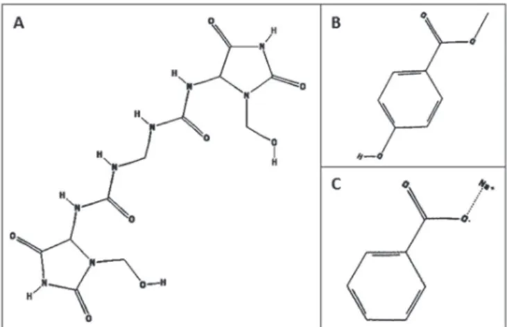
FIGURE 1 - Chemical structures of studied compounds.
(A) Imidazolidinyl urea, (B) methylparaben, and (C) sodium
Evaluation of cytotoxicity using MTT
Cell viability was measured using the standard
methylthiazol tetrazolium (MTT) assay, as previously described by Mosmmann (1983). In brief, 5 × 10⁴ viable
cells were seeded in clear 96-well flat-bottom plates
(Corning, USA) using IMDM supplemented with 10% FBS and incubated with different concentrations of the preservatives for 24 hours. Afterward, 10 μL of MTT was added to each well (5 mg/mL/well) and incubated for 4 hours. Following the incubation, 150 μL of 10% sodium dodecyl sulfate solution in MiliQ H2O was added to each
well and incubated overnight at 37°C to solubilize the
formazan crystals. The absorbance was measured at 595
nm using a FlexStation 3 Multi-Mode Benchtop Reader
(Molecular Devices, Sunnyvale, CA, USA).
Evaluation of cell cycle effects using propidium iodide staining
For cell cycle analysis, 5 × 10⁵ CCD1072Sk cells
were seeded in 6-well plates and incubated with different concentrations of the preservatives for 24 hours. These cells were then harvested, washed with phosphate-buffered saline (PBS), and fixed in 50% ice-cold ethanol for 30 minutes. The fixed cells were resuspended in RNase/PBS solution (20 µg/mL) for 30 minutes, after which propidium iodide (PI) was added to each sample at a concentration of 50 µg/mL. Following this, 105 events were analyzed using
a BD FACSCalibur Flow Cytometry System (Becton-Dickinson, Mountain View, CA, USA). Data was acquired using the CellQuest software (Becton-Dickinson), and the results were analyzed using the FlowJo software (Tree Star, Oregon, USA).
Cell death analysis by flow cytometry using Annexin V-FITC/PI staining
CCD1072Sk cells treated with different concentrations of the preservatives for 24 hours were stained with FITC-conjugated Annexin V and PI according to the manufacturer’s instructions (Annexin V/FITC Apoptosis Detection Kit, BD Pharmingen, CA, USA). The populations of Annexin V−/PI− (viable
cells), Annexin V+/PI− (indicative of apoptosis), Annexin V−/PI+ (indicative of necrosis), and Annexin V+/PI+ (indicative of necroptosis) cells were evaluated by flow
cytometry using a BD FACSCalibur flow cytometer.
Data was acquired using the CellQuest software, and the results were analyzed using the FlowJo software (Tree Star).
Determination of the mitochondrial membrane potential (∆Ψm) using flow cytometric analysis.
The mitochondrial membrane potential (ΔΨm) was
measured in the treated CCD1072Sk cells using flow
cytometric analysis. These cells were collected in FACS
tubes (BD Biosciences Discovery Labware, MA, USA) and stained with TMRE (50 nM) for 20 min. Following
this, 105 events were analyzed using a BD FACSCalibur
flow cytometer. Data was acquired using the CellQuest
software. Log scale fluorescence histograms were
analyzed for the median relative fluorescent unit (RFU)
intensity using the FlowJo software.
Cleaved caspase-3 levels in the treated cells
Caspase-3 activation was evaluated in CCD1072Sk cells treated with the investigated preservatives for 24 hours using the flow cytometric analysis of endogenous levels of the caspase-3 large fragment (17/19 kDa) according to the manufacturer’s instructions (Cell Signaling Technology). These cells were stained with anti-cleaved caspase-3 (Asp175) primary and Alexa Fluor
488-conjugated secondary antibodies for 1 hour in the
dark. Following this, 105 events were analyzed using a BD
FACSCalibur flow cytometer. Data was acquired using the CellQuest software, and the results were analyzed using
FlowJo software.
Statistical analysis
The obtained results were expressed as the mean
± standard error of mean (SEM) from at least three
independent experiments, unless stated otherwise. Paired
data was evaluated by Student’s t-test. One-way analysis
of variance (ANOVA) was used for multiple comparisons. A p value of <0.05 was considered significant.
RESULTS AND DISCUSSION
Preservatives are biocidal chemicals added to cosmetic products, topical medications, and other pharmaceuticals to protect them against microbial spoilage
and to protect the users against infection (Sasseville, 2004). The results of the present study show that only
IMU exhibited cytotoxic activity against the investigated
fibroblasts. According to the results presented in Figure 2A, IMU significantly reduced cell viability at a concentration
of 1%, as shown using the MTT assay, which shows the activity of succinate dehydrogenase and the reduction rate
(Mosmann, 1983). Subsequent testing was performed
using the same concentration of all preservatives to
confirm their activities on different cellular pathways. PI staining is used to label DNA in the investigated
cells, allowing the observation of cell cycle phases as well as the fraction of hypodiploid cells belonging to the
sub-G1 phase. This fraction comprises dead or dying cells in which the DNA content is diminished possibly due to DNA fragmentation, a classical feature of apoptosis
(Riccardi, Nicoletti, 2006). According to Shu et al. (2002),
exposure to ionizing radiation, chemical product treatment,
and oxidative stress represent the possible causes leading
to cellular stagnation in the sub-G1 phase. IMU (1%) treatment led to a significant increase in the percentage of cells in the sub-G1 phase, indicating cell death (Figure 2B and 2C). The observed higher levels of cell death
determined here in contrast to those determined using the MTT assay are due to the PI staining of the genetic
FIGURE 2 – (A) CCD1072Sk cell viability and cell death analyses following the exposure to preservatives. Cells were treated
with imidazolidinyl urea (A), methylparaben (B), and sodium benzoate (C) at the concentrations of 0.01%, 0.1%, and 1.0% for
24 h. (B and C) Percentage of the treated cells in the sub-G1 phase and related histograms. (D) Representative histograms of the
material of the treated cells rather than the assessment of
the mitochondrial enzyme functions (Riccardi, Nicoletti, 2006; Mosmann, 1983; Vermes et al., 1995).
To determine the cell death type induced by
preservatives, we used the Annexin V-FITC/PI double staining test. A significant increase in Annexin V+/PI+ and Annexin V+/PI− cellular fractions, of about 15%
and 10%, respectively, was observed in samples treated with IMU compared with that observed in the untreated
group (Figure 2D). These results indicate that IMU induces programmed cell death in CCD1072Sk cells. In
contrast, cells treated with MP and SB did not undergo
the externalization of phosphatidylserine, a marker of
apoptosis. Finally, the investigated preservatives did
not induce a significant increase in the Annexin V−/PI+ population, which is a fraction of cells with permeabilized
membranes, suggesting the induction of necrotic cell death
(Nikoletopoulou et al., 2013).
Considering the cytotoxicity of IMU and the
induction of different cell death pathways, previous studies
have described that this compound induces apoptosis. Anselmi et al. (2002) reported the induction of apoptosis
and necrosis in human promyelocytic leukemia HL60
cells by four preservatives, including IMU. IMU at low
concentrations (0.01% and 0.1%) led to a significant
reduction in the viability of HL60 cells, together with an
increase in the apoptosis marker levels. However, at higher concentrations (0.5%–1%), necrosis was detected as the
predominant cell death type.
Mitochondria are involved in the processes of cell survival, development, and death due to many intracellular signaling pathways contributing to ATP synthesis, calcium homeostasis, reactive oxygen
species (ROS) production, and others (Tait, Green, 2010). Dewson and Kluck (2009) described that the
receptor binding of extracellular death ligands induces
the oligomerization of proapoptotic BH3-only proteins (Bax, Bak) in the cytosol with the subsequent formation
of a pore in the mitochondrial outer membrane.
Mitochondrial outer membrane permeabilization results
in the release of apoptosis-inducing intermembrane
proteins (e.g., cytochrome c, SMAC/DIABLO, and AIF)
into the cytosol and ultimately leads to apoptosis. (Δψm) is directly linked to the integrity of the mitochondrial
membrane and transition pore opening, leading to the
loss of potential and water influx into the mitochondrial
matrix, which further results in the loss of mitochondrial
function (Gottlieb, Vander Heiden, Thompson, 2000).
In many cell types, this process is crucial for apoptosis
initiation (Ferri, Kroemer, 2001; Fulda, Debatin, 2006). Therefore, Δψm determination allows the
indirect evaluation of mitochondrial integrity, thereby contributing to the initiation and perpetuation of cell death.
To detect the role of mitochondria in
preservative-induced cell death, CCD1072Sk cells were treated with 1% preservative concentrations for 6 h, and Δψm was
assayed by determining the TMRE incorporation. TMRE is a lipophilic cationic dye that readily accumulates inside
the active mitochondria; however, it is not sequestered by depolarized mitochondria (Scaduto Jr., Grotyohann, 1999; Alirol, Martinou, 2006). FCCP, a strong ionophore
uncoupler of oxidative phosphorylation, was used as a
positive control of mitochondrial depolarization. FCCP
interrupts ATP synthesis by transporting protons across the internal mitochondrial membrane, resulting in the complete dissipation of mitochondrial membrane potential
(Zablockaite et al., 2007). Our results demonstrated that
the 6-h IMU treatment leads to a significant reduction of Δψm, indicating the involvement of the mitochondrial cell
death pathway (Figure 3A and 3B).
Activation of caspases is a hallmark of apoptosis.
Caspase-3 is one of the main caspases believed to be a
key enzyme in programed cell death processes (Cohen, 1997; Olsson, Zhivotovsky, 2011; Mcilwain, Berger, Mak, 2013). Here, previous results indicated that caspase-3 is most likely activated in response to the release of mitochondrial intermembrane space proteins. To confirm this, CCD1072Sk cells were treated with the preservatives at the concentrations of 1% for 24 h, and anti-cleaved
caspase-3 antibodies were used for the detection of this protein. IMU significantly activated caspase-3 in
comparison with other preservatives (Figure 3C and 3D), indicating that fibroblasts undergo caspase-dependent cell death following this treatment, most likely induced by the
activation of mitochondrial pathway.
In agreement with our results, Yadav et al. (2016)
showed previously that SB is not cytotoxic on splenocytes
at concentrations of up to 1 mg/mL. However, Park et al.
(2011), using rat cortical neuron cell primary cultures,
reported that the viability of these cells decreases following the preservative treatment. Furthermore, as a hydroxyl scavenger, SB was shown to decrease drug
cytotoxicity in a manner similar to hydroxyurea in L5178Y leukemia cells (Przybyszewski, Kopeć-Szlezak, Malec, 1987), partly explaining the lack of apoptotic marker
activation by SB in our study.
Finally, we showed that MP did not demonstrate cytotoxicity against the studied cells. These results are supported by the results described by Smith and
family (a formaldehyde donor), parabens, and mixtures
of organic acids against BALB/C mouse fibroblast cells after 1 hour of exposure. Parabens were shown to have lower cytotoxicity on these cells than the other preservatives tested. Other studies confirmed the low toxicity of parabens as well, including that of Soni,
Carabin, and Burdock (2005) who showed that methyl and propylparaben at the concentrations of 0.25% and 0.05%,
respectively, did not induce hemolysis in human and rabbit erythrocytes. In contrast, Carvalho et al. (2012) evaluated
the potential of some preservatives for the induction of apoptosis, necrosis, and genotoxicity against HDF cells
(human fibroblasts) following the 24hours of exposure.
Their results showed that methyl and propylparaben have
increased genotoxic potential. These results differ from the results reported in this study most likely because of the use of different cell lines, reagents, methodologies, exposure
times, and concentrations of preservatives.
CONCLUSION
Cytotoxicity and safety assessment of preservatives frequently used in pharmaceutical products are important parameters that need to be evaluated because they can help elucidate the mechanisms of action of products that are regularly consumed by a wide population of users. In
this study, we showed that IMU has cytotoxic effects on human fibroblasts. Moreover, IMU treatment of human
FIGURE 3 – (A) Representative histograms and (B) the respective bar plots showing the loss of ΔΨm in CCD1072Sk cells treated
for 6 h with 1% preservatives: imidazolidinyl urea (A), methylparaben (B), and sodium benzoate (C). FCCP was used as positive
control. **p < 0.01 and ***p < 0.001 compared with the untreated control group (one-way analysis of variance/Tukey test). (C)
Percentage of caspase-3 activation and (D) representative histograms of CCD1072Sk cells treated with the same preservatives.
fibroblasts was shown to result in an increased cell death level, a significant increase in DNA fragmentation, externalization of phosphatidylserine, and activation of caspase-3. Furthermore, the significant depolarization
of mitochondria induced by IMU suggests that the main stimulus for the initiation of programmed cell death is the release of mitochondrial proteins into the cytosol
following outer membrane permeabilization. MP and SB
did not show cytotoxicity on human fibroblasts under the conditions used here. Our data indicate that human
fibroblasts are more resistant to the exposure to MP and
SB in comparison with that to IMU under the same test conditions.
ACKNOWLEDGMENTS
This work was supported by grants from the Conselho Nacional de Desenvolvimento Científico e Tecnológico (CNPq) and CAPES. The authors thank Synth
Laboratory for its material support for this research.
REFERENCES
Alirol E, Martinou JC. Mitochondria and cancer: is there a
morphological connection? Oncogene. 2006;25(34):4706-16.
Anselmi C, Ettorre A, Andreassi M, Centini M, Neri P, Di
Stefano A. In vitro induction of apoptosis vs. necrosis by
widely used preservatives: 2-phenoxyethanol, a mixture of isothiazolinones, imidazolidinyl urea and 1,2-pentanediol. Biochem Pharmacol. 2002;63(3):437-53.
Bordel-Gómez MA, Miranda-Romero A, Castrodeza-Sanz J. Epidemiology of contact dermatitis: prevalence of sensitization
to different allergen and associated factors. Actas
Dermo-Sifiliogr. 2010;101(1):59-75.
Brasil. Agência Nacional de Vigilância Sanitária. Resolução n.162, de 12 de setembro de 2001. Lista de conservantes
permitidos para produtos de higiene pessoal, cosméticos e
perfumes. Diário Oficial da União, 12 set 2001; Seção 1.
Byford JR, Shaw LE, Drew MG, Pope GS, Sauer MJ, Darbre
PD. Oestrogenic activity of parabens in MCF7 human breast
cancer cells. J Steroid Biochem Mol Biol. 2002;80(1):49-60.
Carvalho CM, Menezes PM, Letenski GC, Praes CE, Feferman
IH, Lorencini M. In vitro induction of apoptosis, necrosis and
genotoxicity by cosmetic preservatives: application of flow
cytometry as a complementary analysis by NRU. Int J Cosmet Sci. 2012;34(2):176-82.
Castelain F, Castelain M. Parabens: a real hazard or a scary story? Eur J Dermatol. 2012;22(6):723-7.
Cohen GM. Caspases: the executioners of apoptosis. Biochem J. 1997;326(pt 1):1-16.
Dewson G, Kluck RM. Mechanisms by which Bak and Bax permeabilize mitochondria during apoptosis. J Cell Sci. 2009;122(pt16):2801-8.
European Comission. Scientific Comittee on Consumer Safety. Opinion on parabens. 2011. (COLIPA n.P82) (SCCS/1348/10 Revision 22 March 2011). Available from: http://ec.europa. eu/health/scientific_committees/consumer_safety/docs/ sccs_o_041.pdf.
Ferri KR, Kroemer G. Organelle-specific initiation of cell death pathways. Nat Cell Biol. 2001;3(11):255-63.
Fulda S, Debatin KM. Extrinsic versus intrinsic apoptosis
p a t h w a y s i n a n t i c a n c e r c h e m o t h e r a p y. O n c o g e n e .
2006;25(34):4798-811.
García-Gavín J, González-Vilas D, Fernández-Redondo V,
Toribo J. Allergic contact dermatitis in a girl due to several
cosmetics containing diazolidinyl-urea or imidazolidinyl-urea. Contact Dermatitis. 2010;63(1):49-50.
Gonçalves SD. Razões para preservar cosméticos. São Paulo: Proserv Química;2010. p.22.
Gottlieb E, Vander Heiden MG, Thompson CB. Bcl-XL prevents
the initial decrease in mitochondrial membrane potential and subsequent reactive oxygen species production during tumor necrosis factor alpha-induced apoptosis. Mol Cell Biol.
2000;20(15):5680-9.
Groot AC, Veenstra M. Formaldehyde-releasers in cosmetics in the USA and in Europe. Contact Dermatitis. 2010;62(4):221-4.
Heydaryinia A, Veissi M, Sadadi A. A comparative study of the effects of the two preservatives, sodium benzoate and
potassium sorbate on Aspergillus niger and Penicillium notatum.
Jundishapur J Microbiol. 2011;4(4):301-7.
Jin CY, Zhu BS, Wang XF, Lu QH. Cytotoxicity of titanium
dioxide nanoparticles in mouse fibroblast cells. Chem Res
Kireche M, Gimenez-Arnau E, Lepoittevin J. Preservatives
in cosmetics: reactivity of allergenic formaldehyde-releasers
to-ward amino acids through breakdown products other than
formaldehyde. Contact Dermatitis. 2010;63(4):192-202.
Latorre N, Borrego L, Fernández-Redondo V, García-Bravo B, Giménez-Arnau AM, Sánchez J, Silvestre JF. Patch testing with formaldehyde and formaldehyde-releasers: multicentre study
in Spain (2005-2009). Contact Dermatitis. 2011;65(5):286-92.
Mcilwain DR, Berger T, Mak TW. Caspase functions in cell death and disease. Cold Spring Harb Perspect Biol. 2013;5(4, a008656):1-29.
Mosmann T. Rapid colorimetric assay for cellular growth and survival: application to proliferation and cytotoxicity assays. J
Immunol Methods. 1983;65(1/2):55-63.
Mowad CM. Allergic contact dermatitis caused by parabens: 2 case reports and a review. Am J Contact Dermat. 2000;11(1):53-6.
Nikoletopoulou V, Markaki M, Palikaras K, Tavernarakis N. Crosstalk between apoptosis, necrosis and autophagy. Biochim Biophys Acta. 2013;1833(12):3448-59.
Okubo T, Yokoyama Y, Kano K, Kano I. ER-dependent
estrogenic activity of parabens assessed by proliferation of
human breast cancer MCF-7 cells and expression of ERα and PR. Food Chem Toxicol. 2001;39(12):1225-32.
Olsson M, Zhivotovsky B. Caspases and cancer. Cell Death Differ. 2011;18(9):1441-9.
Orton DI, Wilkinson JD. Cosmetic allergy: incidence, diagnosis and management. Am J Clin Dermatol. 2004;5(5):327-37.
Ozaki H, Sugihara K, Watanabe Y, Fujino C, Uramaru N, Sone T, Ohta S, Kitamura S. Comparative study of the hydrolytic
metabolism of methyl-, ethyl-, propyl-, butyl-, heptyl- and dodecylparaben by microsomes of various rat and human
tissues. Xenobiotica. 2012;43(12):1064-72.
Park HW, Park EH, Yun HM, Rhim H. Sodium
benzoate-mediated cytotoxicity in mammalian cells. J Food Biochem,
2011; 35(4):1034-46.
Przybyszewski WM, Kopeć-Szlezak J, Malec J. Protection of L5178Y cells by vitamin E against acute hydroxyurea toxicity
does not change the efficiency of ribonucleotide reductase-mediated hydroxyurea-induced cytotoxic events. Cancer Lett.
1987; 34(3):337-44.
PubChem. Imidazolidinyl Urea. [cited 2005a Jul 19]. Available
from: https://pubchem.ncbi.nlm.nih.gov/compound/38258.
(accessed Jun 14, 2016).
PubChem. Methyl 4-hydroxybenzoate. [cited 2005b Mar 26]. Available from: https://pubchem.ncbi.nlm.nih.gov/
compound/7456. (accessed Jun 14, 2016).
PubChem. Sodium Benzoate. [cited 2005c Mar 27]. Available
from: https://pubchem.ncbi.nlm.nih.gov/compound/517055.
(accessed Jun 14, 2016).
Rebello T. Guia de produtos cosméticos. 6th ed. São Paulo: Senac; 2005. 161 p.
Riccardi C, Nicoletti I. Analysis of apoptosis by propidium iodide staining and flow cytometry. Nat Protoc. 2006;1(3):1458-61.
Rowe RC, Sheskey PJ, Quinn ME. Handbook of pharmaceutical
excipients. 6th ed. London: Pharmaceutical Press; 2009. 888 p.
Sasseville D, Alfalah M, Lacroix JP. “Parabenoia” debunked, or “Who’s afraid of Parabens?” Dermatitis. 2015;26(6):254-9.
Sasseville D. Hypersensitivity to preservatives. Dermatol Ther.
2004;17(3):251-61.
Scaduto Jr. RC, Grotyohann LW. Measurement of mitochondrial membrane potential using fluorescent rhodamine derivatives. Biophys J. 1999;76(pt 1):469-77.
Shu B, Wu Z, Hao L, Zeng D, Feng G, Lin Y. Experimental study on He-Ne laser irradiation to inhibit scar fibroblast growth in culture. Chin J Traumatol. 2002;5(4):246-9.
Smith CN, Alexander BR. The relative cytotoxicity of personal
care preservative systems in Balb/C 3T3 clone A31 embryonic
mouse cells and the effect of selected preservative systems upon the toxicity of a standard rinse-off formulation. Toxicol In Vitro.
2005;19(7):963-9.
Soni MG, Carabin IG, Burdock GA. Safety assessment of esters of p-hydroxybenzoic acid (parabens). Food Chem Toxicol. 2005;43(7):999-1003.
Statham BN, Smith EV, Bodger OG, Green CM, King CM, Ormerod AD, Sansom JE, English JS, Wilkinson MS, Gawkrodger DJ, Chowdhury MM. Concomitant contact allergy to methylchloroisothiazolinone/ methylisothiazolinone and
formaldehyde-releasing preservatives. Contact Dermatitis.
In vitro cytotoxicity of chemical preservatives on human fibroblast cells
Tait SW, Green DR. Mitochondria and cell death: outer membrane permeabilization and beyond. Nat Rev Mol Cell Biol. 2010;11(9):621-32.
Thyssen JP, Engkilde K, Lundov MD, Carlsen BC, Menné
T, Johansen JD. Temporal trends of preservative allergy in
Denmark (1985-2008). Contact Dermatitis. 2010;62(2):102-9.
Tomankova K, Kejlova K, Binder S, Daskova A, Zapletalova J, Bendova H, Kolarova H, Jirova D. In vitro cytotoxicity and phototoxicity study of cosmetics colorants. Toxicol In Vitro. 2011;25(6):1242-50.
Urcan E, Haertel U, Styllou M, Hickel R, Scherthan H, Reichl FX. Real-time xCELLigence impedance analysis of the
cytotoxicity of dental composite components on human gingival
fibroblasts. Dent Mater. 2010;26(1):51-8.
Vermes I, Haanen C, Steffens-Nakken H, Reutelingsperger
C. A novel assay for apoptosis. Flow cytometric detection of phosphatidylserine expression on early apoptotic cells
using fluorescein labelled Annexin V. J Immunol Methods. 1995;184(1):39-51.
Yadav A, Kumar A, Das M, Tripathi A. Sodium benzoate, a food preservative, affects the functional and activation status of splenocytes at non cytotoxic dose. Food Chem Toxicol. 2016; 88:40-7.
Zablockaite D, Gendiviliene V, Martisiene I, Jurevicius J. Effect
of oxidative phosphorylation uncoupler FCCP and F1F0-ATPase inhibitor oligomycin on the electromechanical activity of human
myocardium. Adv Med Sci. 2007;52:89-93.
Received for publication on 27th October 2016