Performance of Conventional Cytology and
Colposcopy for the Diagnosis of Cervical
Squamous and Glandular Neoplasias
Desempenho da citologia convencional e da colposcopia para o
diagnóstico de neoplasias cervicais escamosas e glandulares
Giselle Fachetti-Machado
1Rosane Ribeiro Figueiredo-Alves
1Marise Amaral Rebouças Moreira
11Health Sciences Postgraduate Program, Universidade Federal de Goiás, Goiânia, GO, Brazil
Rev Bras Ginecol Obstet 2018;40:410–416.
Address for correspondence Giselle Fachetti-Machado, MD, MSc, Universidade Federal de Goiás, Av. T4, esq. com T13, 1478, Salas 91B e 92B, Setor Bueno, Goiânia, GO, 74230-030, Brazil
(e-mail: gfachettimachado@uol.com.br).
Keywords
►
uterine cervical
neoplasms
►
cervical
intraepithelial
neoplasia
►
squamous
intraepithelial lesions
of the cervix
►
adenocarcinoma in
situ
►
papanicolaou test
►
colposcopy
►
sensitivity and
speci
fi
city
Abstract
Objective
To estimate the cytological and colposcopic performances for the diagnosis of
cervical neoplasias.
Methods
Cross-sectional retrospective study with data from patients
’
charts. The
participants underwent colposcopy, guided biopsies, and excision when needed. The
cytological and colposcopic categorization followed the Bethesda System and the
interna-tional colposcopic terminologies. The cytology and colposcopy performances were
evaluated by sensitivity (SE), speci
fi
city (SP), positive predictive value (PPV), and negative
predictive value (NPV) analyses with 95% con
fi
dence interval (95% CI).
Results
From 1,571 participants, a total of 1,154 (73.4%) were diagnosed with
cervical squamous intraepithelial neoplasia grade 2 or worse (CIN 2
þ), 114 (7.2%)
with adenocarcinoma in situ or worse (AIS
þ), 615 (39.2%) presented atypical squamous
cells, cannot exclude high-grade squamous intraepithelial lesion or worse (ASC-H
þ)
cytology, and 934 (59.4%) presented major or suspicious for invasion colposcopic
abnormalities. The SE, SP, PPV, and NPV of ASC-H
þfor diagnoses of CIN 2
þand AIS
þwere, respectively: 44% (95% CI: 41
–
47) and 72% (95% CI: 67
–
76), 79% (95% CI: 77
–
81)
and 79% (95% CI: 75
–
83), 88% (95% CI: 87
–
90) and 55% (95% CI: 50
–
60), and 28% (95%
CI: 26
–
31) and 88% (95% CI: 85
–
91). The SE, SP, PPV, and NPV of major or suspicious for
invasion colposcopic abnormalities for diagnoses of CIN 2
þand AIS
þwere, respectively:
62% (95% CI: 60
–
65) and 86% (95% CI: 83
–
89), 59% (95% CI: 57
–
62) and 59% (95% CI:
55
–
64), 85% (95% CI: 83
–
87) and 44% (95% CI: 40
–
49), and 29% (95% CI: 27
–
32) and
92% (95% CI: 89
–
94).
Conclusion
The SE analyses results of ASC-H
þand major or suspicious for invasion
colposcopic abnormalities were higher for diagnoses of glandular neoplasias. These
results con
fi
rm the role of cytology in identifying women at risk who will have their
fi
nal
diagnoses settled by colposcopy and histology.
received February 3, 2018 accepted May 17, 2018 published online July 11, 2018
DOI https://doi.org/ 10.1055/s-0038-1666995. ISSN 0100-7203.
Copyright © 2018 by Thieme Revinter Publicações Ltda, Rio de Janeiro, Brazil
Introduction
Well organized invasive cervical cancer (ICC) screening pro-grams based on cytology, colposcopically-guided biopsies, and treatment of precursor neoplasias have led to an important decrease in the ICC incidence and mortality. However, none of these programs could eradicate cervical cancer in any part of the globe.1 In fact, evidences have shown that in the last
decades, the incidence of cervical adenocarcinoma (AC) has risen, especially in younger women, denoting the lack of impact of these programs for this particular histological type.2
Although the triage of patients at risk for ICC precursor lesions is based on cytological abnormalities, these do not have adequate specificity to indicate treatment to all women with suchfindings.3Therefore, colposcopically-guided
biop-sy was added to the biop-system aiming to select which women with abnormal cytology would actually need treatment.4
Recently, as expected, a decrease in the prevalence of cervical intraepithelial neoplasias has been observed in developed countries due to high human papillomavirus (HPV) vaccination coverage.5It is possible that in vaccinated
populations, the neoplasias that will still be found will show more subtle appearance and smaller sizes.6At the same time,
a better diagnostic performance has been reached with the introduction of new screening programs with high
sensitiv-ity, which added DNA detection methods to cytology, result-ing in a decrease in false-negatives,6and an improvement in
cytological detection rates, even for more discrete neopla-sias. Nonetheless, knowledge about colposcopy performance is still needed, especially regarding the recognition of subtle neoplasias,6which could be missed since colposcopic
crite-rion has not been updated to the new scenario.
Thehistopathological diagnosis of specimens obtained using colposcopically-guided biopsy has been traditionally consid-ered the gold standard for cytological and colposcopic analyses. However, this assumption has an intrinsicflaw.4Any mistakes
made when choosing the site to take the biopsies, due to a misinterpretation of the colposcopic images, would necessarily lead to a bias, compromising the results of these analyses.
This study estimated cytological and colposcopic perfor-mance to predict thefinal diagnosis of squamous and glan-dular neoplasias and determine the performance of these diagnostic tests used in clinicians’daily practice, considering
that thefinal diagnosis was based on excisional specimens.
Methods
This cross-sectional epidemiological study was based on data collected over a period of 24 years, from April 16, 1991 to November 26, 2015, in a private colposcopy health unit in
Resumo
Objetivo
Estimar o desempenho da citologia e colposcopia no diagnóstico das
neoplasias cervicais.
Métodos
Estudo retrospectivo de corte transversal com dados coletados em
pron-tuários. Foram incluídas participantes que foram submetidas a colposcopia, biópsia e
excisão quando necessário. A categorização da citologia e da colposcopia seguiram a
terminologia de Bethesda e a classi
fi
cação colposcópica internacional. Os
desempe-nhos da citologia e colposcopia foram avaliados por análises de sensibilidade (S),
especi
fi
cidade (E), valor preditivo positivo (VPP) e valor preditivo negativo (VPN), com
intervalos de con
fi
ança de 95% (IC 95%).
Resultados
Das 1.571 participantes, um total de 1.154 (73,4%) foram diagnosticadas
com neoplasia intraepitelial escamosa cervical de grau 2 ou mais grave (NIC 2
þ), 114
(7,2%) com adenocarcinoma in situ ou mais grave (AIS
þ), 615 (39,2%) apresentaram
células escamosas atípicas de signi
fi
cado indeterminado, quando não se pode excluir
lesão intraepitelial de alto grau ou mais grave (ASC-H
þ) e 934 (59,4%) tiveram achados
colposcópicos maiores ou suspeitos de invasão. Os valores de S, E, VPP e VPN das
ASC-H
þpara o diagnóstico de NIC 2
þe AIS
þforam, respectivamente: 44% (IC 95%: 41
–
47) e
72% (IC 95%: 67
–
76), 79% (IC 95%: 77
–
81) e 79% (IC 95%: 75
–
83), 88% (IC 95%: 87
–
90) e
55% (IC 95%: 50
–
60) e 28% (IC 95%: 26
–
31) e 88% (IC 95%: 85
–
91). Os valores de S, E,
VPP e VPN dos achados colposcópicos maiores ou suspeitos de invasão para o
diagnóstico de NIC 2
þe AIS
þforam, respectivamente: 62% (IC 95%: 60
–
65) e 86%
(IC 95%: 83
–
89), 59% (IC 95%: 57
–
62) e 59% (IC 95%: 55
–
64), 85% (IC 95%: 83
–
87) e 44%
(IC 95%: 40
–
49) e 29% (IC 95%: 27
–
32) e 92% (IC 95%: 89
–
94).
Conclusão
Os resultados das análises de S de ASC-H
þe achados colposcópicos
maiores ou suspeitos de invasão foram mais elevados para o diagnóstico das neoplasias
glandulares. Esses resultados con
fi
rmam o papel da citologia na identi
fi
cação de
mulheres em risco que terão seus diagnósticos de
fi
nidos por colposcopia e histologia.
Palavras-chave
►
neoplasias do colo do
útero
►
neoplasia
intraepitelial cervical
►
lesões intraepiteliais
escamosas cervicais
►
adenocarcinoma in
situ
►
exame
colpocitológico
►
colposcopia
Goiânia, GO, Brazil. The project was approved by the Research Ethics Committee of the Hospital das Clínicas of the Universi-dade Federal de Goiás (CAAE no. 58228016.1.0000.5078).
A total of 11,999 medical records of patients referred to colposcopy were reviewed. Among them, 1,527 participants were selected for having their final diagnoses settled by
histopathological analyses of transformation zone excision (TZE) pieces, 7 by analysis of cold knife conization (CKC) pieces, and 37 with invasive cervical neoplasias diagnosed in the initial biopsy fragment. Therefore, thefinal sample was composed of 1,571 participants.
All the patients without a histopathological analysis of an excisional specimen were excluded from the study, even if they had cytological abnormalities, and regardless of wheth-er they had normal or abnormal histopathology. Exceptions were made only in the cases with stromal invasion observed in the initial biopsy fragment, because once invasion is found, no worst diagnosis is possible, making a subsequent excisional procedure unnecessary in most cases.
The data obtained from patients’ charts, colposcopic
reports, and computer software Diagnose Pro 6, Ginecologia e Obstetrícia, prontuário eletrônico e captura de imagens. (LPT4 sistemas de informação, Curitiba, Paraná, Brazil) and Zscan 7 Gineco, version 7.4 (Zscan Software, 2001-2016, Goiânia, Goiás, Brazil) imagefiles were coded and kept on a 2013 Excel spreadsheet (Microsoft Corp., Redmond, WA, USA). The cytological, colposcopic, and histopathological data included referral cytology, colposcopicfindings, visual-ization of squamocolumnar junction (SCJ), the histopatho-logical diagnosis of an excision piece or a hysterectomy piece, and the histopathological report of biopsy fragments.
The cytological abnormalities were classified as proposed by the Bethesda terminology, updated in 2014:7 atypical
squamous cells of undetermined significance (ASC-US), low-grade squamous intraepithelial lesion (LSIL), atypical squa-mous cells, cannot exclude high-grade squasqua-mous intraepithe-lial lesion (ASC-H), high-grade squamous intraepitheintraepithe-lial lesion (HSIL), squamous cell carcinoma (SCC), atypical glandular cells (AGC), adenocarcinoma in situ (AIS), and adenocarcinoma (AC). The ASC-Hþ group had a cut-off point settled in the
cytological results of ASC-H or worse, a threshold in which the patients’management changes to immediate referral to
colposcopy rather than the mere cytological follow-up.8This
group included all patients with cytological abnormalities classified as ASC-H, HSIL, SCC, AIS, and AC.
A single colposcopist performed the exams using atfirst a 5-fold DFV videocolposcope (D. F. Vasconcellos, Valença, RJ, Brazil) and afterward a Medpej PE 7000 MDL videocolpo-scope also withfive levels of magnification (6x, 10x, 16x, 25x and 40x). Initially 5% and 10% acetic acid solutions were applied followed by the spraying of Schiller's solution, at this point, the needed biopsies were taken with Gaylor-Medina forceps. Endocervical currettages with a Kevorkian curette were performed whenever necessary.
The colposcopic images were reviewed by the examiner and the 2011 International Federation of Cervical Pathology and Colposcopy (IFCPC)9 terminology was used to group
them as follows: normal, minorfindings, majorfindings, or
suspicious for invasion. Minorfindings elementary images included:fine mosaic,fine punctuation, and thin acetowhite epithelium with geographic borders. Majorfindings
includ-ed: coarse mosaic, coarse punctuation, and dense acetowhite epithelium with sharp border, with or without ridge and inner border sign, and also cuffed crypt openings. The images considered suspicious for invasion included: atypical or fragile vessels, irregular surface, exophytic lesion, necrosis, necrotic ulceration, and gross neoplasm.9
Moreover, new colposcopic images similar to those de-scribed by Wright et al10were added to the majorfindings
category. It is necessary to emphasize that these images are not accredited by IFCPC terminology.
The cut-off point of colposcopic images was settled at images worse than minor findings, since taking biopsies
from this type offindings is considered needless by many colposcopists. Thus, the colposcopic findings were sorted into two groups: 1) normal and minor findings; 2) major findings and suspicious for invasionfindings.
A Wavetronic 5000 Digital Hf Surgical Unit (Loktal Medical Electronics Ind. Com. Ltda, São Paulo, SP, Brazil) was employed to perform TZE under colposcopic guidance and local anes-tesia, using its handswitch pencil and cord with loop electro-des, at 50% of the coagulation power and the shear power regulated to output 8. A single-fragment resection was per-formed unless a large transformation zone was present.
A single examiner performed all histopathological analy-ses and categorized thefindings following the World Health Organization International Tumors Classification11and the
Richart Classification for cervical intraepithelial
neopla-sias.12Thefinal diagnosis was defined as the most severe
histopathological diagnosis among specimens of excision or hysterectomy, except when invasion was already found in fragments of initial biopsies.
The data analysis was performed using the Statistical Package for Social Sciences (SPSS) for Windows version 21.0 (IBM Corp., Armonk, NY, USA). Descriptive analyses of the sociodemographic, behavioral, and clinical features as well as cytological abnormalities, colposcopicfindings, and histopathological diagnosis were performed.
Diagnostic performance of cytological abnormalities and colposcopic findings to predict the final diagnosis were evaluated by analysis of sensitivity (SE), specificity (SP), positive predictive value (PPV), and negative predictive value (NPV), with the respective 95% confidence interval (95% CI). Values of SE, SP, PPV, and NPV between 0.00 and 0.40 were considered as poor, between 0.40 and 0.60 as low, between 0.60 and 0.80 as moderate, and between 0.80 and 1 as high.
Results
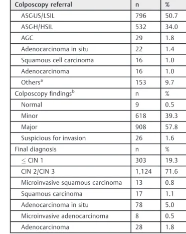
The sociodemographic and behavioral characteristics of the participants in this survey are shown in►Table 1. The mean
age of the participants was 31.7 years and at the first intercourse, it was 18.9 years. A total of 615 (39.6%) patients out of 1,571 were referred to colposcopy due to cytological
Colposcopic images categorized as major or suspicious for invasion were found in 934 (59.4%) patients (►Table 2).
Concerning the final diagnosis, 1,154 (73.4%) participants
had cervical squamous intraepithelial neoplasia grade 2 or worse (CIN 2þ) and 114 (7.2%) had adenocarcinoma in situ or
worse (AISþ) (
►Table 2).
The SE, SP, PPV, and NPV results of ASC-Hþ for the
prediction of CIN 2þ were, respectively: 44% (95% CI 41
–
47), 79% (95% CI: 77–81), 88% (95% CI: 87–90), and 28% (95%
CI: 26–31). The SE, SP, PPV, and NPV results of ASC-Hþfor
the prediction of AISþwere, respectively: 72% (95% CI: 67
–
76), 79% (95% CI: 75–83), 55% (95% CI: 50–60), and 88% (95%
CI: 85–91) (►Table 3).
The SE, SP, PPV, and NPV results of major or suspicious for invasion colposcopicfindings for the diagnosis of CIN 2þwere,
respectively, 62% (95% CI: 60–65), 59% (95% CI: 57–62), 85% (95%
CI: 83–87), and 29% (95% CI: 27–32), whereas to detect AISþthey
were, respectively, 86% (95% CI: 83–89), 59% (95% CI: 55–64),
44% (95% CI: 40–49), and 92% (95% CI: 89–94) (►Table 4).
Discussion
In the present study, the performances of cytological abnor-malities ASC-Hþ and colposcopic major or suspicious for
invasion findings were evaluated to separately predict the
final diagnoses of CIN 2þ and AISþ in a sample of 1,571
participants. The final diagnosis was defined as the most severe report obtained among all biological specimens of
patients who underwent excisional procedure, except when invasion was initially found in biopsy fragments.
The SE of ASC-Hþ(44%) to identify squamous neoplasias
was low; in contrast, SP (79%) and PPV (88%) were moderate and high, respectively. Regarding glandular neoplasias, SE (72%) and SP (79%) of ASC-Hþwere moderate, whereas PPV
(55%) was low. To the best of our knowledge, to date, no studies have simultaneously measured the performance of cytological and colposcopic diagnoses of squamous and glandular neoplasias.
The sensitivity of a test represents its ability to correctly identify unhealthy individuals, while its specificity shows the ability to identify the healthy ones.13Therefore, the low
sensitivity found implies that ASC-Hþ may not sort out a
reasonable number of patients with CIN 2þ, since the
false-negative rate was high (56%). Similarly, the SP of 79% obtained means that absence of ASC-Hþ identi
fies a high number of patients that do not actually have CIN 2þ.
Moderate SE (72%) and SP (79%) of ASC-Hþ to identify
glandular neoplasias mean that ASC-Hþinvolves lower rates
of false-positives (28%) and false-negatives (21%). Analyzed individually, SE of cytology for the detection of glandular
Table 1 Sociodemographic and behavioral profile of 1,571 participants
Variables
Age Years
Range 15–85
Mean (sd) 31.710.8
Marital statusa n %
Single 817 52.0
Married 752 47.9
Age atfirst intercourseb Years
Range 9–47
Mean (sd) 18.93.7
Lifetime sexual partnersc n %
2 624 39.7
>2 756 48.1
Full-term pregnancyd n %
1 1,107 70.5
>1 453 28.8
Tobacco usee n %
Past and current smoker 148 9.4
Never smoker 1,073 68.3
Abbreviation: SD, standard deviation. missing data:a2;b128;c192;d11;
e
350.
Table 2 Cytological, colposcopic, andfinal diagnoses in 1,571 participants
Colposcopy referral n %
ASC-US/LSIL 796 50.7
ASC-H/HSIL 532 34.0
AGC 29 1.8
Adenocarcinoma in situ 22 1.4
Squamous cell carcinoma 16 1.0
Adenocarcinoma 16 1.0
Othersa 153 9.7
Colposcopyfindingsb n %
Normal 9 0.5
Minor 618 39.3
Major 908 57.8
Suspicious for invasion 26 1.6
Final diagnosis n %
CIN 1 303 19.3
CIN 2/CIN 3 1,124 71.6
Microinvasive squamous carcinoma 13 0.8
Squamous carcinoma 17 1.1
Adenocarcinoma in situ 78 5.0
Microinvasive adenocarcinoma 8 0.5
Adenocarcinoma 28 1.8
Abbreviations: AGC, atypical glandular cells; ASC-H, atypical squamous cells, cannot exclude high-grade squamous intraepithelial lesion; ASC-US, atypical squamous cells of undetermined significance; CIN, cervical intraepithelial neoplasia; HSIL, high-grade squamous intraepithelial lesion; LSIL, low-grade squamous intraepithelial lesion.
acervical polip, cervical bleeding, unknown cytological
finding, etc.
neoplasias has been reported in a wide range of values, such as 43.1%14 and 91.2%.15 Those results contrast with the
findings of this study, since most patients with glandular neoplasias were found using ASC-Hþ (72%) cytology, and
most patients without cytological reports of ASC-Hþ(NPV
88%) were truly free of glandular neoplasias. For this reason, when ASC-Hþis found, whether squamous neoplasias have
already been identified or not, it would be safer to exclude the possibility of coexistent glandular neoplasias.
Sensitivity and SP are inherent properties of a test and do not change. However, the predictive values depend on the prevalence of the disease in the study sample.13Therefore,
the PPV will proportionally increase according to the preva-lence of the disease in the studied group. In the present study, on one hand, the high prevalence of squamous neo-plasias implied a high PPV of ASC-Hþfor CIN 2þdetection,
because most participants with ASC-Hþactually had CIN 2þ.
On the other hand, the low prevalence of glandular neo-plasias implied a low PPV of ASC-Hþability to predict AISþ.
Hence, most positive results of ASC-Hþdo not correspond to
patients with glandular neoplasias, but rather to patients with squamous neoplasias.
Evidences involving the performance of cytological ab-normalities for predicting intraepithelial and invasive cervi-cal neoplasias, whether squamous or glandular, were found with SE ranging from 30 to 100%, and SP from 86.8 to 99.3%.16–29This large divergence may be due to the diversity
of cut-off points chosen to consider the tests as positive or negative, as well as to the use of different morphological criteria to interpret cytological smears and classify abnormal
findings. Conversely, a study that used ASC-Hþas a cut-off
point to ascertain the performance for the detection of CIN 2þreported SE of 67.9% and SP of 87%.30
In this study, SE of major or suspicious for invasion colposcopicfindings for the detection of CIN 2þwas
moder-ate (62%), while SP was low (59%). Although these values were different from each other, this difference was not statistically significant, since their confidence interval over-lapped. The major or suspicious for invasion colposcopic
findings failed to identify a substantial number of patients with CIN 2þ. Consequently, the absence of colposcopic
images showing major or suspicious for invasionfindings does not rule out a large number of healthy participants.
These results are in line with a previous study,3and suggest
that guided biopsies are needed by women with cytological abnormalities, even if they present subtle colposcopicfi nd-ings, that is, those colposcopic images classified as minor
findings by the IFCPC. Taking multiple biopsies would also be an acceptable strategy to improve the SE of colposcopy.31
The SE of major or suspicious for invasion colposcopic
findings for the detection of AISþwas high (86%), while the
SP and PPV were low (59% and 44%, respectively). This high SE value found for the detection of AISþimplies that the major or
suspicious for invasion colposcopicfindings express low rates of false-negatives for this singular histopathological type of neoplasia. In view of these colposcopic images, whether squamous neoplasias are suspected or not, the need to exclude a possible coexistent glandular neoplasia still persists.
These results show that the SE values of colposcopic
findings were higher for the detection of squamous (62%)
Table 3 Performance of cytologicalfindings of atypical squamous cells, cannot exclude high-grade squamous intraepithelial lesion or worse (ASC-Hþ
) to predictfinal diagnosis of squamous and glandular cervical neoplasias
Cytological findings
Final diagnosis Estimated performance (%) (95% CI)
ASC-Hþ
CIN 2þ
(n¼1,376)
Positive Negative
Positive 474 63 Sensitivity 44 (41–47)
Specificity 79 (77–81)
Negative 603 236 PPV 88 (87–90)
NPV 28 (26–31)
ASC-Hþ
AISþ
(n¼408) Estimated performance (%) (95% CI)
Positive Negative
Positive 78 63 Sensitivity 72 (67–76)
Specificity 79 (75–83)
Negative 31 236 PPV 55 (50–60)
NPV 88 (85–91)
Abbreviations: AISþ
, adenocarcinoma in situ or worse; ASC-Hþ , atypical squamous cells, cannot exclude high-grade squamous intraepithelial lesion or worse (including ASC-H, HSIL, AIS, SCC, and AC); CIN 2þ
, cervical squamous intraepithelial neoplasia grade 2 or worse; 95% CI, 95% confidence interval; NPV, negative predictive value; PPV, positive predictive value.
Table 4 Performance of major or suspicious for invasion colposcopicfindings to predictfinal diagnosis of squamous and glandular cervical neoplasias
Colposcopic report
Final diagnosis Estimated performance (%) (95% CI)
Major or suspicious for invasion
CIN 2þ
(n¼1,447)
Positive Negative
Positive 713 123 Sensitivity 62 (60–65) Specificity 59 (57–62)
Negative 432 179 PPV 85 (83–87)
NPV 29 (27–32) Major or
suspicious for invasion
AISþ
(n¼416) Estimated performance (%) (95% CI)
Positive Negative
Positive 98 123 Sensitivity 86 (83–89)
Specificity 59 (55–64)
Negative 16 179 PPV 44 (40–49)
and glandular (86%) neoplasias than the cytologicalfindings of ASC-Hþ (44% and 72% for squamous and glandular
neo-plasias, respectively). However, SP values of ASC-Hþ(79% and
79%) were higher than those of colposcopicfindings (59% and 59%) for the detection of these two main histopathological types of neoplasia. Moreover, the SE of major or suspicious for invasion colposcopic findings and ASC-Hþ cytological
findings were higher for glandular (86% and 72%) than for squamous neoplasias (62% and 44%).
The high SE of colposcopy for the detection of glandular neoplasias found in this study was probably due to the inclusion of new subtle images in the major colposcopic
finding category. Such new images are similar to those identified by Wright et al10as suggestive of glandular
neo-plasias and are not described in the International Colpo-scopic Classification of the IFCPC.9They are herein named:
obstructed dilated grouped glands, fused acetowhite villi with invaginated borders, and atypical vessels in cylindrical epithelium area (►Fig. 1).
Considering that the PPV of ASC-Hþ(88%) and major or
suspicious for invasion colposcopic findings (85%) for the detection of CIN 2þwere high, the see and treat protocol,
already endorsed by previous published evidences,32is here
corroborated as a secure option. The sequential use of two tests with high PPV prior to the see and treat protocol substantially decreases the probability of performing unnec-essary excisional procedures in disease-free patients, mainly because both cytological and colposcopic findings must simultaneously be abnormal to indicate such management.
The possibility offinding an unexpected glandular neopla-sia in a see and treat excision piece, estimated to happen in 52% of the patients with final diagnosis of AISþ,33 would not
contraindicate this approach. As a matter of fact, evidences
have shown that outpatient excisional procedures are also suitable for the management of glandular neoplasias.33,34
Conclusion
The performance of cytological abnormalities is somehow different from that of colposcopic findings to predict the diagnoses of AISþand CIN 2þ. Sensitivity of major or
suspi-cious for invasion colposcopicfindings for the diagnoses of
CIN 2þwas moderate. The high PPV values found for ASC-Hþ
and major or suspicious for invasion colposcopicfindings for the detection of CIN 2þendorse the see and treat protocol.
Sensitivity results of ASC-Hþ and major or suspicious for
invasion were higher for the diagnosis of glandular than squamous neoplasias. These results reinforce the role of cytology in sorting out women at risk who should have their diagnosis settled by colposcopy and histopathology.
Contributions
Fachetti-Machado G., Figueiredo-Alves R. R. and Moreira M. A. R. contributed with the project and interpretation of data, writing of the article, critical review of the intellectual content andfinal approval of the version to be published.
Conflicts of Interest
The authors have no conflicts of interest to declare.
References
1 Koss LG. The Papanicolaou test for cervical cancer detection.
A triumph and a tragedy. JAMA 1989;261(05):737–743. Doi:10. 1001/jama.1989.03420050087046
2 Chan PG, Sung HY, Sawaya GF. Changes in cervical cancer incidence
after three decades of screening US women less than 30 years old. Obstet Gynecol 2003;102(04):765–773. Doi:10.1016/S0029-7844 (03)00696-3
3 Massad LS, Jeronimo J, Katki HA, Schiffman M; National Institutes of
Health/American Society for Colposcopy and Cervical Pathology Research Group. The accuracy of colposcopic grading for detection of high-grade cervical intraepithelial neoplasia. J Low Genit Tract Dis 2009;13(03):137–144. Doi:10.1097/LGT.0b013e31819308d4
4 Jordan J, Arbyn M, Martin-Hirsch P, et al. European guidelines for
quality assurance in cervical cancer screening: recommendations for clinical managementofabnormal cervical cytology, part1.Cytopathol-ogy 2008;19(06):342–354. Doi:10.1111/j.1365-2303.2008.00623.x 5 Drolet M, Bénard É, Boily MC, et al. Population-level impact and
herd effects following human papillomavirus vaccination pro-grammes: a systematic review and meta-analysis. Lancet Infect Dis 2015;15(05):565–580. Doi:10.1016/S1473-3099(14)71073-4 6 Jeronimo J, Schiffman M. Colposcopy at a crossroads. Am J Obstet
Gynecol 2006;195(02):349–353. Doi:10.1016/j.ajog.2006.01.091 7 Nayar R, Wilbur DC. The Pap Test and Bethesda 2014.“The reports of my demise have been greatly exaggerated.”(after a quotation from Mark Twain). Acta Cytol 2015;59(02):121–132. Doi:10.1159/0003 81842
8 Ministério da Saúde. Instituto Nacional de Câncer José Alencar
Gomes da Silva. Coordenação de Prevenção e Vigilância. Divisão de Detecção Precoce e Apoio à Organização de Rede. Diretrizes Brasileiras para o Rastreamento do Câncer do Colo do Útero. 2a ed. Rio de Janeiro, RJ: INCA; 2016. http://www1.inca.gov.br/inca/ Arquivos/DDiretrizes_para_o_Rastreamento_do_cancer_do_colo _do_utero_2016_corrigido.pdf. Acessed March 26, 2018 Fig. 1 New colposcopic images are herein named: (A) fused
9 Bornstein J, Bentley J, Bösze P, et al. 2011 colposcopic terminology of
the International Federation for Cervical Pathology and Colposcopy. Obstet Gynecol 2012;120(01):166–172. Doi:10.1097/AOG.0b013e 318254f90c
10 Wright VC, Dubuc-Lissoir J, Ehlen T, Heywood M, Plante M.
Guidelines on adenocarcinoma in situ of the cervix: clinical features and review of management. J Obstet Gynaecol Can 1999;21(07):699–706. Doi:10.1016/S0849-5831(16)30106-9
11 Scully RE, Bonfiglio TA, Kurman RJ, Silverberg SG, Wilkinson EJ. Histological Typing of Female Genital Tract Tumors. 2nd ed. Berlin: Spring Verlag; 1994
12 Buckley CH, Butler EB, Fox H. Cervical intraepithelial neoplasia.
J Clin Pathol 1982;35(01):1–13. Doi:10.1136/jcp.35.1.1
13 Bonita R, Beaglehole R, Kjellström T.Epidemiologia Básica. 2a ed.
São Paulo, SP: Santos; 2010
14 Kietpeerakool C, Srisomboon J, Prompittayarat W, Kanjanavaha P,
Peuwsai R, Dheerakul C. Can adenocarcinoma in situ of the uterine cervix be predicted before cervical conization? Asian Pac J Cancer Prev 2006;7(04):522–524
15 Miller RA, Mody DR, Tams KC, Thrall MJ. Glandular lesions of the
cervix in clinical practice: a cytology, histology, and human papil-lomavirus correlation study from 2 institutions. Arch Pathol Lab Med 2015;139(11):1431–1436. Doi:10.5858/arpa.2014-0633-OA 16 Patil PR, Jibhkate SN. Cytohistopathological correlation of
Papa-nicolaou smears: a hospital based study. Int J Reprod Contracept Obstet Gynecol 2016;5:1695–1699. Doi:10.18203/2320-1770. ijrcog20161424
17 Naik R, Minj AM, Panda R, Satpathi S, Behera PK, Panda KM.
Cytohis-tological correlation and accuracy of the Pap smear test in diagnosis of cervical lesions: a hospital based cross-sectional study from Odisha, India. Med Sci 2015;3:242–249. Doi:10.29387/ms.2015.3.3.242-249 18 Alves RRF, Teixeira TS, Netto JCA. Performance da citologia e
colposcopia frente à histopatologia no rastreamento e diagnóstico das lesões precursoras do câncer do colo uterino. DST J Bras Doenças Sex Transm. 2002;14:33–38
19 Sankaranarayanan R, Thara S, Sharma A, et al; Multicentre Study
Group on Cervical Cancer Early Detection in India. Accuracy of conventional cytology: results from a multicentre screening study in India. J Med Screen 2004;11(02):77–84. Doi:10.1258/ 096914104774061056
20 Wu Q, Zhao X, Fu Y, et al. A cross-sectional study on HPV testing
with type 16/18 genotyping for cervical cancer screening in 11,064 Chinese women. Cancer Med 2017;6(05):1091–1101. Doi:10.1002/cam4.1060
21 Bigras G, de Marval F. The probability for a Pap test to be abnormal
is directly proportional to HPV viral load: results from a Swiss study comparing HPV testing and liquid-based cytology to detect cervical cancer precursors in 13,842 women. Br J Cancer 2005;93 (05):575–581. Doi:10.1038/sj.bjc.6602728
22 Cárdenas-Turanzas M, Nogueras-Gonzalez GM, Scheurer ME, et al.
The performance of human papillomavirus high-risk DNA testing
in the screening and diagnostic settings. Cancer Epidemiol Bio-markers Prev 2008;17(10):2865–2871. Doi:10.1158/1055-9965. EPI-08-0137
23 Yeoh GP, Chan KW. The accuracy of Papanicolaou smear
predic-tions: cytohistological correlation of 283 cases. Hong Kong Med J 1997;3(04):373–376
24 Coste J, Cochand-Priollet B, de Cremoux P, et al; French Society of
Clinical Cytology Study Group. Cross sectional study of conven-tional cervical smear, monolayer cytology, and human papillo-mavirus DNA testing for cervical cancer screening. BMJ 2003;326 (7392):733. Doi:10.1136/bmj.326.7392.733
25 Mayrand MH, Duarte-Franco E, Rodrigues I, et al; Canadian Cervical
Cancer Screening Trial Study Group. Human papillomavirus DNA versus Papanicolaou screening tests for cervical cancer. N Engl J Med 2007;357(16):1579–1588. Doi:10.1056/NEJMoa071430
26 Petry KU, Menton S, Menton M, et al. Inclusion of HPV testing in
routine cervical cancer screening for women above 29 years in Germany: results for 8466 patients. Br J Cancer 2003;88(10): 1570–1577. Doi:10.1038/sj.bjc.6600918
27 Ronco G, Cuzick J, Pierotti P, et al. Accuracy of liquid based versus
conventional cytology: overall results of new technologies for cervical cancer screening: randomised controlled trial. BMJ 2007; 335(7609):28. Doi:10.1136/bmj.39196.740995.BE
28 Moy LM, Zhao FH, Li LY, et al. Human papillomavirus testing and
cervical cytology in primary screening for cervical cancer among women in rural China: comparison of sensitivity, specificity, and frequency of referral. Int J Cancer 2010;127(03):646–656. Doi:10.1002/ijc.25071
29 Arbyn M, Sankaranarayanan R, Muwonge R, et al. Pooled analysis
of the accuracy offive cervical cancer screening tests assessed in eleven studies in Africa and India. Int J Cancer 2008;123(01): 153–160. Doi:10.1002/ijc.23489
30 Kim SH, Lee JM, Yun HG, et al. Overall accuracy of cervical cytology
and clinicopathological significance of LSIL cells in ASC-H cytol-ogy. Cytopathology 2017;28(01):16–23. Doi:10.1111/cyt.12351 31 Underwood M, Arbyn M, Parry-Smith W, et al. Accuracy of
colposcopy-directed punch biopsies: a systematic review and meta-analysis. BJOG 2012;119(11):1293–1301. Doi:10.1111/ j.1471-0528.2012.03444.x
32 Aue-Aungkul A, Punyawatanasin S, Natprathan A, Srisomboon J,
Kietpeerakool C. “See and treat” approach is appropriate in women with high-grade lesions on either cervical cytology or colposcopy. Asian Pac J Cancer Prev 2011;12(07):1723–1726
33 Bryson P, Stulberg R, Shepherd L, McLelland K, Jeffrey J. Is
electrosurgical loop excision with negative margins sufficient treatment for cervical ACIS? Gynecol Oncol 2004;93(02): 465–468. Doi:10.1016/j.ygyno.2004.01.028
34 Jiang Y, Chen C, Li L. Comparison of cold-knife conization versus