Dorian Pierre Jacques Joseph Ponsaillé
Second primary malignancy after curative treatment of oral squamous cell carcinoma
Faculdade Ciências da Saúde Universidade Fernando Pessoa
Dorian Pierre Jacques Joseph Ponsaillé
Second primary malignancy after curative treatment of oral squamous cell carcinoma
Faculdade Ciências da Saúde Universidade Fernando Pessoa
Dorian Pierre Jacques Joseph Ponsaillé
________________________________________________________ Second primary malignancy after treatment of oral squamous cell carcinoma
Trabalho apresentado à Universidade Fernando Pessoa como parte dos requisitos para obtenção de grau de Mestre em Medicina Dentária.
RESUMO
Os carcinomas de células escamosas da cabeça e pescoço foram os sextos cancros mais comuns em Portugal em 2010. Uma detecção precoce, uma melhor estratégia terapêutica levaram a um aumento da sobrevivência e da qualidade de vida dos doentes. No entanto, o risco de desenvolver um segundo cancro primário permanece elevado nesta população, constituindo um importante factor de morbilidade. Aproximadamente um terço das mortes dos carcinomas da cabeça e pescoço são imputáveis aos segundos primários cancros.
Um segundo cancro primário apesar de poder ser considerado uma recaída, é diferente de uma recorrência tumoral, pelo facto de não se originar a partir do tumor primário, apresentando um processo carcinogénico distinto que origina um novo tumor. A sua detecção precoce revela-se um factor fundamental para aumentar as hipóteses de um tratamento com intuito curativo. Para tal, é fundamental um melhor conhecimento dos mecanismos que levam ao aparecimento destes segundos tumores, quais as localizações mais frequentes e qual a probabilidade e tempo necessário para ocorrerem.
O objectivo deste estudo foi comparar os diversos relatos na literatura científica, sobre a realidade da distribuição dos segundos cancros primários e a sua frequência após um tratamento inicial para um carcinoma da cabeça e pescoço. O conhecimento desta realidade será de grande importância para o médico dentista, permitindo uma adequada gestão dos doentes com cancro oral.
A revisão bibliográfica foi realizada a partir das bases PubMed, B-on, Cancer Network, Science Direct e repositório institucional da Universidade Fernando Pessoa.
Palavras chave: “segundo cancro primário”, “cabeça e pescoço”, “carcinoma de células escamosas”, “cancro oral”.
ABSTRACT
Head and Neck squamous cell carcinoma (HNSCC) was the sixth most common cancer in Portugal in 2010. Earlier detection, better therapeutic management has led to an increase survival in patients' lifespan and quality of life. However, the risk of developing second primary malignancy remains elevated in this population and is still an important morbidity factor. Approximately one-third of HNSCC deaths are attributable to second primary cancers.
A second primary cancer may be considered a relapse, but differs from tumour recurrence, by the fact that it doesn’t originate from the primary tumor and presents its own carcinogenic process that will generate a new tumor. Early detection is a fundamental factor to increase the hypotheses of a treatment with a curative objective. For these reasons it is fundamental to have a better knowledge of the appearance mechanisms of secondary tumours, their most frequent locations and how likely and how long they can occur. The purpose of this study is to compare the different relations in the scientific literature and articles, to analyse the reality of the distribution of second primary cancers and their frequency after initial treatment for a head and neck carcinoma. Knowledge of this reality will be important for the dentist, allowing adequate management of patients with oral cancer.
A bibliographic review was carried from PubMed, B-on, Cancer Network, Science Direct and the Fernando Pessoa University institutional repository.
Key words: “second primary malignancy”; “head and neck”; “squamous cell carcinoma”; “oral cancer”.
ACKNOWLEDGMENTS
The following space is dedicated to all those who contribute in one way or another to the realization of this dissertation. Here, I leave deeply and sincerely thank you.
First of all, Professor Carlos Palmeira in his capacity as orientator. For his precious help, his presence and his sympathy which accompanied me throughout the realization of this work.
At the Fernando Pessoa University, its staff and all its professors for their involvement, for their seriousness, for their advice, for their support and for their excellent teaching. To my partner and roommate, who never hesitated to go looking for luvas L during these 2 years and who despite the distance will definitely remain my favourite aspirador.
To my friends from Portugal who have made these four years of study (5 for many) a collection of truly unforgettable moments. We have all started to age together and I thank each of you for that, and you know it.
To my family, my brother and my parents, for your financial support. And for your humour. I wouldn't be here without your help today. You have always supported me in my studies to a point where we forget that it did not necessarily have to. I'm not forgetting that. Flash, I don't forget you either WAF WAF as you would say!
And finally, to Brandy Cooper, with whom these long rainy months have passed much faster than they should have.
GENERAL INDEX
FIGURE INDEX………...v
ABBREVIATION LIST………...vi
I. INTRODUCTION………...1
1. Materials and methods………2
II. DEVELOPMENT………...3
1. Second primary cancer definition………..3
2. Definition’s limitation……….4
3. Second primary cancer risk factors………...4
i. Treatment related………...5
ii. Genetic/Syndromic………6
iii. Etiological exposure………...6
III. ASSOCIATION INDEX TUMOUR/ SECOND PRIMARY CANCER……..7
1. Localisation first primary cancer………...7
2. Synchronous/Metachronous………...9
3. Localisation second primary cancer………11
4. Incidence and survival………..13
IV. DISCUSSION………..13
V. CONCLUSION……….15
VI. REFERENCES……….16
FIGURE INDEX
-Fig 1. Origin of the different risk factors in SPC occurrence………..5
-Fig 2. FPC and SPC distribution based on a study of Schwartz et al., 1994……...8
-Fig 3. Localisation of FOC based on a study of Liu et al., 2013……….9
-Fig 4. Repartition Synchronous/Metachronous in different studies………...10
-Fig 5. Excess absolute risk of second primary malignancy (SPM), by site of index head and neck cancer: (A) index oral cavity, (B) index oropharynx, (C) index larynx, and (D) index hypopharynx cancer (Morris et al., 2011 b)………..12
ABBREVIATION LIST CI: Confidence Interval.
EAR: Excess Absolute Risk. FA: Fanconi Anemia.
FHN: First primary squamous cell carcinoma of the Head and Neck. FLC: First primary squamous cell carcinoma of the Larynx.
FOC: First primary squamous cell carcinoma of the Oral Cavity. FPC: First Primary Cancer.
FTC: First primary squamous cell carcinoma of the Tongue. HN: Head and Neck.
HNSCC: Head and Neck Squamous Cell Carcinoma. HPV: Human Papillomavirus.
LFS: Li-Fraumeni Syndrome. OC: Oral Cavity.
OSCC: Oral Squamous Cell Carcinoma. PYR: Person-Year at risk.
RT: Radiotherapy.
SEER: Surveillance, Epidemiology, and End Results Program. SIR: Standardised Ratio Incidence.
SPC: Second Primary Cancer.
1
I- INTRODUCTION
Head and neck cancers affect the various aerodigestive structures which are the oral cavity (OC), the thyroid and salivary glands, the pharynx, the larynx, the nasal cavity and its sinuses. They derive predominantly from the epithelium: more than 90% of theses cancers are Squamous Cell Carcinomas (Reid et al., 2018).
Incidence of head and neck squamous cell carcinoma (HNSCC) in the United-States is between 11.3-15%, for the period of 2011-2015, according to Noone et al. (2018), based a 30 year follow up database from the Surveillance, Epidemiology, and End Results Program (SEER).
It is the sixth most frequent cancer in the world in 2008 and also in Portugal (Jemal et al., 2011) (RORENO, 2016).
Second primary cancer (SPC) is considered as a relapse but unlike tumour recurrence, SPC does not originate from the first tumour but has its own carcinogenic process, leading to a new tumour (Gleber-Netto et al., 2015). The development of SPC in cancer survivors remains one of the most life-threatening sequelae (Wood et al., 2012). According to Farhadieh et al. (2009) SPC have a reported incidence of 5–30% following successful treatment of the index tumour.
According to Curtis and colleagues (Curtis et al., 2006), survivors of a first cancer have a 14% higher risk of developing SPC than the general population (considering all first and second cancer sites, based on the SEER database program). According to the same authors, the number of diagnoses of SPC continues to increase, reaching 16% of cancers reported in the United States.
After an oral squamous cell carcinoma (OSCC), the incidences of SPC can vary from 10% to 18.4% (Ko et al., 2016).
2
An early detection of SPC increases the chances of successful salvage therapy (Agrawal et al., 2004). A better understanding of the resurgence mechanisms, the probability, the place and the time required before the occurrence of a SPC, seems to be of growing interest.
This monography, based on a bibliographic review and focused on the development of SPC in patients who have developed HNSCC, aims to provide an overview of the reality of SPC in this subpopulation. Dentists are therefore on the front line and have an essential role to play in the management and early diagnosis of possible new malignancies in this at-risk population.
I.1. Materials and methods
For the purpose of this dissertation a bibliographic review was conducted on the theme Second Primary Cancer/ Head and Neck Cancer through the consultation of data from PubMed, B-on, Cancer Network, Science Direct and from the institutional repository of the Fernando Pessoa University.
The Key words used to conduct this research were: second Primary Malignancy, head and neck, squamous cell carcinoma, oral cancer.
A total of 63 references found, including books, scientific articles and journals. The inclusion criteria were the language (articles written in English, Portuguese and French), the possession of complete articles and the types of index tumour (head and neck squamous cell carcinoma). The research period goes from 1990 to 2018, without date limitation for a will to transcribe the evolution of conception and reality of SPC. The exclusion criteria were about articles on first non-squamous cell carcinoma such as Hodkigian lymphomas or lymphoepithelial carcinomas. In accordance with the relevance and framework of the subject, it has been selected 55 references in this dissertation.
3
II-
DEVELOPMENT
1. Second primary cancer definition
SPC is a term used to describe a tumour that develops independently from a first tumour. There are two principal set of rules to define SPC. The North American used in the SEER program cancer registries and the rules developed by the International Agency for Research on Cancer (IARC) used internationally, mainly for reporting. (Coyte, 2014)
Concerning the IARC coding rules, they aim is to suggest a simple set of rules for international comparative purposes. These rules state the following for the squamous cell carcinomas (Muir, 1991): The recognition of the existence of two or more primary cancers does not depend on time. A primary cancer is one which originates in a primary site or tissue and is thus neither an extension, a recurrence nor a metastasis.
The North American classification considers histology, site, laterality and time since initial diagnosis (Coyte, 2014). The diagnosis of a SPC must be done considering several elements, which are the standard criteria based on the Warren and Gates SPC’s definition (cit. in Farhadieh et al., 2009):
1- The new tumour is separated from at least 2 cm from the index tumour or was diagnosed >5 years apart.
2- Its reported as a malignant histology (in both the index and secondary tumours). 3- The index tumour should be completely excised as assessed by histological
examination.
4- The possibility of distant metastasis should be histologically excluded.
It should be noted that there are 2 possible types of SPC, according to Moertel (cit. in Vaamonde et al., 2003): synchronous or metachronous. Synchronous for a cancer diagnosed within 6 months, metachronous for a cancer diagnosed after more than 6 months.
4
2. Definition’s limitation
There is a certain limit in the application of these criteria. Indeed, the malignancy of a lesion can be determined by histopathological examination, but other criteria are problematics and are source of debate.
Concerning differentiation between synchronous and metachronous, reference authors such as Hong et al. (1990) uses the 6 months criteria. However, it should be stressed that the IARC and Europeans studies are using a two month criteria. At last a one-month period could also be used (Liao et al., 2006).
Relating to the distance between the tumours, the studies are divergent. Certain studies like Hong et al. (1990) uses the 2cm criteria. Other studies use a greater distance (3 cm in Tabor et al., 2002), or the contrary, shorter distance (1.5 cm in Scholes et al., 1998). Some authors also point out, between the two close sites, the possible presence of malignant extensions which can collide, below the apparent healthy mucosa. This further complicates diagnosis and differentiation between a potential OSCC synchronous or metachronous and a SPC (Sarode, 2010).
Many studies have been conducted using a molecular approach. For exemple, Gutiérrez et al. (2011) compared the genetic profiles of index tumour, SPC and recurrences. They highlighted the existence of discrepancies between genetic classification and clinical classification, showing that the clinicopathological criteria weren’t always a relevant differentiation tool.
3. Second primary cancer risk factors
The aim is to provide an understanding of the major groups and factors that explain the high rate of SPC in patients with HNSCC, not to realize an exhaustive list. In their study, Wood et al. (2012) classify the risks in function of: treatment related, syndromic, etiologic exposure. These factors can obviously be interrelated. The different risk factors developed in this study are exposed in Fig.1.
5
Fig1. Origin of the different risk factors in SPC occurrence.
i. Treatment related
Concerning impact of radiotherapy (RT) on the SPC’s advent, Rusthoven et al. (2008) showed that RT reduced the risk of SPC in the larynx and pharynx and had no impact on the occurrence of SPC in the OC (positive for patients treated before 1988). However, some other studies have shown a possible increase of SPC after RT for an index tumour. For exemple Gao et al. (2002), found that RT for laryngeal index cancer increases the risk of developing a SPC by 10% and developing a SPC in the Head and Neck (HN) area by 26%.
These two studies, based on the SEER database, provide two diametrically opposed points of view, but both of which are probable. In relation to treatments and more specifically to radiotherapy, there is no consensus. An explanation would be the interrelationship and inability to separate the different risk factors, (Farhadieh et al., 2009). According to Seegobin et al. (2018) the different treatments: RT, chemotherapy and hormonal therapy, are associated to neoplastic development. However, this study shows that there is no significant difference in the SPC rates, after any of these treatments, or after any combination of these treatments.
Some data highlight the fact that children and young adults are most vulnerable to the carcinogenic effects of radio-chemotherapy. However, there is no increase of a subsequent cancer risk, after a cancer therapy for older adults (Bhatia et al., 2005).
6 ii. Genetic/Syndromic
Concerning heredity, Brown et al. (2001) was among the first to suggest that it could exist family factors (genetic) about HNSCC risk. They insisted that studies on this subject include many biases particularly alcohol and tobacco consumption. It was also suggested that HNSCC patients with a family history of HNSCC have an increased risk of SPC (Bongers et al., 1996).
According to Yokoyama and Omori (2002), the inactive forms of aldehyde dehydrogenase-2 present in certain populations (particularly in eastern Asia) are related to a higher risk of HNSCC, increasing with a stronger link with highly alcohol exposure, especially in some subsites like pharynx.
There are many cancer syndromes, caused by mutations in tumour suppressor genes, oncogenes, and genes involved in angiogenesis. Although they are quite rare, it’s essential that general dental practitioner has knowledge about these oral potentially malignant disorders which can be crucial in the prognostic (Sarode et al. 2016). Here we’ll mention the Fanconi anaemia (FA) and the Li-Fraumeni Syndrome (LFS).
According to Adelstein (2005), FA is caused by mutations in DNA repair genes which lead to a higher risk of developing multiple malignancies. A registry-based cohort study noticed that a clear majority of HNSCC patients with FA have no history of alcoholism or tobacco use. This allows to identify FA as a risk factor for HNSCC (Kutler et al.,2003).
LFS is related to germline mutations of the p53 tumour suppressor gene. An equal and high prevalence of p53 mutations, is found in erythroplasia lesions and in leukoplasia lesions related to tobacco use (Quin et al. 1999) which are involved in OSCC genesis. iii. Etiological exposure
Tobacco consumption causes many cancers and is, strongly and scientifically associated with the appearance of primary cancers and SPC of the upper aerodigestive tract (OC, pharynx, larynx, esophagus) and lung (Shiels et al., 2014). Alcohol is also a strong oral carcinogenic factor and has a potential synergistic role of carcinogenesis with tobacco
7
consumption (Petti, 2008). According to Ernani (2015), separately or in association, they are the source of 75% of the HNSCC.
Since 1984, HPV infection has been responsible for an increased incidence of oropharyngeal cancers in the United-States and paradoxically caused an increase in their survival (Chaturvedi et al., 2011). According to Neumann et al. (2016) it also seems that a first cancer caused by HPV is a risk factor for SPC, and his relapse will often occur in other frequent HPV-related areas. The incidence of SPC of the HN is lower in cancer-related HPVs than in non-HPVs cancer-related cancers (Diaz et al., 2015).
In certain regions of the world (Asia, Indian subcontinent) the consumption of betel nuts (often associated with chewing tobacco) is also an important factor in the carcinogenesis of the OC and the upper airways (Asthana et al., 2018). It has been proven that a diet rich in fruits and vegetables plays a protective role of HN cancer development. After the first HN cancer treatment, smoking and alcohol cessation incentives should be accompanied by nutritional advices (Falciglia et al., 2005).
III- ASSOCIATION INDEX TUMOUR /SECOND PRIMARY CANCER
1. Localisation first primary cancer
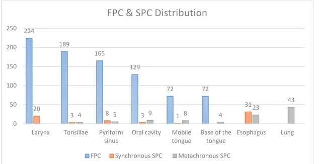
In 1994, Schwartz et al. realised a study with 851 patients who suffered a first head and neck cancer (FHN), diagnosed between 1978 and 1990. The objective was to study the distribution of the first primary cancer (FPC) and SPC. Patients underwent different forms of treatment: surgical, radiotherapy and chemotherapy. Of the 851 FHN (follow up period up to 169 months), 19% (162 cases) developed a SPC. The distribution of FHN and SPC is shown in Fig 2.
The first four localisations of the FHN are larynx with 26.3% (224 cases), followed by tonsillae with 22.2% (189 cases), pyriform sinus with 19.4% (165 cases) and OC with 15.2% (129 cases). It should be noted that anatomic sites of the oral cavity are considered separately in this study (oral cavity, mobile tongue and base of the tongue).
8
Their combination leads the OC to 32.1% (273 cases), that brings it in the first place of FHN localisation.
Fig 2. FPC and SPC distribution based on a study of Schwartz et al., 1994.
In their study, Jégu et al. (2013) realised a retrospective study using the french regional Bas-Rhin database covering the period from 1975 to 2006. They identified 6258 patients with FHN (their follow-up time of 10 years). They found as the main FHN appearance site, the OC 35.5% (2223 cases), followed by hypopharynx 22.8% (1423 cases), larynx 21.9% (1373 cases) and oropharynx 19.8% (1239 cases). Another study based on the SEER registry covering the period from 1975 to 2006, used 75.087 cases of diagnosed FHN (the median follow up was 69.1 months). They noticed in first importance the OC 48.1% (36.107 cases), followed by larynx 34.1% (25.624 cases), oropharynx 11.2% (8440 cases), and hypopharynx 6.5% (4916 cases) (Morris et al., 2011b).
In numerous studies, OC is the first site of FHN development, which is consistent with the literature (Vaamonde et al., 2003, Morris et al., 2011b, Jégu et al.,2013).
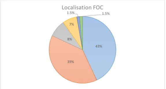
Relating patients with a first oral cavity cancer (FOC), Liu et al. (2013) study was one of the few focusing on FOC and the different subsite of the OC. Out of a total of 72
224 189 165 129 72 72 20 3 8 3 1 31 4 5 9 8 4 23 43 0 50 100 150 200 250
Larynx Tonsillae Pyriform sinus
Oral cavity Mobile tongue Base of the tongue Esophagus Lung
FPC & SPC Distribution
FPC Synchronous SPC Metachronous SPC9
patients with a first oral cavity cancer, they have found the following repartition: tongue 43% (31 cases), buccal mucosa 39% (28 cases), gingiva 8% (6 cases), lip 7% (5 cases), floor of the mouth 1.5% (1 case), retromolar trigone 1.5% (1case). These results are presented in Fig.3.
Fig 3. Localisation of FOC based on a study of Liu et al., 2013.
2. Synchronous/Metachronous
Firstly, it should be noted that all the articles used in this section distinguish synchronous from metachronous using a 6-month criteria.
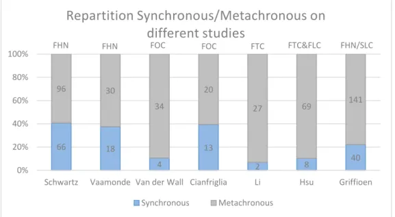
Relating to patients with a first head and neck cancer, the preceding study of Schwartz et al. (1994), out of the 162 cases of SPC, found an appearance of 41% (66 cases) of synchronous and 59% (96 cases) of metachronous (Fig.4).
Another study realised a clinical study on 636 patients with FHN (Vaamonde et al., 2003). In total, 7.5% (48 cases) developed a SPC (the follow up period was up to 122 months). They noticed 37.5% (18 cases) of synchronous and 62.5% (30 cases) of metachronous (Fig.4).
Relating to patients with a first oral cavity cancer and SPC appearance, Van der Wall and De Bree (2010), reported a study from De Vries which included 210 patients with a
43% 39% 8% 7% 1.5% 1.5%
Localisation FOC
10
FOC. Among these patients 18% (38 cases) developed a SPC (during 8.4 years follow up period). These SPC were 10.5% (4 cases) synchronous and 89.5% (34 cases) metachronous (Fig.4). In another study by Cianfriglia et al. (1998) concerning 200 patients with a FOC, 18% (36 cases) developed a SPC. Among these 36 SPC, 39.4% (13 cases) were synchronous and 60.6% (20 cases) were metachronous (Fig.4).
It should be noted that, in the last study, (Cianfriglia et al., 1998) are included patients with FOC, but also patients with a FPC of the oropharynx.
Relating other localisations of the FHN, Li et al. (2011) realised a study including 329 patients with a first primary tongue cancer (FTC). He showed that 8.8% (29 cases) developed a SPC (the minimum follow up period was 36 months). In that study 6.9% (2 cases) were reported as synchronous and 93.1% (27 cases) as metachronous (Fig.4). A retrospective study conducted by Hsu et al. (2008) on 538 patients with FTC and first primary larynx cancer (FLC), showed 14.3% (77 cases) of SPC incidence (follow up median period of 73 months). They found 10.4% (8 cases) of synchronous and 89.6% (69 cases) of metachronous (Fig.4).
In a study about 181 patients with a second primary lung cancer consecutive to an FHN (FH&N/SLC in Fig.4), the synchronous proportion was 22.1% (40 cases) and 77.1% (141 cases) for metachronous (Griffioen et al., 2015).
Fig 4. Repartition Synchronous/Metachronous in different studies.
66 18 4 13 2 8 40 96 30 34 20 27 69 141
FHN FHN FOC FOC FTC FTC&FLC FHN/SLC
0% 20% 40% 60% 80% 100%
Schwartz Vaamonde Van der Wall Cianfriglia Li Hsu Griffioen
Repartition Synchronous/Metachronous on
different studies
11
3. Localisation second primary cancer
Regarding the localisation of SPC after a first head and neck cancer, Scwhartz et al. (1994) found 40.2% (65 cases) for the sum of all the HN subsites, 33.3% (54 cases) for oesophagus e 26.5 % (43cases) for lung. In their study, Vaamonde et al. (2003) found that second primary cancer of the head and neck was the most common (72.9%), followed by lung (14.6%) and oesophagus (8.3%) reminding that it is congruent with the literature.
According to Morris et al. (2011b), based on the analyse of 75,087 cases of FHN, the SPC localisations most frequents were: OC 48.1% (36,107cases), larynx 34.1% (25,624 cases), oropharynx 11.2% (8,440 cases) and hypopharynx 6.5% (4,916 cases).
Concerning the SPC localisation after a first oral cavity cancer, Liu et al. (2013) found that 27.8% (20 patients among the 72 with a FOC) developed SPC. The main SPC localisation was in the oral cavity: 75% (15 cases).
However, according with the literature, the crude incidence of SPC doesn’t reflect the excess risk of cancer due to the FHN. The standardized incidence ratio (SIR) is more accurate. SIR is the ratio of observed to expected SPC with an 95% of confidence interval (CI), it measures the relative risk (RR) of the event on an individual level and does not depend on the frequency of the event in the population (Morris et al., 2011a). The excess absolute risk (EAR) represents the absolute number of additional second cancers attributable to the index HNSCC. EAR is calculated as the excess (observed— expected) number of second cancers in patients with an index HNSCC diagnosis, per 10,000 person-years at risk (PYR). EAR measures the population impact (Morris et al., 2011a).
According to Jégu et al. (2013), after a first oral cancer, the SIR for all sites is 5.4 (95% CI, 5.0 to 5.9) with an EAR equal to 511, for the lung & bronchus, SIR equal to 9.8 (95% CI, 8.3 to 11.6), EAR equal to 159.3. For the HN, SIR equal to 17.7 (95% CI, 15.2 to 20.6), EAR equal to 201. And for oesophagus, SIR equal to 22.6 (95 CI, 17.6 to 28.7), EAR equal to 82.9.
12
After an FOC, according to these results, the risk to develop a cancer in the oesophagus is 22.6 time higher than in the normal population and the FOC is responsible for 201 SPC cases per 10,000 PYR.
In Morris et al. (2011b) study, based on the SEER registry database, showed that SPC risk differed significantly in function of FHN subsite. The SIR of a second solid tumour at any site was highest for patients with an index hypopharynx (3.5; 95% CI, 3.3 to 3.7), followed by oropharynx (3.0; 95% CI, 2.9 to 3.1), oral cavity (2.8; 95% CI, 2.7 to 2.9), and larynx (1.9; 95% CI, 1.9 to 2.0).
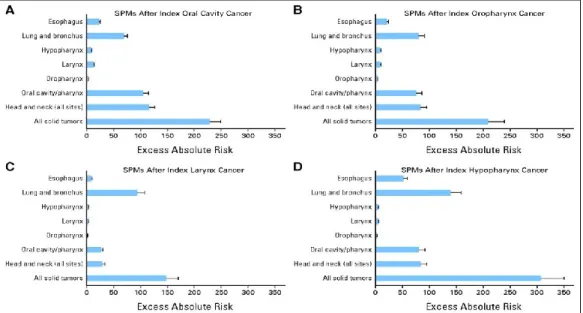
In the Fig.5, (taken from Morris et al., 2011b) is exposed EAR of SPC in function of the first head and neck cancer site. It is observed that the EAR of second solid cancers per 10,000 PYR was highest in patients with an index hypopharyngeal (307.1), and lowest for index larynx (147.8). After an FOC and a FPC of the oropharynx, the highest number of excess SPCs occurred in the HN. For larynx and hypopharynx primary cancers, the highest number of excess SPCs occurred in the lung (Fig. 5, Morris et al., 2011b).
Fig 5. Excess absolute risk of second primary malignancy (SPM), by site of index head and neck cancer: (A) index oral cavity, (B) index oropharynx, (C) index larynx, and (D) index hypopharynx cancer (adapted from Morris et al.,
13
4. Incidence and survival
According to Curtis et al. (2006) SPC diagnosis number is constantly increasing, reaching 16% of reported cancers in United-States, however this number is less important in Europe: 6.3% (Rosso et al., 2009).
Based on the results of Jégu et al. (2013) (with a 10 year follow up period), 1336 patients developed a SPC unlike the 284 expected, giving a SIR of 4.7 (95% CI, 4.4 to 4.9). In this study and during this period, patients with a FHN have 4.7 times a risk to develop a cancer than the normal population.
According to Morris et al. (2011a), SPC’s occurrence in patients treated for a FHN is estimated at 2–6% per year and in a continuous way among years.
According to Vaamonde et al. (2003), more than 50% of the SPC were detected in a period of 1.5 years after the diagnosis of the FHN.
After a first oral cancer treatment, in Liu et al. (2013) study, 80% (16 cases) of the SPC were diagnosed within 5 years. The incidence of SPT was 5% per year and was constant during the follow-up period of at least 7 years.
The overall survival after diagnosis of SPC in the Vaamonde et al. (2003) study, is 87.5% at 6 months, 68.7% at 1 year, 43.5% at 3 years, and 29% at 5 years. According to the same authors the survival rate associated with the SPC is poor, worse if it is in an advanced stage, located outside the HN area or if it is synchronous.
IV- DISCUSSION
Firstly, we could notice that there are few studies realised about the SPC after a FOC. The definition itself shows some limitations and there is obviously a lack of standardisation that difficult the comparison between studies. Different criteria are used, that could lead to misclassification (SEER in North America and IARC internationally and for reporting). The SEER classification includes a greater number of SPC than in the IARC classification (Coyte, 2014).
14
In some aspect, clinical classification seems less precise than the molecular classification.
There is room for improvement, that would affect comparative studies, management and prognostication (Gleber-netto et al., 2015). The research about molecular background of field cancerization, such with the TP53 mutation could bring us new solutions (Morris et al., 2011a).
However, this classification is not yet matured and doesn’t take the total precedence over the clinical classification (Gleber-Netto et al., 2015).
The patients risk behaviours are frequently inter-related, that could bias the studies (Brown et al., 2001).
It also be noted the important heterogeneity of the studies on this theme. The subsites of anatomic regions could be combine or considered separately, for exemple Schwartz et al. (1994), will distinguish the oral cavity from the mobile tongue and the base of the tongue unlike other studies which will regroup them into one site. Some studies like Cianfriglia et al. (1998) and Li et al. (2011) will consider three forms of SPC: simultaneous, synchronous and metachronous. Other studies have defined arbitrarily the FPC as the first tumour detected (Vaamonde et al., 2003).
Limitations of the registries (time, treatment, risk factors) and the unequal follow up period remains also important difficulties for the comparison.Finally, Cianfriglia et al. (1998) reminds us the low power statistics of various small studies.
Despite all these initial adverse factors, it can still be said that, according to many studies, the most frequent FHN location is the oral cavity and within the oral cavity, the tongue is most affected.
Most of the time cancers are metachronous. After a FHN, SPC are located mostly in the HN area, then in the lungs and oesophagus.
Approximately 75% of SPCs after an OSCC emerge in the oral cavity (Liu et al., 2013).
According to Morris et al. (2011b), if the FPC is low in the upper aerogestive tract, the risk of developing a SPC will be higher in the lung and bronchus area.
15
The survival rate among the patients affected by a SPC remains low: 29% at 5 years. The prognosis is even worse if it is in an advanced stage, located outside the region of the head and neck or if it is synchronous (Vaamonde et al., 2003).
There is a lack of evidence-based guidelines and a regular and long-term monitoring surveillance of SPC development is necessary to improve the survival rate (Liu et al.,2013).
New studies should be conducted on the person at risk follow up, focusing on the preclinical detectable phase, timing and risk of a SPC. But also, by considering the cost of the follow up as well as the supportability for the patients (Brands et al., 2018) and the role of the dental practitioner.
Dentist training in early detection of pre-cancerous lesions is important. An information work must also be conducted with patients, too many cancers are diagnosed at an advanced stage. The patient should no longer consult only when he has toothache, but maintain regular follow-up
V- CONCLUSION
Few reports have been made on reality of the SPC after a FHN and even less after a FOC.
There are populational differences and as well specific history for each patient. On the one hand, efforts should be made to standardise the criteria of population registers in order to assess general trends and the emergence of potential new influences. On the other hand, at a more specific level, notably based on genetic research, new studies should be conducted to better adapt the treatment and the follow up to each patient and their history.
The role of the dentist can be crucial in the early detection and in the quality of life after a FHN, and must be aware of the increment of this type of patients in their daily routine.
16
V- REFERENCES
Adelstein, D.J. (2005). Squamous cell head and neck cancer: recent clinical progress and prospects for the future. 1st Ed, Humana Press Inc.
Agrawal, A. et al. (2004). Role of the Physician Versus the Patient m the Detection of Recurrent Disease Following Treatment for Head and Neck Cancer. Laryngoscope, 114, pp. 232-235.
Asthana, S. et al. (2018). Association of Smokeless Tobacco Use and Oral Cancer: A Systematic Global Review and Meta-Analysis. Nicotine & Tobacco Research, Oxford University Press, 00(00), pp. 1-10. Bhatia, S., Tichelli, A. and Shover, L.R. (2005). Cancer survivorship - pediatric issues. American Society of Hematology, pp. 507-515.
Bongers, V. et al. (1996). The relation between cancer incidence and among relatives and the occurrence of multiple primary carcinomas following head and neck cancer. Cancer Epidemiology, Biomarkers and
Prevention, 5, pp. 595-598.
Brands, M.T. et al. (2015). Follow-up after a curative treatment for oral squamous cell carcinoma. A critical appraisal of the guidelines and a review of the literature. European Journal of Surgical Oncology, https://doi.org/10.1016/j.ejso.2018.01.004, pp. 1-7.
Brown, L.M. et al. (2001). Family Cancer History and Susceptibility to Oral Carcinoma in Puerto Rico.
Cancer, 92(8), pp. 2102–2108.
Chaturvedi, A.K. et al. (2011). Human Papillomavirus and Rising Oropharyngeal Cancer Incidence in the United States. Journal of Clinical Oncology, 29(32), pp. 4294-4301.
Cianfriglia, F., Di Gregorio, D.A. and Manieri, A. (1998). Multiple primary tumours in patients with oral squamous cell carcinoma. Oral Oncology, 35, pp. 157-163.
Coyte, A., Morrison, D.S. and McLoone, P. (2014). Second primary cancer risk – the impact of applying different definitions of multiple primaries: results from a retrospective population-based cancer registry study. BioMed Central Cancer, 14(272), pp. 1-15.
Curtis, R.E. et al. (2006). New Malignancies Among Cancer Survivors: SEER Cancer Registries, 1973-2000. National Cancer Institute, 05(5302), pp. 1-7.
17
Diaz, D.A. et al. (2016). Head and neck second primary cancer rates in the human papillomavirus era: A population-based analysis. Head and Neck, 38(1), pp. 873-883.
Ernani, V.I. and Saba, N.F. (2015). Oral Cavity Cancer: Risk Factors, Pathology, and Management.
Oncology, pp. 1-10.
Falciglia, G.A. et al. (2005). A Clinical-Based Intervention Improves Diet in Patients with Head and Neck Cancer at Risk for Second Primary Cancer. Journal of the American Dietetic Association, 105(10), pp. 1609-1612.
Farhadieh, R.D. et al. (2009). Radiotherapy is not associated with an increased rate of Second Primary Tumours in Oral Squamous Carcinoma: A study of 370 patients. Oral Oncology, 45(1), pp. 941-945. Gao, X. et al. (2002). Second primary cancers in patients with laryngeal cancer: a population -based study. International Journal of Radiation Oncology * Biology * Physics, 56(2), pp. 427–435.
Gleber-Netto, F.O. et al. (2015). Molecular events in relapsed oral squamous cell carcinoma: Recurrence vs secondary primary tumor. Oral Oncology, 51(2), pp. 738-744.
Griffioen, G.H.M.J. et al. (2015). Second primary lung cancers following a diagnosis of primary head and neck cancer. Journal Lung Cancer, 88, pp. 94-99.
Gutiérrez, V.F. et al. (2012). Genetic profile of second primary tumors and recurrences in head and neck squamous cell carcinomas. Head Neck, 34, pp. 830-839.
Hisada, M. et al. (1998). Multiple Primary Cancers in Families with Li–Fraumeni Syndrome. Journal of
the National Cancer Institute, 90(8), pp. 606-611.
Hong, W.K. et al. (1990). Prevention of second primary tumors with isotretinoin in squamous cell carcinoma of the head and neck. The New England Journal of Medicine, 323, pp. 795–801.
Hsu, Y.B. et al. (2008). Second Primary Malignancies in Squamous Cell Carcinomas of the Tongue and Larynx: An Analysis of Incidence, Pattern, and Outcome. Journal of the Chinese Medical Association, 71(2), pp. 86–91.
18
Jégu, J. et al. (2013). Trends over three decades of the risk of second primary cancer among patients with head and neck cancer. Oral Oncology, 49, pp. 9-14.
Jemal, A. et al. (2011). Global Cancer Statistics. A Cancer Journal for Clinicians, 61(2), pp. 69-90. Ko, H.H. et al. (2016). Factors influencing the incidence and prognosis of second primary tumors in patients with oral squamous cell carcinoma. Head and Neck, pp. 1459-1462.
Kutler, D.I. et al. (2003). High incidence of head and neck squamous cell carcinoma in patients with
Fanconi anemia.Archives of Otolaryngology - Head and Neck Surgery, 129, pp. 106-112.
Li, Z. et al. (2011). Incidence of second primary tumours in patients with squamous cell carcinoma of the tongue. British Journal of Oral Surgery, 49, pp. 50-52.
Liao, C.T. et al. (2007). Survival of second and multiple primary tumors in patients with oral cavity squamous cell carcinoma in betel quid chewing area. Oral Oncology, 43, pp. 811–819.
Liu, C.H. et al. (2013). Patterns of recurrence and second primary tumors in oral squamous cell carcinoma treated with surgery alone. Kaohsiung Journal of Medical Sciences, 29, pp. 554-559.
Morris, L.G.T et al. (2011a). Anatomic sites at elevated risks of second primary cancer after an index head and neck cancer. Cancer Causes Control, 22(5), pp. 671-679.
Morris, L.G.T. et al. (2011b). Second Primary Cancers After an Index Head and Neck Cancer: Subsite-Specific Trends in the Era of Human Papillomavirus–Associated Oropharyngeal Cancer. Journal of
Clinical Oncology, 29(6), pp. 739-746.
Muir, C.S. and Percy, C. (1991) Classification and coding for neoplasms. In: Jensen, O.M. et al. (eds) Cancer registration: principles and methods, IARC Scientific Publications, Lyon, (95), pp. 64–81. Neumann, F. et al. (2016). Risk of second primary cancer after a first potentially-human papillomavirus-related cancer: A population-based study. Preventive Medicine, 90(2), pp. 52-58.
Noone, A.M. et al. (2018). SEER Cancer Statistics Review, 1975-2015, National Cancer Institute. Bethesda, MD, https://seer.cancer.gov/csr/1975_2015, based on November 2017 SEER data submission, posted to the SEER web site, April 2018. (accessed: 25th Of june 2018)
19
Petti, S. (2008). Lifestyle risk factors for oral cancer. Oral Oncology, 45, pp. 340-350.
Prime, S.S. et al. (2001). A review of inherited cancer syndromes and their relevance to oral squamous cell carcinoma. Oral Oncology, 37, pp. 1-19.
Quin, G.Z. et al. (1999). A high prevalence of p53 mutations in pre-malignant oral erythroplakia. International Journal of Cancer, 80, pp. 345–348.
Reid, P. et al. (2018). Experimental investigation of radiobiology in head and neck cancer cell lines as a function of HPV status, by MTT assay. www.nature.com, Scientific Reports, 8(7774), pp. 1-8.
RORENO. (2016). Registo Oncológico Nacional 2010. Instituto Português de Oncologia do Porto Francisco Gentil - EPE, ed. Porto.
Rosso, S. et al. (2009). Multiple tumours in survival estimates. European Journal of Cancer, 45, pp. 1080-1094.
Rusthoven, K. et al. (2008). Use of external beam radiotherapy is associated with reduced incidence of second primary head and neck: a SEER database analysis. International Journal of Radiation
Oncology * Biology * Physics, 71(1), pp. 192–198.
Sarode, G.S. et al.(2016). Oral Cancer-related Inherited Cancer Syndromes: A Comprehensive Review.
Journal of Contemporary Dental Practice, 17(6), pp. 504-510.
Sarode, S.C., Sarode, G.S. and Patil, A. (2010). Criteria to define true second primary oral squamous cell carcinoma. Oral Oncology, 46, pp. 83.
Scholes, A.G.M. et al. (1998). Synchronous oral carcinomas: independent or common clonal origin?.
Cancer Research, 58, pp. 2003–2006.
Schwartz, L.H. et al. (1994). Synchronous and Metachronous Head and Neck Carcinomas. Cancer, 74(7), pp. 1933-1938.
Seegobin, K. et al. (2018). Pilot study on the occurrence of multiple cancers following cancer-related therapy at the University of Florida, Jacksonville (2011–2016).Journal of Investigative Medicine, 0, pp.
20
Shiels, M.S. et al. (2014). Cigarette Smoking Prior to First Cancer and Risk of Second Smoking-Associated Cancers Among Survivors of Bladder, Kidney, Head and Neck, and Stage I Lung Cancers. Journal of Clinical Oncology, 32(35), pp. 3989-3995.
Tabor, M.P. et al. (2002). Multiple head and neck tumors frequently originate from a single preneoplastic lesion. The American Journal of Pathology, 161, pp. 1051–1060.
Travis, L.B. et al. (2006). Cancer Survivorship_Genetic Susceptibility and Second Primary Cancers: Research Strategies and Recommendations. Journal of the National Cancer Institute, 98(1), pp. 15-26. Vaamonde, P. et al. (2003). Second primary malignancies in patients with cancer of the head and neck.
Otolaryngology–Head and Neck Surgery, 129, pp. 65-70.
Van der Wall, I. and De Bree, R. (2010). Second primary tumours in oral cancer. Oral Oncology, 46, pp. 426-428.
Wood, M.E. et al. (2012). Second Malignant Neoplasms: Assessment and Strategies for Risk Reduction.
American Society of Clinical Oncology, Journal of Oral Oncology, 30(30), pp. 3734-3745.
Yokoyama, A. and Omori, T. (2002). Genetic Polymorphisms of Alcohol and Aldehyde Dehydrogenases and Risk for Esophageal and Head and Neck Cancers. Japanese Journal of Clinical Oncology, 33(3), pp.