Braz. J. of Develop.,Curitiba, v. 6, n. 9, p.71840-71852,sep. 2020. ISSN 2525-8761
Indicators of physical stress during hindlimb suspension in Wistar rats
Indicadores de stress físico durante a suspensão dos membros inferiores em
ratos Wistar
DOI:10.34117/bjdv6n9-578Recebimento dos originais: 08/08/2020 Aceitação para publicação: 24/09/2020
Maurilio Tiradentes Dutra
Doutorado
Instituto Federal de Educação, Ciência e Tecnologia de Brasília - IFB
Área Especial n° 01, Quadra 16, Cidade do Automóvel/SCIA/Estrutural/Brasília, DF mauriliotiradentes@gmail.com
Beshoy Girgis
Mestrado
Centro de Investigação em Actividade Física, Saúde e Lazer. Faculdade de Desporto, Universidade do Porto, Portugal
R. Dr. Plácido da Costa 91, 4200-450 Porto, Portugal drgirgisbeshoy@gmail.com
Antonio Bovolini
Doutorado
Centro de Investigação em Actividade Física, Saúde e Lazer. Faculdade de Desporto, Universidade do Porto, Portugal
R. Dr. Plácido da Costa 91, 4200-450 Porto, Portugal jabovolini@hotmail.com
Catarina C. Costa
Licenciatura
Centro de Investigação em Actividade Física, Saúde e Lazer. Faculdade de Desporto, Universidade do Porto, Portugal
R. Dr. Plácido da Costa 91, 4200-450 Porto, Portugal cardoso.costa@ua.pt
Grace Fernanda Nunes
Mestrado
Centro de Investigação em Actividade Física, Saúde e Lazer. Faculdade de Desporto, Universidade do Porto, Portugal
R. Dr. Plácido da Costa 91, 4200-450 Porto, Portugal gracefernanda@live.com
José Alberto R. Duarte
Doutorado
Centro de Investigação em Actividade Física, Saúde e Lazer. Faculdade de Desporto, Universidade do Porto, Portugal
R. Dr. Plácido da Costa 91, 4200-450 Porto, Portugal jarduarte@fade.up.pt
Braz. J. of Develop.,Curitiba, v. 6, n. 9, p.71840-71852,sep. 2020. ISSN 2525-8761
ABSTRACT
Hindlimb suspension (HS) has been used in animal research as a microgravity simulation model. However, physical stress to the animals during suspension has received little attention. The purpose of this work was to investigate physical stress, oxidative stress, and cross-sectional area (CSA) in rats submitted to hindlimb suspension. Twelve male Wistar rats were allocated into two groups: control (CON, n = 6); hindlimb suspension (HS, n = 6), for ten days. The following signs of physical stress were monitored: bristly hair, spleen hair, edema in the nose, edema or necrosis in the tail, edema in the hind legs, aggressiveness, redness in the dorsal region, body weight loss, and food intake. Samples of the soleus and tibialis anterior were prepared for CSA and carbonylated proteins analysis. The frequency of the signs of stress, except redness in the dorsal region, was significantly higher (P <.05) in the HS group. CSA of soleus (816.6µm2, 95%CI: 866.3-904.0 vs 1158.8mm2, 95%CI: 1171.9-1214.2) and tibialis anterior (1041.5µm2, 95%CI: 1119.1-1185.2 vs 1429.8µm2, 95%CI: 1479.2-1566.4) were lower in the HS group (P <.05). Carbonylated proteins in the soleus (2779.0 ± 1319.8 AU vs 1063.2 ± 289.2 AU) and tibialis anterior (1155.8 AU, 95%CI: 80.0-4064.5
vs 108.4 AU, 95%CI: 35.5-320.7) were higher in the HS group (P <.05). The suspension model
evokes oxidative stress and reduced CSA. However, several signs of physical stress can occur. This indicates that hindlimb suspension models may introduce confounding factors that influence muscle atrophy and should be used cautiously.
Keywords: hindlimb suspension, physical stress, muscle wasting, oxidative damage RESUMO
O protocolo de suspensão de membros posteriores tem sido usado em pesquisas com animais como um modelo de simulação de microgravidade. No entanto, o estresse físico dos animais durante a suspensão tem recebido pouca atenção. O objetivo deste trabalho foi investigar o estresse físico, o estresse oxidativo e a área de secção transversal (AST) em ratos submetidos à suspensão. Doze ratos Wistar machos foram alocados em dois grupos: controle (CON, n = 6); suspensão de membros posteriores por dez dias (SUS, n = 6). Os seguintes sinais de estresse físico foram monitorados: pelos eriçados, edema no nariz, edema ou necrose na cauda, edema nas patas traseiras, agressividade, vermelhidão na região dorsal, perda de peso corporal e redução na ingestão de ração. Amostras do sóleo e tibial anterior foram preparadas para análise de AST e de proteínas carboniladas. A frequência dos sinais de estresse, exceto vermelhidão na região dorsal, foi significativamente maior (P <0,05) no grupo SUS. A AST do sóleo (816,6 µm2, IC 95%: 866,3-904,0 vs 1158,8 µm2, IC 95%: 1171,9-1214,2) e tibial anterior (1041,5 µm2, IC 95%: 1119,1-1185,2 vs 1429,8 µm2, IC 95%: 1479,2- 1566,4) foram menores no grupo SUS (P <0,05). As proteínas carboniladas no sóleo (2779,0 ± 1319,8 UA vs 1063,2 ± 289,2 UA) e tibial anterior (1155,8 UA, IC 95%: 80,0-4064,5 vs 108,4 UA, IC 95%: 35,5-320,7) foram maiores no grupo SUS (P <0,05). O modelo de suspensão gera estresse oxidativo e redução da AST. Além disso, vários sinais de estresse físico podem ocorrer. Isso indica que os modelos de suspensão dos membros posteriores podem introduzir fatores de confusão que influenciam a atrofia muscular e devem ser usados com
cautela na pesquisa com animais.
Palavras-chave: suspensão de membros posteriores, estresse físico, atrofia muscular, dano
Braz. J. of Develop.,Curitiba, v. 6, n. 9, p.71840-71852,sep. 2020. ISSN 2525-8761
1 INTRODUCTION
Hindlimb suspension (HS) has been used for several years in animal research as a microgravity simulation model 1. Also, the model mimics changes related to prolonged bed rest and muscle disuse. Thus, it allows to study and comprehend physiological and morphological consequences of unloading to the musculoskeletal system 2. Hence, a large spectrum of science fields became interested in this kind of research protocol, such as space exploration and flight 1–4.
Previous literature showed that HS results in reduced muscle recruitment, diminished electrical activity of the muscles and, as a consequence, muscle atrophy 1,3. It has been reported that the soleus muscle loss after a week of HS is about 27% in male mice 5, and may reach 35% in Wistar rats 6. Similarly, muscle force production is dramatically reduced after a week of HS. Recently, Mortreux and coll. reported a 50% decrease in grip force of male Wistar rats when compared to baseline 4.
Worthy of note, HS leads to important redox changes. For instance, a recent research showed that HS resulted in greater levels of reactive oxygen species (ROS) in soleus and gastrocnemius mice muscles 5. Of note, it is known that redox alterations in favor of oxidative stress are implicated in several tissues damage 7, as well as in the activation of muscle atrophy pathways during muscle unloading 8,9. Thus, ROS may activate protein degradation while decreases protein synthesis and may be understood as triggers to muscle atrophy during disuse/unloading models 5,8,9.
In addition to force reduction and muscle atrophy, HS may lead to other relevant metabolic alterations. Decreased insulin secretion, as well as the development of insulin resistance and compensatory hyperinsulinemia has been also reported as consequences of HS 2. Also, bone disuse loss secondary to HS was previously reported 2,10. Therefore, previous studies have come to results that highlight the physiological and functional consequences of HS.
Generally, those studies applied one of the two described HS techniques, which are HS by the animal’s tail, or by their pelvis. It is relevant to mention that both techniques may lead to back hyperalgesia and muscle pain to the animals 2, that may represent an additional source of stress to restraint stress. In this sense, it is noteworthy that stress is a crucial factor during unloading models and it is very difficult to eliminate 11. However, to the best of our knowledge, the physical stress that may occur during HS protocols has not been focused on previous studies.
Many signs of physical stress can occur during HS, such as edema in the tail, hair alterations, diminished food intake, weight loss, and others. These signs may lead to metabolic changes associated with increased catabolic activity, such as the activation of the hypothalamic pituitary adrenal axis 11. So, physical stress induced by HS could interfere with the disuse muscle wasting
Braz. J. of Develop.,Curitiba, v. 6, n. 9, p.71840-71852,sep. 2020. ISSN 2525-8761
to investigate signs of physical stress in rats submitted to HS. Also, it aimed to assess cross-sectional area and oxidative stress in soleus and tibialis anterior muscles after ten days of HS.
2 MATERIAL AND METHODS
Experiments were performed on Wistar male rats (Charles River, Barcelona, Spain). A post
hoc analysis was conducted using G*POWER 3.1.9.2 (Universitat Kiel, Germany) software based
on previous investigation 12 to calculate power and effect size d. Then, an a priori analysis was conducted using a 4.4 effect size with α = 0.05. It was determined that 8 animals (4 per group) were needed in the present study for a power of 0.99. The animals came to the laboratory with 4-week-old age. They were housed one per cage and maintained at normal atmosphere (21–22°C; 50–60% humidity). They received ad libitum food (standard laboratory diet 4RF21®, Mucedola, Italy) and water in a 12h light/dark inverted cycle for 24 days of acclimatization. After this, 12 male animals (248.0g ± 26.5g) were allocated into 2 groups: I) control (CON, n = 6); II) hindlimb suspension (HS, n = 6) for ten days. In the first five days, animals were suspended by their tails. In the last five days, the suspension was made by the pelvis. During suspension, the forelimbs maintained in contact with the cage floor, allowing the rat a full range of motion. These model of HS have already been used and described in previous research 2,13. During HS, both groups had ad libitum access to food and water. Immediately after the end of HS, the animals were weighed and anesthetized by intraperitoneal injection with Ketamine (90 mg/kg, Merial, France), Xylazine (10mg/kg, Bayer, German) and euthanized in the laboratory, at morning time. All procedures were performed in accordance with the Guidelines for Care and Use of Laboratory Animals and were approved by the Ethics Committee of the University of Porto.
2.1 PHYSICAL STRESS EVALUATION
During HS, the following signs of physical stress were monitored and registered daily: bristly or spleen hair, edema or bleeding in the nose, edema or necrosis in the tail, edema in the hind legs, aggressiveness, redness in the dorsal region, body weight loss, and food intake. Aggressiveness was monitored subjectively as the animal’s violent reaction during stimuli or approach to weight assessment. The indicators of physical stress, except for food ingestion, were categorized by their presence or absence, and frequency of all of them was analyzed and compared between groups. Food ingestion was categorized based on the percent change from acclimatization to suspension periods. The smaller percent change within the CON group was 22.2% and this was considered the reference value to do the categorization. In other words, all animals were categorized as presenting
Braz. J. of Develop.,Curitiba, v. 6, n. 9, p.71840-71852,sep. 2020. ISSN 2525-8761
or not a lower value than the reference. So, a percent change below 22.2% in food ingestion was assumed to be a sign of stress.
2.2 HISTOLOGICAL ANALYSIS
Soleus (SOL) and tibialis anterior (TA) muscles were harvested, washed in PBS (pH 7.2), and weighed in a precision balance (resolution 0.01mg; Kern 870, Balingen, Germany). The muscles were 24h fixed in 4% paraformaldehyde solution at 4ºC, dehydrated through graded ethanol solutions, cleared in xylene, and mounted in paraffin. 5µm thick sections from both muscles were cut by microtome (Leica, RM 2125) and stained with hematoxylin-eosin (H&E) protocol succeeded by posterior laminar assembly with DPX (dibutyl xylene phthalate; Shandon EZ-Mount, Thermo Electron Corporation, USA). The sections were then analyzed with a light microscope (Axior Imager A1, Carl Zeiss; Germany) and images recorded with a coupled digital camera (Leica DM4000B, Nussloch, Germany) using a 40x objective lens. Cross-sectional area (CSA) of both muscles was assessed using the Image J software (1.50i, National Institutes of Health, Bethesda, Maryland, USA). Five to six photos per animal were used to complete the analysis. A total of 1,168 and 1,280 fibers were analyzed from the SOL in the CON and HS groups, respectively. Meanwhile, 976 and 965 fibers were analyzed from the TA in the CON and HS groups, respectively.
2.3 BIOCHEMICAL ANALYSIS
Immediately after excision and weighing, contralateral samples of the SOL and TA muscles were frozen at -70ºC. Then, portions of around 30mg muscle tissue were homogenized in 1mL buffering solution until used for biochemical assessment. Total protein concentration was determined using the RC DC Protein Assay (Bio-Rad Laboratories). Optical density values were determined at 750nm in a spectrophotometer (Labsystems iEMS Reader MF). Bovine serum albumin known concentrations were used as reference standards. As protein carbonylation can be promoted during oxidative stress 14, the protein carbonyl group was assessed using Slot Blot Protocol. Briefly, the carbonyl groups in the protein side chains were derivatized to 2,4-dinitro- phenylhydrazone. 100μl of sample were transferred to nitrocellulose membrane in the Hybri-slot apparatus. The membranes were then incubated with primary antibody (rabbit IgG, anti-dinitrophenyl antibody, Invitrogen A6430) overnight at 4ºC, followed by incubation with the secondary antibody (goat pAb to Rb IgG – HRP antibody, Abcam ab97051) for one hour under agitation at room temperature. Detection was done by applying ECL solution to the membrane in the ChemiDoc XRS system with Image Lab software (version 6.0, Bio-Rad Laboratories).
Braz. J. of Develop.,Curitiba, v. 6, n. 9, p.71840-71852,sep. 2020. ISSN 2525-8761 2.4 STATISTICAL ANALYSIS
Shapiro-Wilk test was performed to examine data distribution. The chi-square test was used to analyze the signs of physical stress. Independent-T test (SOL and TA weight; SOL carbonyl groups) and Mann Whitney-U test (SOL and TA CSA; TA carbonyl groups) were used to compare variables between groups. To compare body weight and food ingestion between groups at different moments, a multifactorial analysis of variance (ANOVA group*time) was applied. Significance was set at P ≤ .05. Data is expressed as mean ± standard deviation if normally distributed. If not, median 95%CI was employed. All analysis was done using the Statistical Package for Social Sciences software (SPSS 20.0, IBM, Armonk, New York, EUA) for Windows.
3 RESULTS
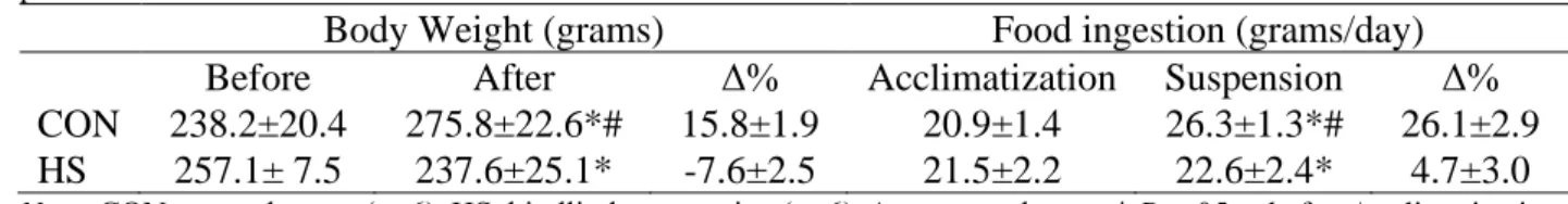
Table 1 presents body weight before and after HS protocol, and mean values of ingestion of food during acclimatization and HS period. The CON group presented body weight increase after the protocol, whereas suspended animals presented a significant weight reduction. During the HS protocol, HS group presented a significantly lower food intake compared to CON group.
Table 1. Body weight before and after HS protocol, and food ingestion mean values during acclimatization and HS period.
Body Weight (grams) Food ingestion (grams/day) Before After Δ% Acclimatization Suspension Δ% CON 238.2±20.4 275.8±22.6*# 15.8±1.9 20.9±1.4 26.3±1.3*# 26.1±2.9 HS 257.1± 7.5 237.6±25.1* -7.6±2.5 21.5±2.2 22.6±2.4* 4.7±3.0
Note: CON: control group (n=6). HS: hindlimb suspension (n=6). Δ: percent change. * P ≤ .05 vs before/acclimatization.
# P ≤ .05 vs HS.
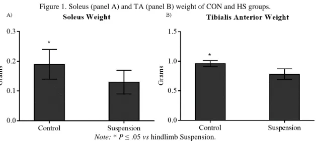
There were significant differences (P ≤ .05) when comparing SOL and TA muscles weight between groups. HS animals showed a significant lower muscle weight compared to CON group. Difference between groups was around 30% to SOL (.19 ± .05g vs .13 ± .04g, control and HS, respectively) and 18% to TA (.96 ± .05g vs .78 ± .09g, control and HS, respectively) (Figure 1).
Braz. J. of Develop.,Curitiba, v. 6, n. 9, p.71840-71852,sep. 2020. ISSN 2525-8761 Figure 1. Soleus (panel A) and TA (panel B) weight of CON and HS groups.
Note: * P ≤ .05 vs hindlimb Suspension.
The signs of physical stress results are presented in Table 2. The frequency of all of them, except redness in the dorsal region, was significantly higher in the HS group (Table 2).
Table 2. Frequency of the signs of physical stress.
Sign of stress Control (n=6) HS (n=6) P
Yes (%) No (%) Yes (%) No (%) Hair 3 (50) 3 (50) 6 (100) 0 (0) .046 Nose 0 (0) 6 (100) 4 (66.7) 2 (33.3) .014 Tail 0 (0) 6 (100) 6 (100) 0 (0) .001 Aggressiveness 0 (0) 6 (100) 6 (100) 0 (0) .001 Redness 3 (50) 3 (50) 1 (16.7) 5 (83.3) .22
Body Weight Loss 0 (0) 6 (100) 6 (100) 0 (0) .001
Food ingestion 0 (0) 6 (100) 6 (100) 0 (0) .001
Hind legs 0 (0) 6 (100) 6 (100) 0 (0) .001
Note: HS: hindlimb suspension. Hair: bristly or spleen. Nose: edema or bleeding. Tail: edema or sign of necrosis. Hind
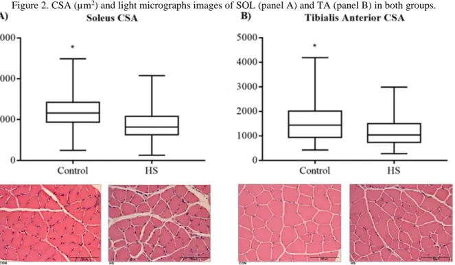
legs: edema. Redness: in the dorsal region of the animal’s body. Food ingestion: percent change lower than 22.2%. Results regarding SOL and TA CSA are presented in Figure 2, panels A and B. Soleus CSA was significantly lower in the HS compared to the CON group (816.6 µm2, 95% CI: 866.3-904.0 vs 1158.8 µm2, 95% CI: 1171.9-1214.2, respectively), representing a 29.5% decrease. A similar result was observed for TA, where CSA was significantly lower in the HS compared to the CON group (1041.5 µm2, 95% CI: 1119.1-1185.2 vs 1429.8 µm2, 95% CI: 1479.2-1566.4), representing a 27.2% reduction.
Braz. J. of Develop.,Curitiba, v. 6, n. 9, p.71840-71852,sep. 2020. ISSN 2525-8761 Figure 2. CSA (µm2) and light micrographs images of SOL (panel A) and TA (panel B) in both groups.
Note: CSA cross-sectional area, HS hindlimb suspension, * P ≤ .05 vs HS.
The protein carbonyl group presented to be significantly higher in the HS group, both in the SOL (2779.0 ± 1319.8 AU vs 1063.2 ± 289.2 AU) and TA (1155.8 AU - 95%CI: 80.0-4064.5 vs 108.4 AU - 95%CI: 35.5-320.7) compared to CON group (Figure 3, panels A and B).
Figure 3. Protein carbonyl group in SOL (panel A) and TA (panel B) in both groups.
Note: HS: hindlimb suspension. * P ≤ .05 vs Control. Panel B is Log 2 scaled.
4 DISCUSSION
The main purpose of the present study was to investigate several signs of physical stress that may occur to animals submitted to hind limb suspension protocol. With this regard, the study shows
Braz. J. of Develop.,Curitiba, v. 6, n. 9, p.71840-71852,sep. 2020. ISSN 2525-8761
that all of the analyzed signs of stress, except for redness in the dorsal region, were significantly higher in the animals submitted to HS when compared to the CON group. Also, the present study aimed to assess CSA and oxidative damage in soleus and tibialis anterior muscles after ten days of HS. As expected, HS evoked a significant reduction of SOL and TA CSA after the protocol along with a concomitant elevation of an oxidative damage marker.
It is reported in the literature that HS leads to protein oxidation 15, as well as bone and muscle atrophy 2,9. So, it was expected that HS protocols applied in the present study would lead to significant reductions of SOL and TA CSA, along with a concomitant increase in carbonyl group expression, used as a marker of oxidative damage. Previous research had shown that one week of HS leads to a 27% SOL loss after a week of HS in male mice 5, and may reach 35% in Wistar rats
6. This is in line with the results of the present investigation, where a 29.5% and 27.2% reduction in
SOL and TA CSA were observed, respectively.
Moreover, carbonyl group expression in the present study was significantly high in both SOL and TA of the HS animals. It is well stablished that HS, as a muscle unloading model, is associated with increased ROS production and, as consequence, increased protein oxidation 5,8,9,15. Thus, muscle CSA reduction reported in the present study may have been caused by oxidative stress associated to HS model. In other words, both protein synthesis and degradation were probably affected by HS induced oxidative stress, leading to muscle atrophy. Of note, atrophy caused by unloading is characterized by reductions in muscle size, weight and function 5, which is consistent with the results of the present study, as not only CSA, but also SOL and TA weight presented to be diminished in HS animals compared to CON.
Besides all of the reported functional, tissue and metabolic changes associated with hindlimb suspension in animals, such as strength reduction 4, bone and muscle loss 5,6,10, and hyperinsulinemia
2, there are some other organic changes associated to the model. For instance, decreased circulating
iron was reported in rats submitted to hindlimb suspension. Indeed, this is a common microgravity response and may be induced by inflammation, causing oxidative stress an anemia 16. Also, the immune system is affected by HS. A few years ago, Li and coll. 17 showed that mice submitted to five days HS and then exposed to bacterial infection failed to elevate blood granulocyte counts and presented a reduced ability to clear the infection with an increase in morbidity (i.e. analyzed as “rough hair coat”, “squinted eyes”, “not eating or drinking” and other parameters). The authors also observed that HS evoked corticosterone elevation. This stress hormone can impair both innate and acquired immune function 17.
In this sense, it was reported previously that blood testosterone levels are dramatically reduced in male adult rats after twelve days of HS, and this reduction was accompanied by a cortisol
Braz. J. of Develop.,Curitiba, v. 6, n. 9, p.71840-71852,sep. 2020. ISSN 2525-8761
elevation 18. It is well known that these hormones play antagonistic functions related to muscle anabolism 19, so that atrophy may also be a result of HS induced testosterone reduction. Furthermore, cortisol and corticosterone are associated with acute and chronic stress 17,20, so that elevation of their concentration shows the stressful characteristic of HS. In combination with the situational restraint stress associated to cage housing 2,21, all these changes contribute to an overall debilitation of the animal.
However, to the best of our knowledge, studies did not fully report other relevant “adverse effects” of HS that may influence and even mask the physiological response to unloading, and this may introduce bias to studies. For instance, although Li and coll. 17 have assessed morbidity parameters in mice submitted to HS, they have reported that only after exposing the animals to a bacterial infection, as mentioned before. So, information about relevant physical stress signs, such as violent reaction or aggressiveness, weight loss, and edema has not been the target of previous investigations and is still lacking. Yet, it is noteworthy that Chowdhury and coll. 2 have reported signs of exaggeration of evoked deep muscle pain in rats submitted to both tail and pelvic models of HS for two weeks. Furthermore, those authors reported that animals submitted to HS presented a retardation in weight gain, with no difference in food intake, when compared to control animals 2. This is partially in line with the present results. We observed that food ingestion during HS was about 20% lower in the suspended animals compared to control (P ≤ .05, Table 1). Apparently, this reflected in weight, once HS animals presented a significant weight reduction that reached almost 8% during suspension (P ≤ .05, Table 1). Although studies use to report that a less than 15% weight loss is not identified as severe stress 11, the changes in weight and food intake observed in this study was accompanied by other indicators of physical stress, as discussed forward.
The present investigation shows that HS promoted significant hair changes (bristly and spleen), edema or bleeding in the nose, edema or necrosis in the tail, and aggressiveness. Altogether, it is plausible to infer that those physical stress indicators may have also influenced muscle tissue plasticity via catabolic induced pathways, such as elevation of many stress related hormones like corticosterone and epinephrine. Moreover, edema in the tail with sign of necrosis is a worrying situation regarding blood flow impairment and because rats use their tail to thermoregulation 22. In this sense, physical stress in the tail was eased when suspension was changed to the pelvic model.
Thus, not only muscle wasting, but a wide variety of physiological alterations may be in course during the protocol with harmful effects on homeostasis. In this scenario, it may be exceedingly difficult to distinguish and explain how much of the muscle atrophy is related to the disuse and how much is related to the stress generated by the suspension. Thus, depending on the duration of the HS protocol, all the reported alterations could lead to the occurrence of confounding
Braz. J. of Develop.,Curitiba, v. 6, n. 9, p.71840-71852,sep. 2020. ISSN 2525-8761
factors regarding the process of muscle wasting and even to the failure of the disuse-induced atrophy model. It is noteworthy that a very recent article showed no evidence of stress in male rats during 28 days of a suspension protocol made by using a pelvic harness. The authors analyzed heart rate, serum glucose, corticosterone levels, tail blood pressure and hind limb oxygen saturation, and they did not observe any change in these parameters 11. So, it is possible that suspension by the pelvis may be less stressful than tail suspension.
This report has limitations. As animals were subsequently submitted to tail and pelvic models of HS, it is hard to fully distinguish and compare stress signs between the protocols. Also, blood metabolic stress markers such as corticosterone levels were not assessed. However, strengths are also recognized in this study. Despite the lack of blood analysis, the association between physical and mental stress with corticosteroids is largely known 23 and several signs of physical stress were
assessed in the present investigation providing a robust evaluation. Moreover, this study is unique regarding a critical physical stress analysis of a common protocol in animal research.
In summary, the present data show that rats submitted to ten days of HS present reduced CSA of SOL and TA muscles, as well as protein oxidation, as expected. Also, HS leads to the occurrence of physical and mental stress, such as edema in the tail, aggressiveness, and others. Altogether, this indicates that HS models may introduce confounding factors to research and may not be appropriate to study muscle wasting induced by disuse, especially when applied for several days. Future investigation is necessary to further elucidate the time course of physical stress signs associated to HS, as well as to compare stress indicators between different suspension models.
ACKNOWLEDGMENTS
The authors thank Ms. Celeste Maria R. dos Santos and Drª. Ana Padrão for their technical assistance. We also thank BSc. Patricia Cord, MSc. Francisco Leite, MSc. Sarah Nobre and MSc Edyla Porto Camelo (in memorian) for their support.
CONFLICTING INTERESTS
Braz. J. of Develop.,Curitiba, v. 6, n. 9, p.71840-71852,sep. 2020. ISSN 2525-8761
REFERENCES
1. American Psychological Association. Resource Book for the Design of Animal Exercise Protocols. Epub ahead of print 2007. DOI: 10.2460/ajvr.68.6.583.
2. Chowdhury P, Long A, Harris G, et al. Animal model of simulated microgravity: A comparative study of hindlimb unloading via tail versus pelvic suspension. Physiol Rep 2013; 1: 1– 11.
3. Fitts RH, Metzger JM, Riley DA, et al. Models of disuse: a comparison of hindlimb suspension and immobilization. J Appl Physiol 1986; 60: 1946–1953.
4. Mortreux M, Riveros D, Bouxsein ML, et al. Mimicking a space mission to mars using hindlimb unloading and partial weight bearing in rats. J Vis Exp 2019; 2019: 1–7.
5. Theilen NT, Jeremic N, Weber GJ, et al. Exercise preconditioning diminishes skeletal muscle atrophy after hindlimb suspension in mice. J Appl Physiol 2018; 125: 999–1010.
6. Appell HJ, Duarte JAR, Soares JMC. Supplementation of vitamin E may attenuate skeletal muscle immobilization atrophy. Int J Sports Med 1997; 18: 157–160.
7. Luciano DMB, Fedato BN, Vieira NM, et al. Hepatoprotective effect of caruru (amaranthus viridis) on the development of experimental hepatic cirrhosis induced by thioacetamide. Brazilian J Dev 2020; 6: 54531–54549.
8. Atherton PJ, Greenhaff PL, Phillips SM, et al. Control of skeletal muscle atrophy in response to disuse: clinical/preclinical contentions and fallacies of evidence. Am J Physiol Metab 2016; 311: E594–E604.
9. Bodine SC. Disuse-induced muscle wasting. Int J Biochem Cell Biol 2013; 45: 2200–2208.
10. Shimano MM, Volpon JB. Biomechanics and structural adaptations of the rat femur after hindlimb suspension and treadmill running. Brazilian J Med Biol Res 2009; 42: 330–338.
11. Mortreux M, Riveros D, Semple C, et al. The partial weight-bearing rat model using a pelvic harness does not impact stress or hindlimb blood flow. Acta Astronaut 2020; 168: 249–255.
12. Vazeille E, Slimani L, Claustre A, et al. Curcumin treatment prevents increased proteasome and apoptosome activities in rat skeletal muscle during reloading and improves subsequent recovery. J Nutr Biochem 2012; 23: 245–251.
13. Ferreira R, Neuparth MJ, Ascensão A, et al. Skeletal muscle atrophy increases cell proliferation in mice gastrocnemius during the first week of hindlimb suspension. Eur J Appl Physiol 2006; 97: 340–346.
14. Nuoc TN, Kim S, Ahn SH, et al. The analysis of antioxidant expression during muscle atrophy induced by hindlimb suspension in mice. J Physiol Sci 2017; 67: 121–129.
Braz. J. of Develop.,Curitiba, v. 6, n. 9, p.71840-71852,sep. 2020. ISSN 2525-8761
15. Brocca L, Pellegrino MA, Desaphy JF, et al. Is oxidative stress a cause or consequence of disuse muscle atrophy in mice? A proteomic approach in hindlimb-unloaded mice: Experimental Physiology-Research Paper. Exp Physiol 2010; 95: 331–350.
16. Cavey T, Pierre N, Nay K, et al. Simulated microgravity decreases circulating iron in rats: role of inflammation-induced hepcidin upregulation. Exp Physiol 2017; 102: 291–298.
17. Li M, Holmes V, Zhou Y, et al. Hindlimb suspension and SPE-like radiation impairs clearance of bacterial infections. PLoS One; 9. Epub ahead of print 2014. DOI: 10.1371/journal.pone.0085665.
18. Wirnalawansa SM, Wimalawansa SJ. Simulated Weightlessness-Induced Attenuation of Testosterone Production May Be Responsible for Bone Loss. Endocrine 1999; 10: 253–260.
19. Schoenfeld BJ. The mechanisms of muscle hypertrophy and their application to resistance training. J Strength Cond Res 2010; 24: 2857–2872.
20. Lee DY, Kim E, Choi MH. Technical and clinical aspects of cortisol as a biochemical marker of chronic stress. BMB Rep 2015; 48: 209–216.
21. Tanaka Y, Nakano J, Hamaue Y, et al. Hindlimb suspension does not influence mechanical sensitivity, epidermal thickness, and peripheral nerve density in the glabrous skin of the rat hind paw. Physiol Res 2013; 62: 119–123.
22. Morey-Holton ER, Globus RK. Hindlimb unloading rodent model: Technical aspects. J Appl Physiol 2002; 92: 1367–1377.
23. Adam EK, Quinn ME, Tavernier R, et al. Diurnal cortisol slopes and mental and physical health outcomes: A systematic review and meta-analysis. Psychoneuroendocrinology 2017; 83: 25– 41.