Original Article
Artigo Original
Change in the nose areas in children with
mouth breathing after nasal cleansing and
massage
Mudança nas áreas nasais em crianças com
respiração oral após a limpeza e massagem
nasal
Ana Carolina Cardoso de Melo1
Adriana de Oliveira Camargo Gomes1
Daniele Andrade da Cunha1
Sandro Júnior Henrique Lima1
Wigna Rayssa Pereira Lima1
Renata Andrade da Cunha1
Hilton Justino da Silva1
Keywords
Mouth Breathing Nasal Cavity Acoustic Rhinometry
Descritores
Respiração Bucal Cavidade Nasal Rinometria Acústica
Correspondence address:
Ana Carolina Cardoso de Melo Universidade Federal de Pernambuco – UFPE
Rua Progresso, 235, Conjunto Betaville, Serraria, Maceió (AL), Brazil, CEP: 570464-20.
E-mail: [email protected]
Received: July 01, 2015
Accepted: November 05, 2015 Study carried out at Laboratório de Multifuncional de Motricidade Orofacial, Departamento de Fonoaudiologia,
Universidade Federal de Pernambuco – UFPE - Recife (PE), Brazil.
1 Universidade Federal de Pernambuco – UFPE - Recife (PE), Brazil. Financial support: nothing to declare.
Conlict of interests: nothing to declare. ABSTRACT
The evaluation and quantiication of possible changes in the nasal cavity can assist in the diagnostics and
treatment in children who breathe predominantly through the mouth. The oral breathing mode can initiate speech disorders, facial deformities, poor positioning of the teeth, improper body posture, and changes in the respiratory system. Purpose: To analyze the changes occurred in the nasal cavity geometry, before and after nasal cleansing, through nasal aeration and acoustic rhinometry in children with oral breathing. Methods: Twenty children aged four to 12 years were included in the study. The gathering of participants was conducted at the Multifunctional Laboratory of the Speech Pathology Department of the Federal University of Pernambuco - UFPE. The following
procedures were conducted: Identiication Index of Signs and Symptoms of Oral Breathing; marking of nasal expiratory airlow using the graded mirror of Altmann, and examination of the Nasal Geometry by Acoustic
Rhinometry. The same procedures were performed after nasal massage and cleansing with saline solution.
Results: Signiicant change was observed in the areas with respect to the nasal airlow on both sides after nasal
cleansing and massage. As for nasal geometry, measured by acoustic rhinometry, comparison between the nostrils showed that the effect of cleansing and massage was discrete. Conclusion: Nasal aeration measures
showed sensitivity to the cleansing and massage technique and measures of nasal geometry conirmed its effect
on respiratory physiology.
RESUMO
A avaliação e quantiicação das possíveis alterações da cavidade nasal são necessárias para o auxílio diagnóstico
e tratamento de crianças que respiram predominantemente pela boca. O modo respiratório oral pode desencadear
distúrbios da fala, deformidades da face, mau posicionamento dos dentes, postura corporal inadequada e alterações
no sistema respiratório. Objetivo: analisar as mudanças ocorridas na geometria das cavidades nasais, antes e depois da limpeza nasal por meio da aeração nasal e da rinometria acústica em crianças com respiração oral.
Método: Foram selecionadas 20 crianças com idade entre quatro e 12 anos. A coleta foi realizada no Laboratório Multifuncional do Departamento de Fonoaudiologia da Universidade Federal de Pernambuco. Foi aplicado
o Índice de Identiicação dos Sinais e Sintomas da Respiração Oral; marcação da aeração nasal por meio do espelho milimetrado de Altmann e o exame da geometria nasal por Rinometria Acústica. Depois da limpeza e massagem nasal com o soro isiológico, foram realizados os mesmos procedimentos. Resultados: Observaram-se
mudanças signiicantes nas áreas relativas ao luxo aéreo nasal em ambos os lados, após limpeza e massagem
nasais. Quanto à geometria nasal, aferida por meio da rinometria acústica, o efeito da limpeza e massagem nasal mostrou-se discreto, quando feita a comparação entre as narinas. Conclusão: As medidas de aeração
INTRODUCTION
Assessment of nasal function and patency is necessary, especially in individuals who breathe predominantly through the mouth. Chronic oral breathing children may develop speech disorders, facial deformities, poor positioning of the teeth, improper body posture, and changes in the respiratory system(1).
There are also negative consequences on the quality of life of these individuals due to the personal, physical, psychological and social impact of oral breathing(2,3).
Currently, studies are conducted with the objective of evaluating and quantifying the possible changes and characteristics of the nasal cavity(4-6) that interfere with nasal patency and,
consequently, with the respiratory mode. These instruments assist in the diagnosis of nasal breathing(5,6), as well as in the
treatment and monitoring of nasal deformities and disorders(7).
In addition to the medical and surgical treatments, which aim at improving nasal aeration, there are maneuvers that allow near-normal breathing function in these patients. One of these maneuvers consists of a clinical procedure called nasal
cleansing, which allows bilateral airlow, with better balance
between both nostrils. However, the results of nasal cleansing on the patency of the nose are usually evaluated subjectively; therefore, assessments of nasal patency, using the Altmann graph mirror, and of nasal geometry, by acoustic rhinometry, before and after nasal cleansing, provide subsidies to analyze the
eficacy of this procedure on nasal aeration and allow correlation
between the different results(8,9).
Considering the importance of a quantitative evaluation of this dysfunction, the objective of this study is to analyze changes in the nasal cavity geometry, before and after nasal cleansing and massage, through nasal aeration and acoustic rhinometry, as well as the correlation between these measures in mouth breathing children.
METHODS
Children diagnosed with oral breathing, who did not present an otorhinolaryngologic diagnosis of nasal obstruction, aged four to 12 years were selected for this study. Study participants were referred to the Speech-language Pathology Clinic of the Federal University of Pernambuco - UFPE, where signs and symptoms of the oral respiratory mode were observed and clinically evaluated by a speech-language therapist specialized in Orofacial Motricity.
Prior to initiation of the study, the parents/guardians of the participating children signed an Informed Consent Form (ICF).
Next, we applied the Index of Identiication of Signs and Symptoms of Oral Breathing questionnaire developed by the Research Group
on Pathophysiology of the Stomatognathic System of the UFPE. This is a practical, effective instrument for the clinical diagnostics
of oral breathing in the ield of research and clinical practice
comprising two parts: Field 1 - Information on the respiratory mode: Breathe through the mouth, Breathe through the mouth during the day, Breathe through your mouth at night, Present frequent colds, People notice that you breathe through mouth,
Restless sleep, Snoring, Drooling, Wake up with a dry mouth, Present a dry throat sensation during sleep, Dificulty in tasting, Dificulty in smelling, Dificulty in chewing (with two response ields - companion or patient over 18 years); Field 2 - Signs and symptoms related to the respiratory mode: Present dark circles
under the eyes, Present altered body posture (anterior head, head tilted to the right, head tilted to the left, anterior shoulder rotation), Keep your lips parted, Keep your mouth open, Have
a long face, Nose wing (symmetrical/asymmetrical), Cheeks
(symmetrical/asymmetrical), Protrusion of the upper arch, Lip commissure (symmetrical/asymmetrical), Shortened upper lip, Everted lower lip, Dry lips, Whitish tongue, Present drowsiness throughout the day, Fatigue when performing physical activities
or sport, Present adequate school performance, Dificulty in paying attention, Tiredness when speaking, Reduced appetite.
All responses were of the “yes or “no” type. The following percentages were established for diagnosis: from 51% to
60% - mixed respiratory mode; from 61% to 70% - mild oral
breathing; from 80% to 90% - moderate oral breathing; above 90% - severe oral breathing.
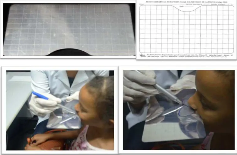
Measures of nasal aeration were obtained using the Altmann
graph mirror by marking the haze area with a blue marker pen before nasal cleansing and with a red marker pen after nasal
cleansing. The measures were recorded on special millimeter
paper sheets alike the mirror (Figure 1). The images were imported to the computer using a scanner (HP - Photosmart C3100) and then analyzed using the software program Image J 1.46r (http://imagej.nih.gov/ij). In this program, the area was
calculated according to the transformation from pixels to cm2
scales.
After collection of the nasal aeration, examination of the
nasal internal geometry was conducted by Acoustic Rhinometry, whose analysis enables the measure of the nasal cross-sectional areas (CSA) separately on both sides, corresponding to the
delections in the rhinogram, generally related to the nasal
valve region (CSA1) and the anterior and (CSA2) posterior (CSA3) portions of the middle and inferior turbinates, as well as to their respective distances (DIST1, DIST2, DIST3) to the nostrils (Figure 2); it also allows the measure of nasal volumes,
thus favoring the identiication of the sites of constriction that
contribute to nasal resistance(4,11). For analysis of the volume
measures, the region from 0 to 8 cm in relation to the entrance of the nostril was considered in the children; this region is equivalent to the portion of the nose that goes from the nostril
to the nasopharynx(12-14). The tests were performed using the
Eccovision Acoustic Rhinometer (HOOD Laboratories) system.
To conduct the examination, a rhinometry tube, attached to a
nasal adapter, was placed against one of the nostrils; lubricating gel was used to seal the nasal adapter to the nostril (Figure 3).
Proper methodological care was taken to avoid interference from
the environment in the rhinometric assessment, minimizing the study bias(4,11).
To this end, room temperature and noise level were controlled,
to position the rhinometry tube correctly, avoid sound losses,
and keep the patient’s head always stable(4,11). The children were
requested to look at the computer screen steadily so that head position was maintained throughout the examination (Figure 4).
After that, the nasal cleansing and massage procedure was conducted with the instillation of 2.5 ml of 0.9% saline solution at room temperature in each nostril with the aid of a needleless syringe. Immediately after serum instillation, circular massage
Figure 1. Altmann graph mirror, record sheet, and nasal aeration marking
Caption: CSA 3 = cross-sectional area of the posterior portion of the middle and inferior turbinate
with the thumb was performed on the lateral nasal region, 10 times
on each side. Next, the child blew one side of the nose at a time
onto tissue to remove all the secretion(12). After nasal cleansing
and massage, the same examination procedures were performed.
For data analysis of the nasal aeration measure, the areas obtained in each nasal cavity, separately (left and right sides), and the total area were considered, that is, the area corresponding to
the airlow of the two nasal cavities: left nasal cavity (LNC) plus
right nasal cavity (RNC). For the measures of nasal geometry, each cavity was analyzed separately, totaling 40 nasal cavities of 20 children.
To compare the results obtained before and after the cleansing technique application and between the sides of the nasal cavity, the
Wilcoxon signed-rank test was used: analysis of the relationship between variables, assigning a signiicance level of 5% (p<0.05). This study was approved by the Research Ethics Committee of the Health Sciences Center of the UFPE under protocol no. 15860213.5.0000.5208 according to Resolution CNS 466/12.
RESULTS
The population of the present study showed responses
above 60% in the Identiication Index of Signs and Symptoms
of Oral Breathing questionnaire, characterizing the functional diagnosis of oral breathing.
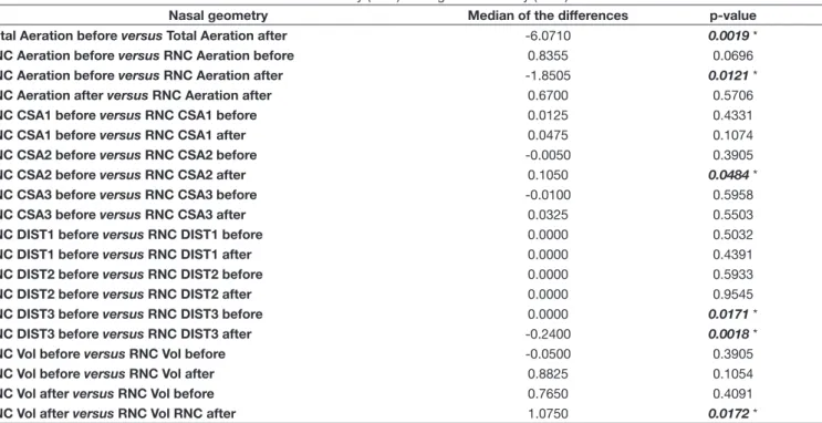
Tables 1 and 2 show the results obtained from the analysis of the correlation of nasal geometry before and after nasal
cleansing, by means of the Wilcoxon signed-rank test, in the
20 investigated children with speech-language pathology clinical diagnosis of oral breathing.
Table 1 shows the mean values of the nasal cavities and their respective standard deviations (±SD), the medians of the differences, and the p value of the aeration and internal geometry of the nose before and after nasal cleansing and massage.
Figure 4. Acoustic rhinometry examination
The values of 8.60±2.04 cm2 and 11.84±2.14 cm2 were observed,
respectively, before and after nasal cleansing, showing signiicant
increase in the area of nasal aeration (p=0.00025). Regarding the variables of the cross-sectional areas, their respective distances (CSA1-DIST1, CSA2-DIST2, and CSA3-DIST3) and volumes, no
signiicant differences were observed for the values obtained
before and after nasal cleansing and massage.
Table 2 shows the medians of the differences between total nasal aeration (LNC+RNC) before and after nasal cleansing and massage, right nasal aeration vs. left nasal aeration, and of the geometry of the left and right nasal cavities, separately, before
and after nose cleansing and massage. Signiicant differences
were observed between the following variables: Total Aeration
before versus Total Aeration after with md=-6.071 (p=0.0019); LNC Aeration before versus RNC Aeration after with md=-1.8505 (p=0.0121); CSA2 of LNC before and CSA2 of RNC after with
md=0.105 (p=0.0484); DIST3 of LNC before and DIST3 of RNC before with md=0 (p=0.0171); DIST3 of LNC before and DIST3 of RNC after with md=-0.24 (p=0.0018); VOL of LNC after and VOL of RNC after with md= 1.075 (p=0.0172).
DISCUSSION
The nasal cavity and its structures are complex, from the embryological point of view, beginning with the irst pharyngeal
arch, which originates the formation of the nasal fossae through the
Table 2. Correlation between the variables of the left nasal cavity (LNC) and right nasal cavity (RNC)
Nasal geometry Median of the differences p-value
Total Aeration before versus Total Aeration after -6.0710 0.0019 *
LNC Aeration before versus RNC Aeration before 0.8355 0.0696
LNC Aeration before versus RNC Aeration after -1.8505 0.0121 *
LNC Aeration after versus RNC Aeration after 0.6700 0.5706
LNC CSA1 before versus RNC CSA1 before 0.0125 0.4331
LNC CSA1 before versus RNC CSA1 after 0.0475 0.1074
LNC CSA2 before versus RNC CSA2 before -0.0050 0.3905
LNC CSA2 before versus RNC CSA2 after 0.1050 0.0484 *
LNC CSA3 before versus RNC CSA3 before -0.0100 0.5958
LNC CSA3 before versus RNC CSA3 after 0.0325 0.5503
LNC DIST1 before versus RNC DIST1 before 0.0000 0.5032
LNC DIST1 before versus RNC DIST1 after 0.0000 0.4391
LNC DIST2 before versus RNC DIST2 before 0.0000 0.5933
LNC DIST2 before versus RNC DIST2 after 0.0000 0.9545
LNC DIST3 before versus RNC DIST3 before 0.0000 0.0171 *
LNC DIST3 before versus RNC DIST3 after -0.2400 0.0018 *
LNC Vol before versus RNC Vol before -0.0500 0.3905
LNC Vol before versus RNC Vol after 0.8825 0.1054
LNC Vol after versus RNC Vol before 0.7650 0.4091
LNC Vol after versus RNC Vol RNC after 1.0750 0.0172 *
* p ≤ 0.05: statistically significant difference
Table 1. Measures of aeration and nasal geometry before and after nasal cleansing and massage
Nasal Aeration (cm2) N=40
CSA1 (cm2) N=40
CSA2 (cm2) N=40
CSA3 (cm2) N=40 DIST1 (cm) N=40 DIST2 (cm) N=40 DIST3 (cm) N=40 VOL (cm3) N=40
Measures and SD
Before 8.60 ± 2.04 0.40 ± 0.06 0.75 ± 0.18 1.01 ± 0.28 1.73 ± 1.20 2.90 ± 0.21 5.51 ± 0.09 6.96 ± 2.20 After 11.84 ± 2.14 0.41 ± 0.11 0.69 ± 0.20 0.99 ± 0.25 1.72 ± 0.20 3.79 ± 0.22 5.61 ± 0.12 6.47 ± 1.40 Median of the
differences 0.56 -0.00 0.02 0.02 0.78 0.79 0.33 0.37
p values 0.00025 * 0.8373 0.3617 0.7977 0.3662 0.7225 0.8473 0.3135
* p ≤ 0.05: statistically significant difference
proliferation of the ectoderm of the frontal process. Knowledge on its development can assist in the understanding of the pathologies found in this anatomical region, considering that, after birth, several factors can interfere in the normal breathing pattern, such as: anatomical predispositions, environmental factors - such as
climatic conditions - sleeping position, artiicial feeding, and
oral habits(15,16). Until the age of several months, the child is an
obligate nasal breather, considering that, in infants, laryngeal descent has not yet occurred and the soft palate necessarily rests on the epiglottic vallecula, precluding the natural patency of the oral airways(17).
With growth, the nasal cavities may undergo anatomical changes, such as deviated septum and hypertrophies of the nasal turbinates(18-25). The nasal breathing mode can become
predominantly oral and, thereby, bring functional changes not only to the respiratory process, but also to the stomatognathic system(19,20). Etiologies such as allergic rhinitis and nasal turbinate
hypertrophy are generally the main causes of nasal obstruction and oral breathing. As a consequence of this altered respiratory
pattern, children may present dificulties in speaking, chewing
and swallowing, in addition to dental and postural alterations,
which directly inluence their development(20-22). Therefore, it
is important to perform prior, accurate diagnoses for adequate speech-language pathology prevention and treatment in children with mouth breathing.
The age range in the sample of 20 patients diagnosed
with oral breathing was four to 12 years. It is known that
craniofacial growth undergoes changes in this period; however, no interference in the results was observed because there were no control individuals. The present study presents results after the quantitative analysis of aeration and nasal cavity geometry before and after the application of the nasal cleansing and massage technique, widely used in speech-language pathology practice in the treatment of oral breathing.
Analysis of the nasal cavity using the Altmann graph mirror
and the Image J software program showed signiicant increase
of the area after the cleaning and nasal massage, in both nasal
cavities. This inding corroborates those of another study(7),
which also used the Altmann millimeter mirror to verify changes in the nasal aeration areas after cleansing and massage,
quantitatively conirming improvement after the application of the speech-language pathology technique, whose beneits
of greater respiratory freedom(23) and shift from oral to nasal
breathing pattern(24) had already been perceived subjectively.
With respect to the measures taken by acoustic rhinometry
in this study, it was possible to observe that the cross-sectional areas (CSA1, CSA2, and CSA3) did not present signiicant increase when compared, on the same side, at the pre- and post-technique
application moments. This outcome can be justiied by the
fact that the applied technique, in spite of interfering in nasal aeration and favoring change of the respiratory mode, does
not expressively interfere in the nasal structure, that is, it does not provoke signiicant changes in the nose mucosa that can
interfere in the internal nasal area.
This result allows us to understand the nasal respiratory
physiology better, to the extent that it illustrates the response
of the nose to the applied technique: nasal aeration is probably increased due to the elimination or reduction of secretions
present in the nasal cavity and not owing to signiicant effects
on the mucosa structure. Moreover, it is possible to infer that
the tactile-kinesthetic stimulation caused by massage promotes
sensitization of the nasal cavity, thus promoting the routing of
airlow to the region, increasing nasal aeration(22).
This hypothesis can be further reinforced by comparing the present survey with other studies conducted with children
in which the CSAs presented signiicant increase after the use
of nasal vasoconstrictor(23,25), opposing the present study; that
is, the effect of the medicine on the nasal mucosa changes the measures of the cross-sectional areas, which does not occur with nasal cleansing.
This indicates that the improvement achieved with the use of vasoconstrictors, in spite of increasing nasal geometry and favoring patency, does not necessarily increase the functional gain in terms of aeration, considering that aeration is favored
by tactile-kinesthetic stimuli in oral breathers. Therefore,
although the effect of the cleansing and massage technique on nasal patency, related to the nasal mucosa in structural terms,
is not signiicant, its functional effect on aeration was, and this
may prove to be satisfactory to adapt the respiratory mode(26)
in chronic oral breathers.
It is also worth mentioning that the children in this study had already received medical treatment for nasal obstruction, corroborating the fact that they were referred to speech-language
pathology care for nasal function adequacy. This may explain the fact that the nasal geometry had not been signiicantly altered.
Thus the importance of applying this same study to individuals with nasal obstruction of allergic origin should be highlighted, so that the effect of massage on nasal obstruction could be tested.
In contrast, signiicant differences were observed between
the two cavities when the pre- and post-cleansing and massage moments were compared in two situations: CSA2 – corresponding to the cross-sectional area of the anterior portion of the middle and inferior turbinate and DIST3 – corresponding to the distance to the nostril from the third nasal constriction area (3rd delection
of the rhinogram curve) related to the posterior portion of the middle and inferior turbinate(14).
Considering that the measures of the CSA2 before the application of the technique did not show difference between the sides, it is possible to infer that there was a change in the area corresponding to the anterior portion of the inferior turbinate after the technique was applied. This may justify the effect of the technique on this region of the nasal cavity, where
greater reaction of the nasal mucosa is expected and where the
point of greatest constriction of the nasal cavity is located in cases without obstruction, after the nasal valve(12), being more
susceptible to the functional effects of the mucosa - considering that the nasal valve is less affected(4) – and, therefore, the main
Nevertheless, the nasal cycle phenomenon must be considered,
which can also explain the difference between the sides, mainly
because this phenomenon was not neutralized(26) taking into
account that nasal vasoconstrictors were not used in this study. The nasal cycle is characterized by the alternation of periods of greater resistance between the nasal cavities. This occurs due to the variation of predominance of the sympathetic or parasympathetic systems on the mucosa of the right and left nasal cavities, alternately. This physiological alternation persists during oral breathing, nasal occlusion, and even under the effect of topical anesthesia(27)
. Studies indicate that children up
to eleven years of age present a reciprocal nasal cycle, though not always in the classic way, as most adults do(25,28).
It is essential that studies on aeration and nasal geometry that aim to quantitatively analyze the patency of the nasal cavities value this aspect of the nasal cycle(28). The passage of airlow
through the nose is usually asymmetric and it is necessary to understand how this physiological process occurs in order to evaluate the internal nasal geometry.
Regarding distance, the signiicant difference observed in
the DIST3 in the comparison between the sides was maintained at the pre- and post-technique application moments. Because this measure refers to the topographic location of the third area of nasal constriction(4), it should be considered that nasal
cleansing and massage do not signiicantly interfere with this
characteristic.
With respect to volume, in the present study, we chose to analyze the total segment of 8 cm from the nostril entrance
to the nasopharynx (0-8 cm), corresponding to the nasal
(from 0 to 5 cm) and nasopharyngeal cavities in children,
considering the nasopharynx region as of 6 cm(12-14). The choice
for this segment was due to the fact that children within this
age group still suffer from pharyngeal tonsil inluence in an
increased volume, mainly because they are allergic.
The outcomes of the present study did not show signiicant
differences in nasal volume, in both cavities, when compared in the same side, after the cleansing and massage technique application. Comparison with other studies(14,25) that analyzed
the effects of vasoconstrictors on the nasal volume also showed that the improvement in nasal aeration observed in this study is
not determined by a signiicant structural change.
However, it is necessary to consider the segment chosen for
analysis, whose distance includes the nasopharynx region, which
does not directly receive the effects of the nasal cleansing and massage technique. In addition, in the present study, it was not possible to control the movement of the palatine veil, which may interfere with the measure of nasopharyngeal volume(29).
Therefore, it is suggested that a subsequent study evaluate the volumes in other segments, especially in the region between 1 and 5 cm, that is, the nasal cavity segment before
the nasopharynx(12-14).
In contrast, signiicant difference in nasal volume was
observed when the sides were compared after the technique was applied. In the same way that in the area and distance the values obtained for the right and left cavities, before the
cleansing and massage, did not differ signiicantly; therefore,
it is also possible to consider that there may have been some effect on the nasal volume, after the technique application, but in this case the nasal cycle effect should also be considered, as previously discussed.
Therefore, nasal cleansing and massage have proved to be effective in improving nasal patency, in relation to nasal aeration, in children with physiological oral breathing. Regarding its effect on nasal geometry, we suggest that further studies be conducted with individuals with nasal obstruction diagnosed by otorhinolaryngology.
CONCLUSION
Nasal cleansing and massage positively inluence nasal
aeration in children with physiological oral breathing. After
the application of this technique, signiicant improvement was
observed in nasal aeration, as well as in the cross-sectional area corresponding to the anterior portion of the middle and inferior turbinate (CSA2) and in the volume, in the comparison between the two nostrils. Measures of the area of nasal aeration and of the area and volume between the two cavities were sensitive to changes after the application of the cleansing and massage technique. It is suggested that further studies be conducted with children with nasal obstruction and with volumes of 0-5 cm, which correspond to the segment between the entrance of the
nasal cavity and the inal portion of the nasal turbinate.
REFERENCES
1. Díaz MJE, Fariñas CMM, Pellitero RBL, Álvarez IE. La respiración
bucal y su efecto sobre la morfologia dentomaxilofacial. CCM [Internet]. 2005;9(1) [citado em 2011 Mar 27]. Disponível em: http://www.cocmed.
sld.cu/no91/n91ori6.htm
2. Carr AJ, Gibson B, Robinson PG. Is quality of life determined by expectations
or experience? BMJ. 2001;322(7296):1240-3. PMid:11358783. http://
dx.doi.org/10.1136/bmj.322.7296.1240.
3. Abreu RR, Rocha RL, Lamounier JÁ, Guerra AFM. Etiology, clinical manifestations and concurrent findings in mouth-breathing children. J Pediatr. 2008;84(6):529-35. PMid:19060979. http://dx.doi.org/10.1590/ S0021-75572008000700010.
4. Gomes AOC, Sampaio-Teixeira ACM, Trindade SHK, Trindade IEK. Áreas seccionais nasais de adultos sadios aferidas por rinometria acústica. Rev Bras Otorrinolaringol. 2008;74(5):746-54. http://dx.doi.org/10.1590/ S0034-72992008000500017.
5. Trindade IEK, Conegliam PCP, Trindade SHK, Dias NH, Sampaio-Teixeira ACM. Internal nasal dimensions of adults with nasal obstruction. Braz J Otorhinolaryngol. 2013;79(5):575-581. http://dx.doi.org/10.5935/1808-8694.20130103.
6. Uzun A, Ozdemir F. Morphometric analysis of nasal shapes and angles in young adults. Braz J Otorhinolaryngol. 2014;80(5):397-402. PMid:25303814.
http://dx.doi.org/10.1016/j.bjorl.2014.07.010.
repaired unilateral cleft lip and palate after rhinoseptoplasty. J Craniofac Surg. 2009;20(2):308-14. PMid:19258909. http://dx.doi.org/10.1097/ SCS.0b013e3181992287.
8. Silva AML, Gamboa T. Rinometria acústica: valores de referência numa
população de estudantes universitários. [dissertação]. Lisboa: Universidade
Nova de Lisboa; 2012.
9. Melo FMG, Cunha AD, Silva HJ. Avaliação da aeração nasal pré e pós realização de manobras de massagem e limpeza nasal. Rev CEFAC. 2007;9(3):375-82. http://dx.doi.org/10.1590/S1516-18462007000300011.
10. Sistema Respiratório. Faringe [Internet]. 2014 [citado em 2014 Dez 26]. Disponível em: http://sistema-respiratorio36.webnode.com/faringe/
11. Hilberg O. Objective measurement of nasal airway dimensions using acoustic rhinometry: methodological and clinical aspects. Allergy. 2002;57(Supl 70):5-39. PMid:11990714. http://dx.doi.org/10.1046/j.0908-665x.2001.
all.doc.x.
12. Millqvist E, Bende M. Two-year follow-up wiyh acoustic rhinometry in children. Am J Rhinol. 2006;20(2):203-4. PMid:16686389.
13. Qian W, Chen W, Chen JM, Haight J. Acoustic rhinometry in preschool
children. Otolaryngol Head Neck Surg. 2007;137(1):39-42. PMid:17599562.
http://dx.doi.org/10.1016/j.otohns.2007.02.007.
14. Nigro CEN, Goto E, Nigro JFA, Junior JFM, Mion O, Voegels R. L.
Avaliação da cavidade nasal e nasofaringe através da rinometria acústica
antes e após adenoidectomia. Rev Bras Otorrinolaringol. 2003;69(3):333-6.
http://dx.doi.org/10.1590/S0034-72992003000300006.
15. Santos-Neto ET, Barbosa RW, Oliveira AE, Zandonade E. Fatores associados ao surgimento da respiração bucal nos primeiros meses do desenvolvimento infantil. Rev Bras Crescimento Desenvolv Hum. 2009;19:237-48.
16. Barbosa C, Vasquez S, Parada MA, Gonzalez JC, Jackson C, Yanez ND, et al. The relationship of bottle feeding and other sucking behaviors with speech disorder in Patagonian preschoolers. BMC Pediatr. 2009;9(1):66. PMid:19845936. http://dx.doi.org/10.1186/1471-2431-9-66.
17. Trindade IEK, Gomes AOC, Sampaio-Teixeira ACM, Trindade SHK. Adult nasal volumes assessed by acoustic rhinometry. Rev Bras Otorrinolaringol. 2007;73(1):32-9. PMid:17505596. http://dx.doi.org/10.1016/S1808-8694(15)31119-8.
18. Lee WT, Koltai PJ. Nasal deformity in neonates and young children. Pediatr Clin North Am. 2003;50(2):459-67. PMid:12809334. http://dx.doi. org/10.1016/S0031-3955(03)00036-1.
19. Mello JF Jr, Mion O. Rinite alérgica. In: Campos CAH, Costa HOO. Tratado de otorrinolaringologia. São Paulo: Rocca; 2002. p. 68-87.
20. Barbosa RW, Oliveira AE, Zandonade E. Fatores associados ao surgimento da respiração bucal nos primeiros meses do desenvolvimento infantil. Rev Bras Crescimento Desenvolv Hum. 2009;19(2):237-48.
21. Lemos CM, Wilhelmsen NSW, Mion OG, Mello JF Jr. Functional alterations of the stomatognathic system in patients with allergic rhinitis: case-control study. Braz J Otorhinolaryngol. 2009;75(2):268-74. PMid:19575115. http://
dx.doi.org/10.1016/S1808-8694(15)30789-8.
22. Marson A, Tessitore A, Sakano E, Nemr K. Effectiveness of speech and language therapy and brief intervention proposal in mouth breathers. Rev. CEFAC. 2012;14(6).
23. Cunha DA, Silva HJ. Terapia fonoaudiológica em respiração oral: como eu
trato. In: Marchesan IQ, Silva HD, Berretin-Felix G. Terapia fonoaudiológica em motricidade orodacial. São José dos Campos: Pulso Editorial; 2012. p.
87-94.
24. Krakauer LH. Terapia do respirador oral. In: Krakauer LH, Di Francesco
RC, Marchesan IQ. Respiração oral: abordagem interdisciplinar. São José
dos Campos: Pulso. 2003. p. 119-125.
25. Trindade IEK, Gomes AOC, Fernandes MBL, Trindade SHK, Silva Filho
OG. Nasal airway dimensions of children with repaired unilateral cleft lip and palate. Cleft Palate Craniofac J. 2015;52(5):512-6. PMid:25210862.
http://dx.doi.org/10.1597/14-103.
26. Melo ACC, Gomes AOC, Silva HJ. Correlação de três variáveis na descrição
da permeabilidade nasal (HD, MCA, escala NOSE) de pacientes saudáveis:
resenha. Dist Comunic. 2014;26:417-9.
27. Kayser R. Die exakte Messung der Luftdurchgängigkeit der Nase. Arch. Laryng. Rhinol. 1895;8:101.
28. Gallego AJ, Cavallari FEM, Valera FCP, Demarco RC, Anselmo-Lima WT. Study of nasal cycles in children by acoustic rhinometry. Am J Rhinol. 2006;20(6):560-2. PMid:17181092. http://dx.doi.org/10.2500/ ajr.2006.20.2951.
29. Gomes AOC. Dimensões nasais e nasofaríngeas de adultos sem obstrução
nasal, aferidas por rinometria acústica [dissertação]. Bauru: HRAC-USP;
2004.
Author contributions