w w w . r b o . o r g . b r
Original Article
Randomized clinical trial on percutaneous
minimally invasive osteosynthesis of fractures of
the distal extremity of the radius
夽
,
夽夽
Marcio Aurélio Aita, Carlos Henrique Vieira Ferreira, Daniel Schneider Ibanez
∗,
Rafael Saraiva Marquez, Douglas Hideki Ikeuti, Rodrigo Toledo Mota,
Marcos Vinicius Credidio, Edison Noboru Fujiki
Ortopedia e Traumatologia, Faculdade de Medicina do ABC, Santo André, SP, Brazil
a r t i c l e
i n f o
Article history:
Received 16 June 2013 Accepted 21 June 2013 Available online 18 April 2014
Keywords:
Fractures of the radius Internal fracture fixation Bone plate
a b s t r a c t
Objectives:the purpose of this study was to compare the postoperative radiological and clin-ical outcomes with minimally invasive percutaneous osteosynthesis using three implants: volar locking plate, intramedullary nail system and nonbridging external fixator for distal radius fractures.
Methods:forty-eight patients (A group, 16; B group 16; C group 16) underwent minimally invasive percutaneous osteosynthesis of reductible and unstable displaced (Type IIB by Ray-hack Classification) distal radius fractures. In B group intramedullary nail system was used, in A group the patients were treated with volar locking plate and in C group the patients were treated by nonbridging external fixator from January 2011 to December 2012. The mean follow-up period was 12 months. Radiologic parameters, range of motion, grip strength, and disability of the arm, shoulder, and hand score were evaluated at each examination (3rd and 6th week, and 12th months). The visual analog scale of wrist pain and complications were assessed at the final follow-up.
Results:the groups did not differ significantly in radiological outcomes after 12 months, but the clinical results, VAS scale and dash score in group A (volar locking plate) and B (nail intramedullary) were statistically significantly better than that of C group (nonbridg-ing external fixator). One patient underwent an osteosynthesis with nail intramedullary and another with external fixator (C group) developed persistent pain near the site of the superficial radial nerve because of the distal’s screw and pins, respectively.
Conclusion:in clinical parameters, significant differences in outcomes were found between groups A and B after six weeks versus C group.
© 2014 Sociedade Brasileira de Ortopedia e Traumatologia. Published by Elsevier Editora Ltda. All rights reserved.
夽Please cite this article as: Aita MA, Vieira Ferreira CH, Schneider Ibanez D, Saraiva Marquez R, Hideki Ikeuti D, Toledo Mota R, et al. Ensaio clínico randomizado de osteossíntese percutânea e minimamente invasiva das fraturas da extremidade distal do rádio. Rev Bras Ortop. 2014;49:218–226.
夽夽
Work performed by the Hand and Microsurgery Group, ABC School of Medicine, Santo André, SP, Brazil. ∗ Corresponding author.
E-mail: [email protected] (D. Schneider Ibanez).
Ensaio clínico randomizado de osteossíntese percutânea e minimamente
invasiva das fraturas da extremidade distal do rádio
Palavras-chave:
Fraturas do rádio
Fixac¸ão interna de fraturas Placas ósseas
r e s u m o
Objetivos: comparar o resultado clínico funcional dos pacientes com diagnóstico de fratura com desvio, redutível e instável da extremidade distal do rádio submetidos ao tratamento cirúrgico pela técnica de osteossíntese percutânea e minimamente invasiva com o uso de três tipos de implante: placa volar bloqueada, haste intramedular bloqueada e fixador externo. Comparar os resultados quanto à melhoria da qualidade de vida pelo questionário Dash e ao tempo de retorno ao trabalho.
Métodos: divididos em três grupos (A, placa – 16; B, haste – 16; C, fixador externo – 16), 48 pacientes foram submetidos ao tratamento cirúrgico da fratura da extremidade distal do rádio, redutível e instável, classificac¸ão Rayhack (Tipo IIB), pelo método minimamente invasivo, com três tipos de implante: haste intramedular bloqueada, placa volar bloqueada e fixador externo radio-radial. Estudo feito de janeiro de 2011 a dezembro de 2012. O tempo de seguimento foi de 12 meses. Parâmetros radiográficos, dor (escala VAS), medida do arco de movimento, forc¸a de preensão palmar e o questionário Dash foram avaliados na terceira e sexta semana e no sexto mês de pós-operatório.
Resultados: numa análise vertical dos valores apresentados, observamos uma melhoria estatística significativa em todos os parâmetros clínicos analisados no estudo, nos três gru-pos. Em relac¸ão à análise horizontal, ou seja, na comparac¸ão dos grupos entre si, não houve diferenc¸as estatísticas significativas quanto aos parâmetros radiográficos após o 12◦mês de
seguimento. O grau de forc¸a de preensão palmar, a medida do arco de movimento, VAS e Dash apresentaram, na terceira e sexta semana de pós-operatório, valores estatísticos sig-nificativos superiores nos grupos A e B. Um paciente do grupo B apresentou dor no punho, por provável proximidade do parafuso com o primeiro túnel extensor, que foi removido; e outro do C apresentou dor no punho, no trajeto do ramo sensitivo do nervo radial, pela presenc¸a do pino de Schantz.
Conclusão: a técnica minimamente invasiva é eficaz e segura, com melhoria clínica e funcional em todos os momentos do estudo. Ambos os três implantes são estáveis. Há supe-rioridade estatística significativa dos resultados clínico-funcionais (grau de forc¸a e arco de movimento, Dash e VAS) até a sexta semana, para os grupos A (placa) e B (haste). No fim de 12 meses não há diferenc¸as estatísticas significativas entre os grupos.
© 2014 Sociedade Brasileira de Ortopedia e Traumatologia. Publicado por Elsevier Editora Ltda. Todos os direitos reservados.
Introduction
Fractures that affect the distal radius are among the most fre-quent fractures of the upper limbs and account for 74.5% of the fractures of the forearm, i.e. an incidence of 1:10,000 people.1
There is greater incidence in the dominant arm.2
Currently, fractures of the distal extremity of the radius are regarded as complex, with a prognosis depending on the type of fracture and/or the treatment used. In seeking better clin-ical and functional results, surgclin-ical methods are increasingly indicated, and there has been great evolution in the implants developed for the distal extremity of the radius. Nonetheless, there is still space for classical conservative treatment, partic-ularly for fractures without displacement.3
Surgical methods have also evolved, with minimally inva-sive osteosynthesis techniques and approaches toward soft tissues that are more biological, i.e. less aggressive toward the covering of soft tissues.4
The implants that are most often used in our setting for treating these fractures are Kirschner wires, volar locking plates and transarticular external fixators.4
Stabilization of fractures of the distal extremity of the radius, using Kirschner wires placed separately or using the transarticular external fixation method, does not allow early rehabilitation of the wrist. Kirschner wires are a method with minimal osteosynthesis and require plaster-cast immobiliza-tion throughout the treatment, while external fixaimmobiliza-tion using Schanz pins placed in the second or third metacarpals blocks flexion-extension of the wrist.
However, these and new implants developed for fractures of the distal extremity of the radius, such as intramedullary locking nails5and nonbridging external fixators can be used
percutaneously.6
The possibility of indirect open reduction of such fractures, i.e. without viewing the focus of the fracture, allows ever-earlier rehabilitation, given that the implants and the surgical approach are also less aggressive and more stable.6
Since there are no statistically significant results in the studies so far published with regard to surgical treatment of these fractures, and these systematic reviews and meta-analyses7 state that it is necessary to conduct studies with
implants: intramedullary locking nail, volar locking plate and nonbridging external fixator. All the implants allow the same rehabilitation protocol, with early mobilization recommended immediately after the surgery.
Objectives
To compare the functional clinical results (palm grip strength, wrist range of motion and pain) among patients with a diag-nosis of an unstable but reducible displaced extra-articular fracture (type IIB of Rayhack’s classification)8 of the distal
extremity of the radius, who underwent surgical treatment by means of a minimally invasive percutaneous osteosynthesis technique, using three different implants: volar locking plate, intramedullary locking nail and external fixator.
To compare the results with regard to quality-of-life improvement through applying the DASH questionnaire.9
Material and methods
Between January 2011 and December 2012, 100 patients who presented at the outpatient clinics of the Hand and Micro-surgery Group, ABC School of Medicine, with a diagnosis of fracture of the distal extremity of the radius were evaluated. Forty-eight of these patients were included in the study and underwent physical examination and posteroanterior (PA) and lateral (L) radiographic examinations.
The inclusion criteria were that the patients should be adults aged 18–65 years, of either sex, with a clinical and imag-ing diagnosis of this fracture (type IIB), and needed to fill out the voluntary free and informed consent statement and the protocol of conflicts of interest, as specified by the Research Ethics Committee (Appendix A).
Patients were excluded according to the following crite-ria: presentation of associated diseases of the wrist, such as osteometabolic diseases; having undergone any previous sur-gical procedure; or presentation of conditions that affected the wrists bilaterally.
The functional evaluation was conducted by professionals in the Hand Occupational Therapy Sector of the Mario Covas State Hospital, who did not have access to information on the group to which the patients belonged.
Functional measurements of wrist range of motion were made in degrees using a single specific goniometer. Palm grip strength was measured in kilogram-force (kgf) by means of a hydraulic dynamometer (Jamar®).
The clinical analysis on pain was performed using a visual analog scale (VAS) from zero to ten, for subjective evaluation. Quality of life was assessed using the DASH questionnaire,9
which is the instrument used in the majority of studies pub-lished on treatments for fractures of the distal extremity of the radius.
The randomization consisted of making matches in groups of three, by means of a draw, using standardized tickets marked A, B or C (A, volar locking plate; B, intramedullary nail; C, external fixator). The tickets were placed in a bag and mixed, and then the secretary of the sector pulled out a single ticket and placed it on the table. This ticket did not participate fur-ther in the draw until the group of three had been completed. This was done in the outpatient clinic, before the surgery.
Thus, the patients were divided into three groups: A, B and C.
The patients underwent percutaneous minimally invasive osteosynthesis using one of the three different implants. The patients in group A received a volar locking plate,6,10those in
group B received an intramedullary nail and those in group C received an external fixator.11
Operative technique for osteosynthesis with volar locking plate (group A)
A volar access was used, under the radial flexor tendon of the carpus, 1 cm distally to the fracture, with dissection in layers down to the radius bone (Table 1).
The volar plate was slid in with the aid of radioscopy, through which the ideal positioning could be viewed.
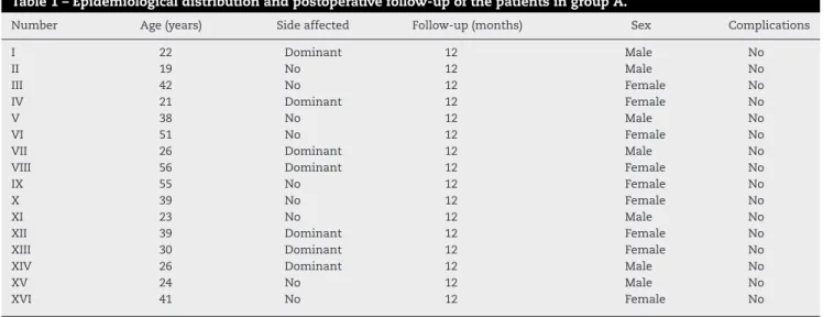
Table 1 – Epidemiological distribution and postoperative follow-up of the patients in group A.
Number Age (years) Side affected Follow-up (months) Sex Complications
I 22 Dominant 12 Male No
II 19 No 12 Male No
III 42 No 12 Female No
IV 21 Dominant 12 Female No
V 38 No 12 Male No
VI 51 No 12 Female No
VII 26 Dominant 12 Male No
VIII 56 Dominant 12 Female No
IX 55 No 12 Female No
X 39 No 12 Female No
XI 23 No 12 Male No
XII 39 Dominant 12 Female No
XIII 30 Dominant 12 Female No
XIV 26 Dominant 12 Male No
XV 24 No 12 Male No
XVI 41 No 12 Female No
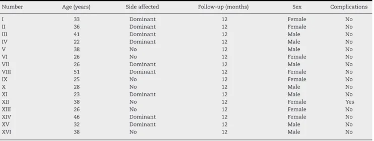
Table 2 – Epidemiological distribution and postoperative follow-up of the patients in group B.
Number Age (years) Side affected Follow-up (months) Sex Complications
I 33 Dominant 12 Female No
II 36 Dominant 12 Female No
III 41 Dominant 12 Male No
IV 22 Dominant 12 Male No
V 38 No 12 Male No
VI 26 No 12 Female No
VII 26 Dominant 12 Male No
VIII 51 Dominant 12 Female No
IX 25 No 12 Female No
X 28 No 12 Male No
XI 23 Dominant 12 Male No
XII 38 No 12 Female Yes
XIII 26 No 12 Female No
XIV 46 Dominant 12 Female No
XV 32 Dominant 12 Male No
XVI 38 No 12 Male No
Source: medical filing service of the hospitals of the ABC School of Medicine.
Closed reduction of the fracture was performed, with pro-visional placement of one Kirschner wire.
A distal volar access was made in the wrist, at the exact location of the first hole of the plate, with dissection in layers down to the bone.
The plate was fixed using a 2.5 mm proximal cortical screw and a 2.5 mm distal locking screw, with viewing of the reduc-tion with the aid of radioscopy.
The remaining screws were then put in: six distal and three proximal screws.12
The layers and the skin were then sutured.
Postoperative radioscopy and radiography were performed on the wrist to check on the surgery.
A dressing was then placed around the wrist.
Operative technique for osteosynthesis with intramedullary locking nail (group B)
Closed reduction was performed on the fracture and it was sta-bilized provisionally using one or two Kirschner wires inserted in the region of the distal ulna, as far as the medial column of the radius, with the aid of radioscopy (Table 2).
The access route was minimal, dorsal and radial, measur-ing 2 cm, usmeasur-ing the first extensor tunnel as the anatomical parameter. A single longitudinal opening was made in the dor-sal retinaculum, with exposure of the extensor tendons of the first tunnel, which were pushed away dorsally.
A standard Kirschner wire and guide were then implanted in the region proximal to the radial styloid, which was pegged in the metaphyseal intramedullary region of the radius, just below the first extensor tunnel.
Intramedullary milling was performed using a specific can-nulated drill bit, with a guidewire as an essential parameter.
Intramedullary milling tools of the size of the nail (ranging from one to five) were then emplaced manually. For perfect adjustment of the milling tool with the medulla of the bone, the size of the implant to be used was measured.
Finally, the intramedullary nail was emplaced, with the aid of radioscopy.
The fracture was stabilized distally using three 2.7 mm lat-eral locking screws, which enabled angular stability, using a guide adjacent to the nail.
The system was stabilized proximally using two 2.4 mm dorsal cortical screws, with the aid of a specific guide.
The layers and skin were sutured.
Postoperative radioscopy and radiography were performed on the wrist to check on the surgery.
A dressing was then placed around the wrist.
Operative technique for osteosynthesis with nonbridging external fixator (group C)
Percutaneous accesses measuring 0.5 mm were made in the radial styloid and in the distal region of the medial column of the radius, in order to insert Kirschner wires (Table 3).
Closed reduction was performed on the fracture and two crossed Kirschner wires were emplaced: one entering through the radial styloid and the other through the distal region of the medial column of the bone.
Two 3 mm Schanz pins were placed dorsally and distally to the fracture and two Schanz pins were placed dorsally radially and proximally to the fracture.
The external fixator was assembled on the Schanz pins, which allowed mobility of the wrist and enabled secure stabi-lization of the fracture.
The skin was sutured.
Postoperative radioscopy and radiography were performed on the wrist to check on the surgery.
A dressing was then placed around the wrist.
Postoperative period for groups A, B and C
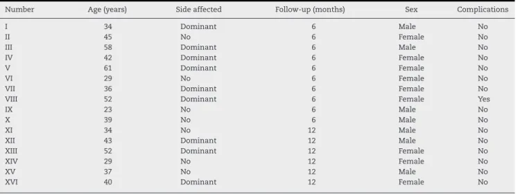
Table 3 – Epidemiological distribution and postoperative follow-up of the patients in group C.
Number Age (years) Side affected Follow-up (months) Sex Complications
I 34 Dominant 6 Male No
II 45 No 6 Female No
III 58 Dominant 6 Male No
IV 42 Dominant 6 Female No
V 61 Dominant 6 Female No
VI 29 No 6 Female No
VII 36 Dominant 6 Female No
VIII 52 Dominant 6 Female Yes
IX 23 No 6 Male No
X 39 No 6 Male No
XI 34 No 12 Male No
XII 43 Dominant 12 Male No
XIII 52 Dominant 12 Female No
XIV 29 No 12 Female No
XV 37 No 12 Male No
XVI 40 Dominant 12 Female No
Source: medical filing service of the hospitals of the ABC School of Medicine.
functional gain, from the first week after the surgery until discharge.
This study was approved by the Medical Research Ethics Committee of the ABC School of Medicine in July 2007, through CEP/FMABC Protocol Registration No. 160/2007.
All the data were sent for statistical analysis. The statisti-cal level of 5% (0.050) was used for applying statististatisti-cal tests, i.e. when the calculated significance value (p) was less than 5% (0.050), a difference that was said to be “statistically signif-icant” was observed (marked in red); and when the calculated significance value (p) was greater than or equal to 5% (0.050), a difference that was said to be “statistically non-significant” was observed. The SPSS software (Statistical Package for the Social Sciences), version 13.0, was used to obtain the results.
Results
The results from the patients were presented at three study times: third week (Table 4), sixth week (Table 5) and twelfth month (Table 6).
Table 4 – Clinical and functional results in the third week.
Parameters Group
A – plate
Group B – nail
Group C – fixator
Pain (VAS) 3.8 2.6 3.75
DASH 36.4 23.6 68.13
Strength 53.6 66.67 29.03
ROM 86.1 89.72 71.0
Complications – 6.25 6.25
Source: medical filing service of the outpatient clinic of the ABC School of Medicine.
ROM, range of motion; DASH, disability of arm, shoulder and hand. % of ROM and strength in relation to the values of the contralateral side.
% complications in relation to the total number of patients.
Table 5 – Clinical and functional results in the sixth week.
Parameters Group
A – plate
Group B – nail
Group C – fixator
Pain (VAS) 1.80 1.00 1.70
DASH 14.7 10.4 29.5
Strength 78.44 88.89 60.00
ROM 94.60 98.01 83.00
Complications – 6.25 6.25
Source: medical filing service of the outpatient clinic of the ABC School of Medicine.
ROM, range of motion; DASH, Disability of Arm, Shoulder and Hand. % of ROM and strength in relation to the values of the contralateral side.
% complications in relation to the total number of patients.
Statistical study
The three groups studied were described and compared (hori-zontal statistical analysis) by applying the Kruskal–Wallis test,
Table 6 – Clinical and functional results in the 12th month.
Parameters Group
A – plate
Group B – nail
Group C – fixator
Pain (VAS) 0.0 0.0 0.0
DASH 1.1 1.2 1.50
Strength 97.00 98.90 94.50
ROM 99.95 100 99.4
Complications – – –
Source: medical filing service of the outpatient clinic of the ABC School of Medicine.
ROM, range of motion; DASH, disability of arm, shoulder and hand. % of ROM and strength in relation to the values of the contralateral side.
with the aim of ascertaining possible differences between the three groups studied, when compared concomitantly for the variables of interest.
Since some differences that were said to be statistically dif-ferent were encountered, the Mann–Whitney test was applied, with adjustment using the Bonferroni correction, in order to attempt to identify which groups differed from each other when compared pair by pair.
The data were described and compared between the observation times, for each study group (vertical statistical analysis).
The Friedman test was applied, with the aim of ascertain-ing possible differences between the three observation times, for each study group and for the variables of interest.
Since there were statistically significant differences in all of the comparisons made, the Wilcoxon signed-rank test was applied, with adjustment using the Bonferroni correction, in order to attempt to identify which observation times differed from each other when compared pair by pair.
Complications
One patient in group B presented pain in the wrist, probably due to proximity of the distal locking screw to the first exten-sor tunnel. Just after the sixth month after the operation, this screw was removed and the pain ceased.
Another patient, in group C, presented wrist pain on the path of the sensory branch of the radial nerve, due to proximity of the presence of the Schanz pin. This pain also improved with removal of the pin in the sixth month after the surgery.
Discussion
Evolution of the treatments for fractures of the distal extremity of the radius has taken place along two lines: technologically, through development of implants that enable angular stability, and biologically, also through stabilizing the bone, but preserving the covering of soft tissues and thus enabling earlier functional return for this joint and leaving patients with less economic and social damage.
New designs for volar plates with a minimum thickness of only 2 or 3 mm, and new designs for instruments, with guides coupled to the intramedullary nail, along with the use of radiotransparent materials for external fixators, which have become established for treating these fractures over the last ten years, have made it possible to use minimally invasive techniques.
From vertical analysis, i.e. making comparisons between the data obtained within each group, our study showed statis-tically significant functional clinical results, irrespective of the implant, at all assessment times of the study, which showed that the minimally invasive technique was effective.
From the analysis on the radiographic parameters, all the patients presented the same initial reduction of the fracture, which showed that all the implants used in this study were safe and stable, thus enabling effective bone consolidation at the fracture.
Horizontal comparison of the functional clinical results between the groups (range of motion, palm grip strength, DASH and VAS) showed that there were statistically signifi-cant differences between the values for group C and those for groups A and B, in the third and sixth weeks after the surgery. From this, we can affirm that the patients who were treated with an intramedullary nail or a volar plate presented bet-ter quality of life and fewer pain symptoms than did those who were treated with an external fixator, up to the sixth week. However, after the sixth week, the clinical-functional parameters became statistically similar between the three groups.
The data obtained in this study were also compared with those from studies in the literature. Our results were similar to those of Schønnemann et al.11The palm grip strength results
were better among patients treated with an intramedullary nail than among patients treated with a nonbridging exter-nal fixator,12–15 and the results relating to the radiographic
parameters analyzed were similar, without any loss of the initial reduction, after consolidation of the fracture. We also used Kirschner wires in association with external fixators, in order to ensure greater fracture stability.14
Comparing our values with those of Cui et al.,16there were
similarities in the results, with better evidence among the patients treated with a volar plate than among those treated with an external fixator.
Although the study by Zenke et al.17did not show any
sig-nificant differences between the conventional method and the minimally invasive method using volar plates for treat-ing these fractures, we observed better clinical-functional and DASH results in our study, in relation to the study by Orbay et al.,10with palm grip strength of 97% versus 77% and DASH
of 1.1 versus 8.28.
Regarding the number of screws for stabilizing the volar plate, better fixation was observed with three screws inserted proximally to the fracture focus (diaphyseal location) and at least six locking screws distally to the fracture focus. This was also observed in the biomechanical study by Mehling et al.12
In evaluating complications, the studies by Richard et al.18
and Rampoldi and Marsico19showed better functional results
and a lower rate of complications among patients treated with volar plates than among those with external fixation. This was also observed in the present study, with values of 6.25% in group C and 0% in group A.
In a systematic review, Espósito et al.20found a lower DASH
score, better restoration of the length of the radius and lower infection rate in the group of patients with fractures who were treated by means of internal osteosynthesis using a plate than in those treated using an external fixator. These results resem-ble those of the present study.
Both in our study and in that of Xie et al.21 in 2013
(which was a systematic review on surgical treatment of these fractures), there were better functional results (fore-arm supination, restoration of palmar tilt and radial slope and a lower complication rate) in the group of patients treated with a volar plate than in those treated with an external fixa-tor.
Sando et al.22 conducted a study that showed the
of the distal extremity of the radius and found that out of the 215 studies analyzed, 75% presented positive results, 20% neutral results and 5% negative final values. These authors concluded that these articles presented characteristics that would lead to positive results and that these results may have facilitated publication of these studies. They also sug-gested that a standardized clinical register covering the entire United States should be created for the results to be followed up and assessed. In our study, we attempted to use mini-mally invasive methods, cases in which the fractures had the same characteristics and recognized assessment and reha-bilitation protocols in order to minimize these sources of bias.
Although using intramedullary nails to treat these frac-tures is not common in our setting, it could be seen in the present study that the results obtained were excellent and that the learning curve was short.
Conclusion
The minimally invasive technique is effective and safe, with clinical and functional improvements at all observation times of the study. The three implants were stable, given that there were no alterations in the radiographic parameters. There were statistically significantly better clinical-functional results (degree of strength, range of motion, DASH and VAS) in group A (plate) and group B (nail), up to the sixth week. However, after the 12th month, there were no statistically significant differences in the clinical-functional parameters analyzed, between the three groups.
Conflicts of interest
The authors declare no conflicts of interest.
Anex 1. Voluntary free and informed consent
statement
Study: a randomized comparative study using external fixa-tors, percutaneous volar plates and intramedullary nails to surgically treat fractures of the distal extremity of the radius. Principal investigator: Walter Yoshinori Fukushima; Medi-cal Registration Number (CRM): 109969-SP.
Address: Rua Morvan Dias de Figueiredo, 155, apto 12, CEP 09732-580, São Bernardo do Campo, São Paulo.
Discipline of Diseases of the Locomotor System (Ortho-pedics and Traumatology), ABC School of Medicine, Avenida Lauro Gomes, 2000, Bairro Vila Sacadura Cabral, Santo André – SP, CEP 09060-870, Brazil. Tel: +55 011 4993 5400.
This study was assessed and approved by the institution’s Research Ethics Committee on July 4, 2007 (registration no. 160/2007). This committee is a body that has the aim of pro-tecting your well-being. It is responsible for assessing and following up the ethical issues of all studies involving human beings so that the dignity, rights, safety and well-being of the research subjects are ensured. If you have any doubts and/or questions about your rights as a participant in this study, or if you are dissatisfied with the way in which the study is being
conducted, you can get in touch with the Research Ethics Com-mittee of the ABC School of Medicine at the following address: Avenida Príncipe de Gales, 821, 1st floor, CEPES Building, Santo André, SP, or by telephone: (11)-4993-5453. The hours of atten-dance are from Monday to Friday from 07:00 to 17:00.
Name of subject or person with legal responsibil-ity:
No. of identity document: Sex: M ()/F ()
Date of birth: / /
Telephone: () Address:
I, Mr/Ms
exchange of the implant(s) will be performed if necessary. Discomfort, pain, loss of strength, diminished range of joint motion, motor incapacity and other risks are inherent to the surgical procedure. Current diseases compatible with the degree of surgical complexity may progress to definitive motor incapacity and death. I will be benefited directly through the opportunity to receive adequate treatment for the disease, in its present state, at the institution that I have voluntarily chosen, from trained and qualified professionals. Regarding alternative procedures that could be advantageous, which I might have chosen, I am rejecting these and placing this treatment as the best option for the present moment of the disease. I have had the opportunity to ask all the questions that I deemed fundamental, important and necessary, and all of these were answered fully and satisfactorily. I am aware of the confidentiality, secrecy and privacy of the study. Nonetheless, at any stage of the treatment, clarifications for possible doubts are assured and I can ask such questions by appointment. I will have the right to be kept up to date with regard to the partial results from the treatment, complica-tions and favorable or unfavorable evolution of the present disease, with support for intercurrences from the Hand Surgery Outpatient Clinic of the Padre Anchieta Teaching Hospital. I have the obligation to declare all previous diseases, diabetes, hypertension, contagious diseases, hematological diseases, allergies, drug use, smoking, medications in use, psychiatric treatment, etc.; and to rigorously follow all the guidance, make return visits to the outpatient clinic, take medications in accordance with prescriptions and follow the rules of the hospital and the ABC School of Medicine. There will not be any financial compensation in relation to my participation in the surgery and in the study. I will have the right to the surgical treatments proposed, anesthesia and medical follow-up, in conformity with the fees paid for these procedures, which are expressed in the contract with the Brazilian National Health System (SUS), to which I have a right. If a health problem is detected prior to the start of the study, I would expect to be referred to SUS for treatment. I believe that I have received sufficient explanations regarding the information that I have received and read, relating to the treatment, risks and benefits to which I have a right, and all the obligations. The purposes of the treatment and the study are clear to me; it is also clear that this form does not encompass all the complications. I am aware of making my choice and decision, and I voluntarily agree to submit myself to the treatment proposed. Likewise, I acknowledge that, regardless of the efforts of the doctor and the team, there is no guarantee or absolute assurance that the results will bring a cure for my disease. The guidance regarding anesthesia, the blood bank, the intensive care unit and other factors remains under the responsibility of each hospital sector, and the surgical team does not hold these responsibilities. I declare for legal purposes that I have obtained information relating to voluntary free and informed consent in a clear, assured and appropriate manner. I have had the opportunity to ask all my questions and obtain all the responses. All the information will be confidential; I am free to withdraw from the study at any time without prejudicing my medical care; I may be excluded from the study; and the data will only be used for research.
Place:
Signature of patient or legal representative Date / /
Signature of witness Date / /
Investigator Date / /
r e f e r e n c e s
1. Alffram PA, Bauer GC. Epidemiology of fractures of the forearm. A biomechanical investigation of bone strength. J Bone Joint Surg Am. 1962;44:105–14.
2. Angelini LC, Grecco MAS. Tratamento da pseudartrose do terc¸o distal do rádio. Acta Ortop Bras. 2005;13(2):95–9. 3. Fernandez DL, Palmer AK. Fractures of the distal radius. In:
Green DP, Hotchkiss RN, Pederson WC, editors. Green’s operative hand surgery. 4th ed. New York: Churchill Livingstone; 1999. p. 925–85.
4. Wei DH, Poolman RW, Bhandari M, Wolfe VM, Rosenwasser MP. External fixation versus internal fixation for unstable distal radius fractures: a systematic review and meta-analysis of comparative clinical trials. J Orthop Trauma.
2012;26(7):386–94.
5. Nishiwaki M, Tazaki K, Shimizu H, Ilyas AM. Prospective study of distal radial fractures treated with an intramedullary nail. J Bone Joint Surg Am. 2011;93(15):1436–41.
6. Sen MK, Strauss N, Harvey EJ. Minimally invasive plate osteosynthesis of distal radius fractures using a pronator sparing approach. Tech Hand Up Extrem Surg. 2008;12(1):2–6. 7. Handoll HH, Madhok R. Withdrawn: surgical interventions for
treating distal radial fractures in adults. Cochrane Database Syst Rev. 2009;(3):CD003209.
8. Rayhack JM. The history and evolution of percutaneous pinning of displaced distal radius fractures. Orthop Clin North Am. 1993;24(2):287–300.
9. Jester A, Harth A, Wind G, Germann G, Sauerbier M. Disabilities of the arm, shoulder and hand (Dash) questionnaire: determining functional activity profiles in patients with upper extremity disorders. J Hand Surg Br. 2005;30(1):23–8.
10. Orbay JL, Badia A, Indriago IR, Infante A, Khouri RK, Gonzalez E, et al. The extended flexor carpi radialis approach: a new perspective for the distal radius fracture. Tech Hand Up Extrem Surg. 2001;5(4):204–11.
11. Schønnemann JO, Hansen TB, Søballe K. Randomised study of non-bridging external fixation compared with intramedullary fixation of unstable distal radial fractures. J Plast Surg Hand Surg. 2011;45(4–5):232–7.
12. Mehling I, Müller LP, Delinsky K, Mehler D, Burkhart KJ, Rommens PM. Numberand locations of screw fixation for volar fixed-angle plating of distal radius fractures: biomechanical study. J Hand Surg Am. 2010;35(6):885–91. 13. McQueen MM. Redisplaced unstable fractures of the distal
radius. A randomised, prospective study of bridging versus non-bridging external fixation. J Bone Joint Surg Br. 1998;80(4):665–9.
14. Wolfe SW, Austin G, Lorenze M, Swigart CR, Panjabi MM. A biomechanical comparison of different wrist external fixators with and without K-wire augmentation. J Hand Surg Am. 1999;24(3):516–24.
16. Cui Z, Pan J, Yu B, Zhang K, Xiong X. Internal versus external fixation for unstable distal radius fractures: an up-to-date meta-analysis. Int Orthop. 2011;35(9):1333–41.
17. Zenke Y, Sakai A, Oshige T, Moritani S, Fuse Y, Maehara T, et al. Clinical results of volar locking plate for distal radius fractures: conventional versus minimally invasive plate osteosynthesis. J Orthop Trauma. 2011;25(7):
425–31.
18. Richard MJ, Wartinbee DA, Riboh J, Miller M, Leversedge FJ, Ruch DS. Analysis of the complications of palmar plating versus external fixation for fractures of the distal radius. J Hand Surg Am. 2011;36(10):1614–20.
19. Rampoldi M, Marsico S. Complications of volar plating of distal radius fractures. Acta Orthop Belg. 2007;73(6):714–9. 20. Esposito J, Schemitsch EH, Saccone M, Sternheim A, Kuzyk
PR. External fixation versus open reduction with plate fixation for distal radius fractures: ameta-analysis of randomised controlled trials. Injury. 2013;44(4):409–16. 21. Xie X, Xie X, Qin H, Shen L, Zhang C. Comparison of internal
and external fixation of distal radius fractures. Acta Orthop. 2013;84(3):286–91.