REVISTA DE
ODONTOLOGIA
DA UNESP
Rev Odontol UNESP. 2013 Nov-Dec; 42(6): 454-457 © 2013 - ISSN 1807-2577
CLINICAL REPORT
Diagnosis of the nasolabial cyst: case report
Diagnóstico do cisto nasolabial: relato de caso
Ana Rafaela Motta MOITINHO
a, Cristiano Leal REZENDE
a, Sarah Mascarenhas SOUZA
a,
Arlei CERQUEIRA
a, Bruno Andrade Cantharino de CARVALHO
a, Márcio Campos OLIVEIRA
aaUEFS – Universidade Estadual de Feira de Santana, Feira de Santana, BA, Brasil
Resumo
Introdução: O cisto nasolabial está classificado no grupo dos cistos epiteliais de desenvolvimento, não odontogênicos. De ocorrência rara, situa-se sob o sulco nasolabial próximo à inserção da asa do nariz externamente ao tecido ósseo maxilar. Caracteriza-se por uma tumoração flutuante, geralmente assintomática que promove a elevação da asa do nariz. Objetivo: Relatar um caso clínico de cisto nasolabial abordando aspectos clínicos, histopatológicos e radiográficos, de modo a alertar o profissional quanto à sua responsabilidade no diagnóstico. Material e método: Paciente do sexo feminino com um aumento de volume na região de lábio superior e asa do nariz. Após exame clínico, exame radiográfico e realização da punção aspirativa, realizou-se a enucleação cirúrgica total do cisto e encaminhamento do material coletado para análise histopatológica. Conclusão: O cirurgião-dentista deve estar atento quanto ao diagnóstico precoce, pois, a inobservância da lesão nos estágios iniciais pelo paciente não é incomum.
Descritores: Cistos; diagnóstico; estomatologia.
Abstract
Introduction: The nasolabial cyst is classified in the group of non-odontogenic epithelial developmental cysts. Their occurrence is rare, however, they are located in the nasolabial sulcus, close to the alar insertion in the nose, external to the maxillary bone tissue. It is characterized as a floating tumor, generally asymptomatic, which promotes elevation on the nasal ala. Objective: To report a case of nasolabial cyst addressing clinical, histopathological and radiological aspects, in order to alert professionals as regards their responsibility in diagnosis. Material and method: Female patient with a swelling in the region of the upper lip and nasal ala. After clinical examination, radiographic examination, puncture and aspiration, total surgical enucleation of the cyst was performed and the material collected was sent for histopathologic analysis. Conclusion: the dentist must be alert in order to make an early diagnosis, because it is not uncommon to overlook the lesion in the early stages.
Descriptors: Cysts; diagnosis; oral medicine.
INTRODUCTION
he nasolabial cyst, also known as the nasoalveolar cyst, is a rare non odontogenic cyst, of origin speciically in the maxillary sot tissues, and is situated in the region of the upper lip right below the ala of the nose1. It was described for the irst time by
Zuckerkandl in 1882, and since then many theories have been proposed with respect to its etiopathogenesis, however, two main theories have been recorded. he irst considers the nasolabial cyst to be “issural”, originating from epithelial remnants retained along the line of fusion of the lateral, median and maxillary nasal processes2. he second, more plausible and currently
accepted theory suggests a possible embryonic origin from embryonic remainders of the inferior and anterior portion of the nasolacrimal duct3.
Its most relevant clinical characteristic is a tumefaction in the superior region of the labial sulcus, lateral to the median line,
close to the base of the nostril, which promotes elevation of the ala nasi and deformation of the top lip, with obliteration of the nasolabial sulcus. Diiculty in breathing through the nose and disturbances in the adaptation of maxillary dental prostheses are rarer symptoms, and unless infected, exhibit no painful symptomatology. In the majority of episodes, the nasolabial cyst is unilateral, and may be bilateral in 10% of cases4. It is slow
growing and on palpation, is shown to be laccid and loating1.
Puncture is a fundamental maneuver for preparing the diagnosis.
his lesion has a strong predilection for women5, in a ratio of
approximately 3:13, with a mean age between the fourth and ith
decades of life, and higher incidence in individuals of the black race4.
Rev Odontol UNESP. 2013; 42(6): 454-457 Diagnosis of the nasolabial cyst: case report 455
radiographic contrast is injected into the hollow passage of the cyst to facilitate its visualization. However, its growth may determine compression of palatine structures, especially in the lateral inferior limits of the nasal fossae, allowing observation of deviation of the line demarcating the loor of the nasal fossa in the occlusal radiographic exam6. In addition to this, the pressure
exerted by the nasolabial cyst may produce supericial erosion of the external surface of the maxilla7,8.
Histologically, the nasolabial cyst is characteristically limited by pseudostratiied columnar-type epithelium with caliciform and ciliated cells. Areas of cuboidal epithelium and squamous metaplasia are not uncommon. he cyst capsule is composed of ibrous conjunctive tissue with adjacent muscle tissue3,9.
With regard to the form of treatment, the literature is unanimous in indicating complete surgical excision of the cyst3,6,8,9, with simple enucleation being preformed through
intraoral access, with an incision in the gingival labial sulcus and rhombic dissection of the lesion. As the cyst walls are intimately related to the mucosa of the nasal vestibule loor, in the majority of cases, the surgical maneuver may result in laceration of this mucosa. In this case, suturing must be performed with absorbable threat in order to prevent the development of an oronasal istula. Recurrence is rare, and the prognosis excellent. Nevertheless, one must be careful in order to avoid perforation and collapse of the lesion10.
CASE REPORT
he patient, a 38-year-old woman, melanoderma, presented to the reference center for oral lesions, having been referred for evaluation with reference to an increase in volume in the region of the top lip and ala nasi on the right side, ater a routine dental consultation at a municipal health clinic in the suburb where she lived.
On extraoral physical exam, asymptomatic elevation of the ala nasi and obliteration of the right nasolabial sulcus was observed (Figure 1). he patient related absence of trauma or infection in
the region, and that the swelling was perceived approximately six months previously, an progressively increased in size.
During the intraoral exam, a single, mobile, sot, loating mass was detected, which extended from the superior vestibule fornix in the direction towards the nose, with a pink coloring (similar to that of the mucosa) measuring approximately 2.5 cm. No relevant data were found during the occlusal radiographic evaluation. However, in the panoramic radiograph a radiolucent area was identiied in the region referred to (Figure 2), and puncture and aspiration was shown to be positive for viscous yellow liquid. he patient demonstrated no systemic alterations, or base diseases that would contra-indicate the proposed treatment; in this case surgical excision of the lesion via an intraoral approach.
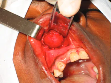
he surgical procedure was performed with extra- and intraoral antisepsis, under iniltrative administration of local anesthetic agents injected into infraorbital and perilesional areas. he incision was made in the gingival labial sulcus between the right maxillary central incisor and the maxillary irst molar on the same side. he lesion was individualized by means of rhombic dissection. Nevertheless, although the lesion was individualized, the capsule and nasal mucosa were ruptured due to its intimate relationship with the nasal mucosa (Figure 3).
he material that was collected and sent for histopathological exam consisted of a fragment of sot tissue measuring 1.7x1.5x0.4 cm.
Figure 1. Extraoral aspect of lesion in which elevation of right ala nasi and disappearance of nasolabial sulcus on same side is observed.
Figure 2. Panoramic radiograph identifying bone rarefaction
suggestive of lesion in maxillary right canine region.
Figure 3. Transoperative aspect: Tissue separation and exposure of
456 Moitinho, Rezende, Souza et al. Rev Odontol UNESP. 2013; 42(6): 454-457
Histological exam revealed the presence of ibrous cystic wall lined with pseudostratiied cylindrical ciliated epithelium, at times with a few layers of cells and at others with a prickle cell layer without atypical cells, exhibiting caliciform cell and mucosal cells, with a inal diagnosis of nasolabial cyst (Figure 4).
During follow-up, good healing without phlogiston signs were veriied in the post-operative period of seven days, with the suture being maintained and having a good aspect. At the clinical follow-up consultation six months ater surgical treatment, there was absence of facial asymmetry and no recurrence of the lesion was observed.
DISCUSSION
Nasolabial cysts are usually unilateral, and may be bilateral in only 10% of cases. hey have predilection for the female sex3,11,12,
and their occurrence in women is three times higher4,5. hey
frequently occur during middle age, with a possible prevalence in persons of the black race4. Although we have related only once
case, the patient revealed the same characteristics as those present in the literature, viz: female sex, middle age, black race, and an asymptomatic lesion.
Clinically the indings of nasolabial cysts are typical, and are characterized by the presence of tumefaction, slow growth at the bottom of the superior vestibular fornix, causing obliteration of the nasolabial sulcus, protrusion of the top lip and elevation of the ala nasi, facts that leave the patient with a certain deformity and facial asymmetry, in addition to discomfort with the use of maxillary dental prosthesis and nasal obstruction1,3,11. hese
indings are in agreement with the manifestations found in the case here related. his emphasizes the importance of a well performed clinical exam, and the dentist’s role at this stage is to prevent the lesion from developing too far, by making an adequate diagnosis.
Diagnosis of the nasolabial cyst is fundamentally clinical9.
By means of intraoral bidigital palpation, it presents as a loating
tumefaction of sot consistency which, associated with puncture and aspiration, helps to conirm the diagnosis; this fact was also observed in the present study.
As this is a sot tissue lesion, the nasolabial cyst is only detected radiographically if deformation of the lateral and anterior limit of the nasal fossa, with convexity in the posterior direction is detected in the occlusal radiograph10 or if it causes
important bone erosion in the maxillary bone7. In this study, a
radiolucent area to the right of the anterior nasal spine could be observed in the panoramic radiograph, indicating bone erosion of the maxillary bone.
As stomatological conduct in the treatment of cysts, it is recommended that fragments of the cystic membrane should be sent for histopathological exam, which normally reveals a ibrous cystic wall limited by pseudostratiied columnar epithelium with calciform and ciliated cells, which may have areas of cuboid epithelium and squamous metaplasia. hese indings were revealed during the histopathological exam in the case referred to in this study.
he treatment indicated included complete surgical enucleation of the cyst by means of intraoral access11,12, because
these lesions rarely reach large proportions. It is not rare for the need for removal of part of the nasal mucosa to occur, in order to achieve complete removal of the lesion1, which in fact occurred
in the present case. Its prognosis is favorable and its recurrence is rare8,9,11,12.
In the present case, the oral and nasal structures involved presented excellent healing, without any report of complications or recurrence of the lesion. he correction of facial asymmetry with consequent esthetic restoration was also observed. Nevertheless, we point out that nasal hemorrhage is a relatively common inding in the immediate post-operative period, as a result of rupture of the nasal mucosa, because it is sometimes necessary to remove part of it during the surgical procedure, fundamental for performing adequate therapy in such cases.
FINAL CONSIDERATIONS
Nasolabial cysts are frequently underdiagnosed in their initial stages, and end up leading to progressive facial asymmetry with subsequent esthetic-emotional imbalance. It is fundamental for the dentist to be alert with regard to early diagnosis, because it is not uncommon for the patient to fail to observe the lesion in the initial stages, as may be observed in the present case. Adequate diagnostic conduct and the correct establishment of the possible diferential diagnoses allow the adoption of suitable therapeutic measures for each case. he treatment of choice is complete surgical removal of the lesion. he objectives of this strategy is related to the prevention of infection, establishment of histopathological diagnosis and the correction of possible esthetic deformations.
Rev Odontol UNESP. 2013; 42(6): 454-457 Diagnosis of the nasolabial cyst: case report 457
REFERENCES
1. Pereira Filho VA, Silva AC, Moraes M, Moreira RWF, Villalba H. Nasolabial cyst: case report. Braz Dent J. 2002; 13(3): 212-4. http:// dx.doi.org/10.1590/S0103-64402002000300015
2. Aikawa T, Iida S, Fukuda Y, Nakano Y, Ota Y, Takao K, et al. Nasolabial cyst in a patient with clet lip and palate. Int J Oral Maxillofac Surg. 2008; 37: 874–6. http://dx.doi.org/10.1016/j.ijom.2008.04.016
3. Nixdorf DR, Peters E, Lung KE. Clinical presentation and diferential diagnosis of nasolabial cyst. J Can Dent Assoc. 2003; 69 (3): 146-9. 4. El-Hamd KEAA. Nasolabial cyst: a report of eight cases and a review of the literature. J Laryngol Otol. 1999; 113: 747-9.
5. Ben Slama L, Zaghbani A, Hidaya S. Nasolabial cyst. Rev Stomatol Chir Maxillofac. 2009; 110: 338–9. http://dx.doi.org/10.1016/j. stomax.2009.09.004
6. Castro AL. Estomatologia. 2a ed. São Paulo: Santos; 1995.
7. Choi JH, Cho JH, Kang HJ, Chae SW, Lee SH, Hwang SJ, et al. Nasolabial cyst: a retrospective analysis of 18 cases. Ear Nose hroat J. 2002; 81: 94–6.
8. Enoki, AM, Pizarro GU, Morais MS, Fernandes DPP, Oliveira PRG. Cisto nasolabial bilateral como causa de obstrução nasal: Relato de
caso e revisão de literatura.Arq Int Otorrinolaringol. 2012; 16 (1): 121-5. http://dx.doi.org/10.7162/S1809-48722012000100018
9. Felix JAP, Ferreira PJF, Correa R, Cantini R, Neto RM, Felix F. Cisto nasolabial bilateral: relato de dois casos e revisão da literatura. Rev Bras Otorrinolaringol. 2003; 69 (2): 279-82. http://dx.doi.org/10.1590/S0034-72992003000200021
10. Cantisano, MH, Soubhia AMP, Tucci R, Zambon RLD. Cisto nasolabial: revisão de literatura e relato de um caso clínico. Rev Ciênc Odontol. 1998; 1(1): 27-30.
11. Soldatelli MV, Maschmann RA; Wobido FB, Pinto JGS, Isolan TMP, Hernandez PAG, Silva Júnior ANN, Santos MA. Cisto nasolabial unilateral: relato de caso clínico. Rev Ciênc Méd Biol. 2008; 7(1): 90-5.
12. Tiago RSL, Maia MS, Nascimento GMS, Correa JP, Salgado DC. Cisto nasolabial: aspectos diagnósticos e terapêuticos. Rev Bras Otorrinolaringol. 2008; 74(1): 39-43. http://dx.doi.org/10.1590/S0034-72992008000100007
CONFLICTS OF INTERESTS
he authors declare no conlicts of interest.
CORRESPONDING AUTHOR
Márcio Campos Oliveira
Departamento de Saúde, UEFS – Universidade Estadual de Feira de Santana, 44036-900 Feira de Santana - BA, Brasil e-mail: [email protected]