566 Letter to the Editor
ambulation since the patients have less postoperative pain.
The article by Nakamura et al. asks what to do when patients do not comply with the IPCD. My suggestion would be to emphasize to the nurses and the patient the importance of the devise and in addition use a spinal anesthetic. Our patients remove the devices the morning after surgery and begin ambulation.
References
1. Geerts WH, Pineo GF, Heit JA, Bergqvist D, Lassen MR, Colwell CW, et al: Prevention of venous thromboembolism: the seventh ACCP conference on
antithrombotic and thrombolytic therapy. Chest. 2004; 126: 338S-400S.
2. Sved P, Nieder A, Manoharan M, Gomez P, Meinbach DS, Kim SS, et al: Evaluation of analgesic requirements and postoperative recovery after radical retropubic prostatectomy utilizing long acting spinal anesthesia. Urology. 2005; 65: 509-12.
3. Koya MP, Manoharan M, Kim S, Soloway MS: Venous thromboembolism in radical prostatectomy: is heparanoid prophylaxis warranted? BJU Int. 2005; 96: 1019-21.
4. Rodgers A, Walker N, Schug S, McKee A, Kehlet H, van Zundert A, et al: Reduction of postoperative mortality and morbidity with epidural or spinal anesthesia: results from overview of randomised trials. BMJ. 2000; 321: 1493.
Dr. Mark S. Soloway
Professor & Chair, Department of Urology Miller School of Medicine, University of Miami Miami, Florida, USA E-mail: [email protected]
Re: Inflammatory Atrophy on Prostate Needle Biopsies: Is There Topographic
Relationship to Cancer?
Athanase Billis, Leandro L.L.Freitas, Luis A. Magna, Ubirajara Ferreira
Departments of Anatomic Pathology (AB,LLLF), Genetics and Biostatistics (LAM), and Urology (UF),
School of Medicine, State University of Campinas (Unicamp), Campinas, São Paulo, Brazil
Int Braz J Urol 2007;33:355-63
To the Editor:
The editorial comments of our paper by Dr.H.Samaratunga, Dr. Rodolfo Montironi, and Dr. Liang Cheng were very informative on a lesion that is one of the most frequent mimics of prostatic ad-enocarcinoma. It occurs most frequently in the pos-terior lobe or peripheral zone (1-3) and gained
initi-567 Letter to the Editor
ated during the 5th decade and continues as a pro-gressive process into the 8th decade. It is a frequent lesion: 85% in autopsies and 83.7% in needle biop-sies (5,6).
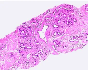
Why this lesion mimics adenocarcinoma? Histologically prostatic atrophy may be partial or complete. The latter is subtyped in simple, hyperplas-tic (or post-atrophic hyperplasia), and sclerohyperplas-tic (5). It seems that the subtypes represent a morphologic con-tinuum of a single lesion (4). Partial atrophy and hy-perplastic (or postatrophic hyperplasia) most fre-quently mimic adenocarcinoma. Hyperplastic atrophy shows small acini closely packed together and lined by atrophic epithelium. Fibrosis is present or not in the stroma. When present, the proliferation is irregu-lar and can result in distortion of the acini simulating stromal infiltration (Figure-1). Partial atrophy was de-scribed by Oppenheimer et al. (7). The name is due to the fact that there is partial preservation of the cy-toplasm simulating neoplastic micro-acini (Figure-2). An additional pitfall for the surgical pathologist is the fact that in partial atrophy the basal cells may be scattered and in some acini may be completely ab-sent (Figure-3).
There are some findings associated to the etio-pathogenesis of the lesion. Atrophy is clearly associ-ated to advanced age (1,5). Radiotherapy and hor-monal deprivation are associated with diffuse
atro-phy. Inactive or active inflammation is a frequent cause for the lesion (8) and based on a study on au-topsies there is evidence that chronic local ischemia may also be a cause of atrophy (5). However, many examples of atrophy are still considered idiopathic in nature. Both inflammation and ischemia are associ-ated with focal forms of atrophy.
The relation of prostatic atrophy to neopla-sia is exciting and controverneopla-sial. This topic was thor-oughly commented in our study and discussed in the editorial comments by Dr.H.Samaratunga, Dr. Rodolfo Montironi, and Dr. Liang Cheng (6). Figure 1 – Hyperplastic atrophy (or postatrophic hyperplasia).
Figure 2 – Partial atrophy.
568 Letter to the Editor
In diagnostic practice it is not rare to find patients with serum prostate- specific antigen (PSA) elevation and several biopsies showing no atypical, preneoplastic or neoplastic lesions, except prostatic atrophy. Regardless of the cause, we hypothesized that damaged epithelial cells in atrophic acini could be a source of the elevation of PSA. Our study was based on 131 needle prostatic biopsies correspond-ing to 107 patients. The only diagnosis in all biopsies was focal prostatic atrophy without the presence of cancer, high-grade prostatic intraepithelial neoplasia, or areas suspicious for cancer. A positive and signifi-cant association was found between the extent of at-rophy and the total or free serum PSA elevation (9). All patients showing 35mm or higher linear extent of atrophy in the biopsy cores, had serum PSA above 4ng/mL. The findings suggest that damaged epithe-lial cells in atrophic acini, regardless of cause, could be a source of serum PSA elevation.
Prostate-specific antigen is a single chain gly-coprotein with proteolytic enzyme activity mainly di-rected against the major gel-forming protein of the ejaculate (semenogelin). PSA induces liquefaction of semen with release of progressively motile sperma-tozoa (10). There are several efficient physiologic barriers to prevent the escape of any significant amounts of PSA from the prostatic ductal system: basement membrane of the acini, basal cells lining the acini, prostatic stroma, basement membrane of capillary endothelial cells, and endothelial cells. These barriers normally prevent PSA from entering the gen-eral circulation at concentrations of more than 3ng/ mL (10).
Focal prostatic atrophy represents a form of adaptive response to injury most commonly to inflam-mation and/or local ischemia. It is intriguing that atro-phic acini may produce an excess of serum PSA. In-flammation and/or ischemia are injurious stimuli re-sulting in diminished oxidative phosphorilation, mem-brane damage, influx of intracellular calcium, and accumulation of oxygen-derived free radicals (oxi-dative stress) (11). We speculate that these injurious stimuli may interfere in the physiologic barrier that prevent the escape of any significant amounts of PSA to the general circulation.
Atrophy is a frequent, exciting, intriguing le-sion and a relevant subject for further research.
Pa-thologists should include the presence and extent of the lesion in the pathology report.
References
1. Moore RA: The evolution and involution of the pros-tate gland. Am J Pathol. 1936;12:599-624.
2. Franks LM: Atrophy and hyperplasia in the prostate proper. J Pathol Bacteriol. 1954;68:617-21.
3. Liavag I: Atrophy and regeneration in the pathogen-esis of prostatic carcinoma. Acta Pathol Microbiol Scand [A]. 1968;73:338-50.
4. Cheville JC, Bostwick DG: Postatrophic hyperplasia of the prostate. A histologic mimic of prostatic adeno-carcinoma. Am J Surg Pathol. 1995;19:1068-76. 5. Billis A: Prostatic atrophy: An autopsy study of a
his-tologic mimic of adenocarcinoma. Mod Pathol. 1998;11:47-54.
6. Billis A, Leandro LL Freitas, Luis A Magna, Ubirajara Ferreira: Inflammatory atrophy on prostate needle bi-opsies: Is there topographic relationship to cancer? Int Braz J Urol. 2007;33:355-63.
7. Oppenheimer JR, Wills ML, Epstein JI: Partial atro-phy in prostate needle cores: another diagnostic pitfall for the surgical pathologist. Am J Surg Pathol. 1998;22:440-5.
8. Srigley JR: Benign mimickers of prostate cancer. Mod Pathol. 2004;17:328-48.
9. Billis A, Meirelles LR, Magna LA, Baracat J, Prando A, Ferreira U. Extent of prostatic atrophy in needle biopsies and serum PSA levels: is there an associa-tion? Urology. 2007;69:927-30.
10. Oesterling JE, Lilja H: Prostate-specific antigen. The value of molecular forms and age-specific reference ranges. In: Vogelzang NJ, Scardino PT, Shipley WU, Coffey DS (eds.), Comprehensive textbook of geni-tourinary oncology. Baltimore, Williams & Wilkins. 1996; pp.668-80.
11. Kumar V, Abbas AK, Fausto N: Robbins and Cotran Pathologic Basis of Disease, 7th ed. Philadelphia, Elsevier Sanders.2005; pp.3-46.