Vesicoenteric fistula caused by bladder diverticulitis:
MRI Findings
_______________________________________________
Ihsan Yuce
1, Hayri Ogul
2, Suat Eren
2, Ummugulsum Bayraktutan
2, Mecit Kantarci
2, Ahmet Yalcin
11
Department of Radiology, Erzurum Regional Training and Research Hospital, Erzurum, Turkey;
2Department of Radiology, Ataturk University Medical Faculty, Erzurum, Turkey
CASE REPORT
_______________________________________________________________________________________
598
RADIOLOGY PAGE
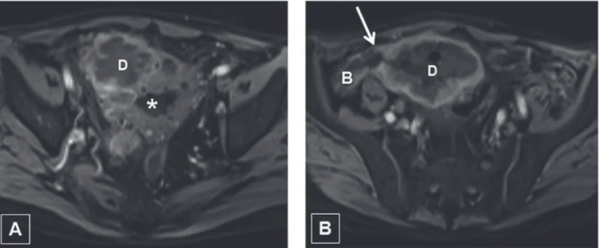
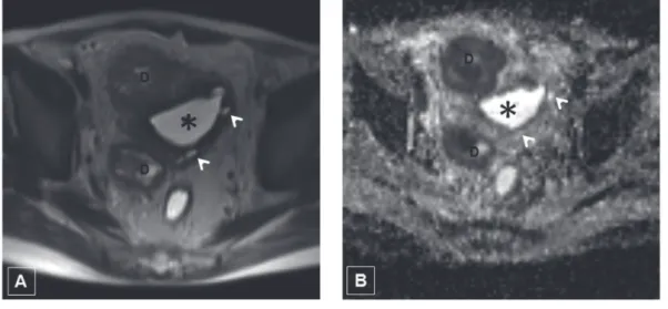
The images presented below were from a man, at the age of 80, who was admitted to hospital with recurrent uri-nary tract infection. Magnetic resonance imaging (MRI) was performed after initial examination. Conventional MRI showed multiple diverticulum in bladder and one of them had contact sigmoid colon via fistula tract (Figure-1). Diffusion weighted images (DWI) showed restricted diffusion in a few diverticulum (Figure-2). Sagittal T2 weighted MR image shows diverti-culum and bladder (Figure-3). Sistoscopic evaluation confirmed imaging findings. Pathological findings were in favor of diverticulitis and exclude malignancy.
Vesicoenteric fistulas are generally the consequences of inflammatory processes of the bowel, mainly colonic di-verticulitis; colorectal cancer and, more rarely, carcinoma of the bladder; trauma; and iatrogenic fistulas between bladder and sigmoid colon as a result of prostatic surgery (1). In a study, Alapont Pérez et al. showed acquired vesicoenteric fistula in 23 patients: 48% of these were of inflammatory etiology, 35% were caused by neoplasms and 17% were iatrogenic (2). Bladder diverticula are usually asymptomatic and found incidentally. Large diverticula may present with symptoms inclu-ding hematuria, urinary tract infection, urinary retention, neoplasm formation, or even acute abdomen due to rupture (3). Conventional MRI with DWI is an important imaging modality for evaluation malignancies and inflammatory conditions.
Figure 1 - Enterovesical fistula secondary to diverticulitis. T1-weighted axial MR images (A-B) with intravenous contrast medium shows thick-walled diverticulum (D) and fistula track (arrow) between diverticulum (D) and bowel (B). T2-weighted axial MR images (C-D) shows bladder diverticulum (D) and large fistula track (arrow) between diverticulum (D) and bowel (B). Asterisk: bladder.
IBJU| RADIOLOGY PAGE
599
ARTICLE INFO
Int Braz J Urol. 2014; 598-9
_____________________
Submitted for publication: October 29, 2013
_____________________
Accepted after revision: June 01, 2014
Figure 2 - Axial T2-weighted MR image (A) shows multiple bladder diverticula (D) and pseudodiverticula (arrowheads). Axial ADC image (B) shows that the diverticula (D) have a low ADC value. Asterisk: bladder.
Figure 3 - Sagittal T2 weighted image shows diverticulum (D) and bladder (asterisk). P: prostate.
REFERENCES
1. Melekos MD, Asbach HW, Barbalias GA. Vesical diverticula: etiology, diagnosis, tumorigenesis, and treatment. Analysis of 74 cases. Urology. 1987;30:453-7.
2. Alapont Pérez FM, Gil Salom M, Esclapez Valero JP, Marti Obiol R, Santamaría Meseguer J, Rafie Mazketly W, et al. Acquired enterovesical fistulas. Arch Esp Urol. 1994;47:973-7.
3. Mićić S, Ilić V. Incidence of neoplasm in vesical diverticula. J Urol. 1983;129:734-5.