*Correspondence: M. E. L. Consolaro. Departamento de Análises Clínicas, Universidade Estadual de Maringá. Av. Colombo, 5790 - 87020-900 - Maringá – PR, Brasil. E-mail: melconsolaro@uem.com.br
A
rti
Pharmaceutical Sciences vol. 47, n. 1, jan./mar., 2011
Effects of depomedroxyprogesterone acetate on the development
and maintenance of
Candida albicans
in the vagina of
oophorectomized Wistar rats (
Rattus norvegicus
)
Agenor Storti-Filho
1, Edilson Damke
1, Márcia Aparecida Carrara
1,
Marcia Regina Batista
1, Lucélia Donatti
2, Cinthia Gandoli Boer
1,
Terezinha Inez Estivalet Svizdinski
1, Márcia Edilaine Lopes Consolaro
1*1Department of Clinical Analysis, State University of Maringá, Paraná, 2Department of Biology, Federal University of Paraná, Curitiba, Paraná
The objective of this study was to determine the effect of medroxyprogesterone acetate (DMPA) on the development and maintenance of Candida albicans in the vagina of oophorectomized Wistar rats. The animals were divided into negative control groups (NCG), which received injections of sterile saline; positive control groups (PCG), which were given injections of estradiol valerate; and progesterone groups (PG), which were given injections of Depo-Provera®. After one week of hormonal induction, vaginal infection by C. albicans was induced in all the groups and detected by vaginal yeast culture and Papanicolaou smear. In addition, scanning and transmission electron microscopy images were obtained to conirm the vaginal infection by yeast in PG. A difference in progesterone levels in PG was observed between the basal level and after hormonal induction (P<0.0001). In this group, 100% of the rats acquired vaginal infection in the irst week, but did not maintain it until the third week. The pharmaceutical brand of DMPA was effective for inducing the metestrus or diestrus phase of the estrous cycle in rats, similar to the use of pure progesterone. In contrast to estrogen treatment, progesterone alone could not support an experimental vaginal infection by C. albicans for any signiicant period of time.
Uniterms: Depomedroxyprogesterone acetate/effects/experimental study. Vulvovaginal candidiasis.
Candida albicans.
O objetivo do presente estudo foi determinar os efeitos do acetato de depomedroxyprogesterona (ADMP) no desenvolvimento e manutenção de Candida albicans na vagina de ratas Wistar ooferectomizadas. Os animais foram divididos em grupos controle negativos (GCN), que receberam injeções de salina estéril; grupos controle positivos (GCP), que receberam injeções de valerato de estradiol; e grupos progesterona (GP), nos quais foram feitas injeções de Depo-Provera®. Após uma semana da aplicação hormonal, foi induzida a infecção vaginal por C. albicans em todos os grupos, detectada por cultura para leveduras vaginais e esfregaço de Papanicolaou. Foram feitas ainda imagens por microscopia eletrônica de varredura e transmissão para conirmar a infecção pela levedura no GP. Foram observados diferentes níveis de progesterona em GP, entre os valores basais e após a indução hormonal (P<0,0001). Neste grupo, 100,0% das ratas contraíram a infecção vaginal na primeira semana, mas não a mantiveram até a terceira semana. A forma farmacêutica de ADMP foi efetiva em induzir as fases de metaestro e diestro do ciclo estral das ratas, da mesma forma que usando progesterona pura. Em contraste com o que ocorre no tratamento com estrógeno, a progesterona não pôde manter a infecção vaginal experimental por C. albicans por um período signiicativo de tempo.
Unitermos: Acetato dedepomedroxyprogesterona/efeitos/estudo experimental. Candidiase vulvovaginal.
INTRODUCTION
Vulvovaginal candidiasis (VVC) is a pathology caused by the abnormal growth of yeast-like fungi in the mucosa of the female genital tract. It is an infection of both the vulva and the vagina caused by commensal yeasts, especially Candida albicans that inhabits the va-ginal mucosa (Sobel, 1993; Sobel, 2007). This infection is characterized by itching, burning, dyspareunia, and a grumous cream-like vaginal discharge. The symptoms worsen during the pre-menstrual period, when vaginal acidity increases (Sobel, 1993).
VVC is sometimes dificult to treat, and represents a worldwide health problem of indisputable importance (Corsello et al., 2003; Patel et al., 2004). The diagnosis and treatment of VVC, together with lost productivity, result in an estimated cost of US$ 1 billion per year in the USA, where this infection is the second most common cause of vaginal infections (close to 25%), after bacterial vaginosis. Recently, the incidence of VVC has increased markedly worldwide. It is estimated that 75% of adult women have at least one episode of VVC during their lives, 40-50% of these will experience recurrences, and 5% will reach the recurrent character (RVVC), which is deined as the oc-currence of three to four episodes of VVC in a period of 12 months in the absence of a recognized predisposing factor (Corsello et al., 2003).
The attachment of C. albicans to mucosal surfaces has been shown to be an important step in the infectious process, particularly in the oral cavity and vaginal muco-sa (Irie et al., 2006). The pathogenesis of this infection involves the initial adherence of the yeasts to the vaginal mucosa, followed by asymptomatic colonization, from which the yeasts may reach the status of infectious agent (symptomatic vaginitis). This occurs when the coloniza-tion site of the host becomes favorable for the development of yeasts, normally as a consequence of some predisposing factor, such as previous colonization by yeast, the dimi-nished immunological response observed in immunosup-pressive diseases, diabetes mellitus, pregnancy, or chronic use of corticoids. The use of antibiotics, estrogen therapy, minor traumas such as the sexual act, the habit of wearing tight or synthetic clothing, and diet also seem to contribute (Corsello et al., 2003; Patel et al., 2004; Sobel, 2007). In the absence of these factors, clinical observations show that VVC occurs predominantly during the luteal phase of the menstrual cycle, when the levels of estrogen and prin-cipally progesterone are high. In contrast, pre-menstrual girls and post-menopausal women who are not receiving hormone replacement therapy, rarely exhibit this infection (Kalo-Klein, Witkin, 1989). However, the mechanisms
by which these hormones, principally progesterone, act in VVC are not fully known (Fidel et al., 2000; Miller et al., 2000).
The behavior of the epithelia and of the cervical and vaginal stroma of rodents in response to sexual steroids is similar to that of humans, i.e., changes take place in the cellular pattern of the vaginal epithelium, depending on the phase of the estrous cycle (Mandal, Zuckerman, 1951). Therefore, experimental models of VVC in female rats have been extremely useful in the identiication of factors concerning the inluences of hormones on the infection, the virulence of the yeasts, and the susceptibility to and the treatment of the infection (Kalo-Klein, Segal, 1988; Kalo-Klein, Witkin, 1989; Junqueira et al., 2005). In these models, the key to obtaining a persistent infection is the state of pseudo-estrus, which is often induced through the expensive subcutaneous administration of estradiol benzoate (Clemons et al., 2004; Junqueira et al., 2005). Models using progesterone are scarce.
The purpose of the present study was to determine the effects of medroxyprogesterone acetate (Depo-Pro-vera®) (DMPA) on the development and maintenance of Candida albicans in the vagina of oophorectomized rats.
MATERIALS AND METHODS
Animals
Oophorectomized Wistar rats (Rattus norvegicus) weighing 200 to 300 g and 70 days old, supplied by the Central Animal House of the State University of Maringá-PR (UEM), were employed. The animals were kept at a temperature of 22 ºC, under a light-dark cycle of 12 hours, and received a standard ration of chow and water ad li-bitum. This research was approved by the Committee on Ethical Conduct in the Use of Animals of UEM (Protocol No. 013/2006, statement No. 050/2006). The rats were used in the experiments after a period of adaptation of about seven days at the animal house.
Groups of rats per experiment
oil at 0.2 mg/wk/rat, both administered as fractions three times/wk for four weeks. The infection was induced in all groups as described below.
Induction of metestrus and diestrus in the progesterone groups (PG)
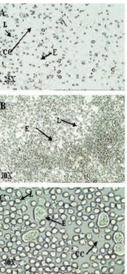
The model of infection was carried out as described for mice (Fidel et al., 2000), but instead using rats and a pharmaceutical brand of depomedroxyprogesterone ac-etate. Briely, subcutaneous injections of Depo-Provera® (Aventis)were given at a concentration of 100 mg/wk/ rat, administered as fractions three times/wk for four weeks, to obtain the state of metestrus and diestrus. This schedule was used because in the estrous cycle, progesterone secretion increases during metestrus and diestrus, decreasing thereafter (Mandal, Zuckerman, 1951; Fidel et al., 2000). The hormonal activity was checked through the analysis of the cells of the vaginal luid. Vaginal material was collected every three days after hormonal induction for four weeks. The vaginal luid was obtained with the aid of a plastic pipette, after administration of 20 µl sterile saline in the vagina of the rats. A drop of this material was placed between a glass slide and coverslip and observed fresh by ordinary opti-cal microscopy under 10X and 40X objectives. Three types of cells were identiied: round and nucleated cells are epithelial cells; irregular cells without a nucleus are the corniied cells; and the small round cells are the leu-kocytes. The proportion among these types was used to determine the estrous cycle phases. A metestrus smear consists of the same proportion of leukocytes, corniied, and nucleated epithelial cells whereas a diestrus smear primarily consists of a predominance of leukocytes (Mandal, Zuckerman, 1951).
Hormonal activity was also checked by determin-ing the blood level of progesterone in PG and NCG, and estrogen in PGC. Three days after hormonal induction, and weekly for four weeks, soon after the collection of the vaginal luid, blood was collected for the determina-tion of hormones, after 12-h fasting. The rats’ tails were cut, and the blood was collected in tubes containing an anti-clotting agent (luoride EDTA). The tails were immediately cauterized to accelerate healing. The rats were then returned to their cages, and the ad libitum supply of water and chow was reestablished. Blood hor-mones were determined through solid-phase competi-tive chemiluminescent enzymatic immunoassay. The procedure was totally automated (IMMULITE 2000 – DPC MEDLAB Equipment) and results expressed as pg/mL (NCCLS, 1998).
Experimental infection
A human vaginal isolate of C. albicans identiied at the Medical Mycology section of the Laboratory of Teach-ing and Research in Clinical Analyses (LEPAC) of UEM was used. This isolate had been stored since identiication in 10% glycerinated water at -20 °C. The yeast isolate was identiied through classical (Kurtzmann, Fell, 1998) and molecular methods (Sugita et al., 2002). For the prepara-tion of the suspension, the yeast was reactivated through seeding onto SDAC- Sabouraud Dextrose Agar (Sigma Chemical, St. Louis, USA) supplemented with 50 mg/mL of chloramphenicol (Sigma Chemical, St. Louis, USA) and incubated at 25 °C/48-72 h. An inoculation of the yeast at a concentration of 5x10⁸ CFU/mL (colony-forming units), counted in a Neubauer chamber, was used.
To induce the infection, 80 µL of the yeast suspen-sion was introduced intra-vaginally one week after the be-ginning of hormonal induction. The animals were followed weekly to monitor the infection, from the irst week after infection until week three (PG, NCP, PCG). Samples from the vaginal luid, collected as described previously, were placed in Petri dishes containing SDAC, and incubated at 25 °C/ 48-72 h. After fungal growth, the CFU was counted. The infection was considered positive if at least one yeast colony grew. The same procedure was followed in the rats from the control groups. In addition to the culture, the vaginal samples were prepared as smears on glass slides, and stained for Papanicolaou cytology (Bibbo, 1997).
Scanning (SEM) and transmission (TEM) electron microscopy
After one week of infection, one rat each from the PG and the PCG, was sacriiced with an overdose of anes-thetics (Ketamine and Xylazine) and the vaginas removed. For SEM, after the washes these were ixed in 2.5% glu-taraldehyde solution dissolved in 0.1 M cacodylate buffer (Sigma Chemical, ST. Louis, USA) and dehydrated in an ascending series of alcohol. The critical point was obtained using a Balzers CPD-010 (Balzers Instruments, Balzers, Liechtenstein) with carbonic gas. Metallization in gold was done on a Balzers SCD-030 (Balzers Instruments, Balzers, Liechtenstein). Documentation was carried out on a JEOL-JSM 6360 LV scanning electron microscope (Jeol Ltda, Tokyo, Japan) at the Center of Electron Microscopy – Federal University of Paraná.
material was post-ixed in 2% osmium tetroxide in 0.1 M cacodylate buffer pH 7.2 for one hour. The contrasting in the blocks was done in 2% uranyl acetate for two hours. The material was dehydrated in an ascending series of alcohols and then placed in acetone. The impregnation and inclusion occurred in Epon-812 resin. The sections were produced on a Sorval Porter Blum MT-2 ultra-microtome using glass and diamond blades. The ultra-thin sections were contrasted in aqueous solution of 2% uranyl acetate and lead nitrate/acetate. The observation of the material was made under a JEOL 1200EX II transmission electron microscope at the Center of Electron Microscopy of the Federal University of Paraná.
Statistical analysis
The results were analyzed using a t test and analysis of variance for comparisons between the hormone con-centration at the basal level, and after the injections of the hormone, and for the percentage of the rats infected for one experimental week.A p value of less than 0.05 was consi-dered signiicant. The tests were carried out with Graph Pad Prism® version 4.0 software (Graph Pad Software Inc.).
RESULTS AND DISCUSSION
In PG, 90.0% of the rats had smears of the diestrus type, and 10.0% metestrus whereas in PCG, 100% were in pseudoestrus. In NCG, the smears showed that the rats were in various phases of the estrous cycle. Figure 1 shows an unstained native vaginal smear from PG rats, visualized under a light microscope.
Therefore, the pharmaceutical brand of DMPA pro-ved effective for inducing the metestrus or diestrus phase of the estrous cycle in rats, similarly to other studies using pure progesterone (Kinshiman, Collard, 1986; Fidel et al., 1993).This experimental model is both economically and operationally accessible, and may help to clarify important aspects in the pathogenesis of infection.
In the NCG, the blood progesterone (as mean values) ranged from 9.0 to 11.5 pg/mL (P> 0.05) (Figure 2). These rats, although infected with the yeast, did not develop vaginal infections.
In the PCG, the basal levels of estrogen were betwe-en 44.9 and 55.9 pg/mL; and after hormonal administra-tion they ranged from 1384.0 to 1579.0 pg/mL (P< 0.05) (Figure 3 (A). Figure 3 (B) shows that 100% of these rats acquired a vaginal infection in the irst week, and retained it for the three weeks of the experiment.
FIGURE 1 - Photomicrographs of unstained native vaginal
smear from rats, observed under a light microscope with 25, 10, and 40Xobjective lenses, respectively, after administration of depomedroxyprogesterone acetate 100.0 mg/wk/rat. The proportion of the three types of cells was used for determination of estrous cycle phases. The round and nucleated cells are epithelial cells (E); irregular cells without a nucleus are the corniied cells (CC); and the small round cells are the leukocytes (L). A metestrus smear consists of the same proportion of leukocytes, corniied, and nucleated epithelial cells (A); and a diestrus smear primarily consists of a predominance of leukocytes (B).
FIGURE 2 - Blood progesterone concentration (pg/ml)
In the PG, the basal levels of progesterone were between 7.8 and 16.9 pg/mL; after hormonal administra-tion, values ranged from 188.0 to 211.0 pg/mL (P< 0.05) (Figure 4A). In this group, 100% of the rats acquired a vaginal infection in the irst week, and 0% retained it after the third week (P< 0.05) (Figure 4B). The CFU/mL of C. albicans in PG rats ranged from 275 to 5000 in the irst week, and from 19 to 66 in the second week. No growth was observed in the third week (P< 0.05) (Figure 4C).
In contrast to estrogen (Figure 3B), progesterone treatment alone could not support an experimental vaginal infection for any signiicant period of time (Figure 4B, C). Other investigators have reported similar results, although these used pure progesterone rather than a pharmaceutical brand for human treatment (Kinshiman, Collard, 1986; Fidel et al., 1993). This phenomenon also occurs in women taking progesterone contraceptives such as Depo-Provera. Miller et al. (2000) reported a decreased proportion of posi-tive Candida vaginal cultures (from 32% to 8%) after only
3 months of using DMPA as a contraceptive. Similarly in this study, there were no symptomatic Candida infections among women who had used DMPA for longer than 1 year. Fidel et al. (2000) also suggested that progesterone treat-ment in women and rodents alone could not support a vagi-nal infection by Candida, because of a lack of, or reduced, inluence of endogenous estrogen. This theory was studied by Cundy et al. (1998), who reported that serum estradiol
FIGURE 3 - (A) Blood estrogen concentration (pg/mL)
determinations (as mean values) of the positive control group (PCG) at basal level and seven days after subcutaneous injections of estradiol valerate 0.2 mg/wk/rat, administered as fractions three times/wk for four weeks; •P < 0.05 for
estrogen concentration compared at the basal level and after injections of the hormone. (B) Percentage of rats acquiring and retaining vaginal infection by Candida albicans weekly, after
administration of 0.2 mg/wk/rat estradiol valerate. FIGURE 4 - (A) Blood progesterone concentration (pg/ml) determinations (as mean values) of the progesterone groups (PG) at basal level and seven days after injection (■P < 0.05). PG=
subcutaneous injections of depomedroxyprogesterone acetate at 100.0 mg/week/rat, administered as fractions three times/wk for four weeks. (B) Percentage of rats acquiring and retaining vaginal infection by Candida albicans, at each week after administration of progesterone; ▲P < 0.05 for percentage of rats
infected in the irst week compared to the second/third weeks. (C) Mean number of yeast colonies counted (colony-forming units - CFU/ml) after three weeks of infection by Candida albicans in hyper-progesterogenic rats; ◊P < 0.05 for CFU/ml
(E2) levels are markedly reduced in human DMPA users. The Papanicolaou staining of the rat vaginal smears showed the presence of yeasts and pseudohyphae only in the PCG and PGs, as illustrated in Figure 5C. It was also possible to observe the phases of the estrous cycle (Figure 5). This staining may be used for infection and hormonal control.
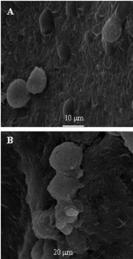
In addition to ordinary optical microscopy of the vaginal fluid, the scanning and transmission electron microscopy images conirmed the vaginal infection by C. albicans in PG. However, this methodology cannot be used in many research laboratories because it is dificult to use in experimental models of VVC in female rodents, where cultures and Papanicolaou smears are more feasi-ble. In Figure 6, SEM showing the surface of the vaginal epithelium of female rats in diestrus infected with C. albicans. In (A) and (B), a few blastoconidial yeasts can be seen, adhered as colonies. The yeasts adhered in an external location relative to the epithelial surface.
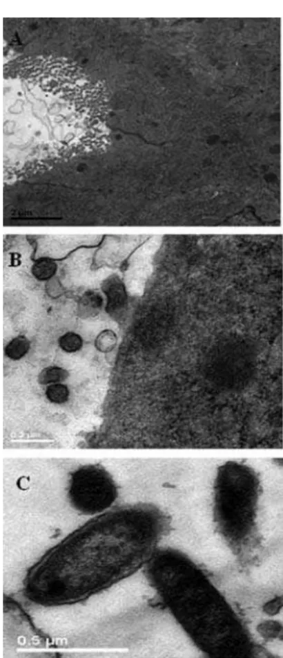
Kinsman, Collard (1986) suggested that host factors in the rat which predispose toward vaginal infection with C. albicans, include the presence of a cornified epithelium and the absence of leukocytes. These factors were also observed in the PCG smears in the present study (Figure 5C). In PG however, several leukocytes were observed in the lumen of the vaginal canal (Figure 5A, B; Figure 7A, B), as described by Medonça et al. (2007). Fidel et al. (2000)reported that leukocytes have little effect against C. albicansin vivo, irrespective of whether estrogen or progesterone is pre-sent. More studies are necessary to assess the speciic role of leukocytes in VVC.
Concerning the hypothesis that the presence of a corniied epithelium predisposes to VVC, our results also demonstrated the presence of abundant corniied cells in PCG, as previously described by other authors (Smith et al., 1975; Kinsman, Collard, 1986).However, because
FIGURE 5 - Photomicrographs of Papanicolaou staining in
vaginal smears from rats observed under a light microscope with 10X (A), 20X (B) and 40X (C ) objectives, after administration
of depomedroxyprogesterone acetate 100.0 mg/wk/rat and infection by Candida albicans. Positive infection was demonstrated in the PCG and PGs (yeasts-Y and pseudohyphae-PY). Leukocytes (L), epithelial (E), and corniied cells (CC).
FIGURE 6 - Scanning electron micrographs (SEM) showing
some authors hold that adherence is lesser in corniied epi-thelial cells and greater in intermediate epiepi-thelial cells (IC) of the vaginal epithelium Segal et al., 1984; Kalo-Klein, Segal, 1988; Irie et al., 2006), further studies to assess these results should be undertaken. In addition, some cli-nical observations have established that VVC occurs more frequently during the luteal phase of the menstrual cycle, when progesterone is secreted in a greater quantity than estrogen (Kalo-Klein, Witkin, 1989; Miller et al., 2000). Recent research efforts have revealed that yeast cells can respond to human steroids (Smith et al., 1975; Banerjee et al., 2008).Banerjee et al., (2008)listed a total of 99 genes of C. albicans that are regulated by progesterone, and their correlation with drug resistance, virulence, mor-phogenesis, and general stress response. This explained, in part, the mechanisms by which progesterone acts in VVC pathogenesis.
FIGURE 7 - Yeasts and vaginal epithelium of rats observed by
transmission electron microscopy (TEM), after establishment of diestrus and induced vulvovaginal candidiasis. In (A) and (B), the integrity of the epithelium can be seen, as well as several leukocytes in the lumen of the vaginal canal. In (C), TEM showing several Candida albicans yeasts in the lumen of the vaginal canal (bar = 0.5 μm).
CONCLUSIONS
Our results showed that the pharmaceutical brand of DMPA was effective for inducing the metestrus or diestrus phase of the estrous cycle in rats, similarly to other studies using pure progesterone.In contrast to estrogen, proges-terone treatment alone could not support an experimental vaginal infection for any signiicant period of time. The Papanicolaou staining of the rat vaginal smears showed the presence of yeasts, and also revealed the phases of the estrous cycle. Further in vivo studies are necessary to assess the speciic role of progesterone and other hormonal methods of contraception, and yeast colonization and in-fection. The experimental model used in the present study can be applied in this research.
REFERENCES
BANERJEE, D.; LELANDAIS, G.; SHUKLA, S.; MUKHOPADHYAY, G.; JACQ, C.; DEVAUX, F.; PRASAD, R. Responses of pathogenic and nonpathogenic yeast species to steroids revel the functioning and evolution of multidrug resistance transcriptional networks. Eukaryot. Cell., v.7, n.1, p.68-77, 2008.
BIBBO, M. Comprehensive cytopathology. Philadelphia: W.B. Saunder, 1997. p.101-124.
CLEMONS, K. V.; SPEAROW, J. L.; PARMAR, R.; ESPIRITU, M.; STEVENS, D. A. Genetic susceptibility of mice to
Candida albicans vaginites correlates with host estrogen sensitivity. Infect. Immun., v.72, p.4878-4880, 2004.
CORSELLO, S.; SPINILLO, A.; OSNENGO, G.; PENNA, C.; GUASCHINO, S.; BELTRAME, A.; BLASI, N.; FESTA, A. An epidemiological survey of vulvovaginal candidiasis in Italy. Eur. J. Obstet. Gynecol. Reprod. Biol., v.110, p.66-72, 2003.
CUNDY, T.; CORNISH, J.; ROBERTS, H.; ELDER, H.; REID, J. R. Spinal bone density in women using depomedroxyprogesterone contraception. Obstet. Gynecol., v.92, p.563-579, 1998.
IRIE, M. M. T.; CONSOLARO, M. E. L.; GUEDES, T. A.; DONATTI, L.; PATUSSI, E. V.; SVIDZINSKI, T. I. E. A simpliied technique for evaluating the adherence of yeasts to human vaginal epithelial cells. J. Clin. Lab. Anal., v.20, p.195-203, 2006.
JUNQUEIRA, J. C.; DIAS, C. E.; MARTINS, J. S.; ITO, C. Y. K.; CARVALHO, Y. R.; JORGE, O. C. Experimental candidosis and recovery of Candida albicans from the oral cavity of ovariectomized rats. Microbiol. Immunol., v.49, p.199-207, 2005.
KALO-KLEIN, A.; SEGAL, E. Interaction of Candida albicans
with genital mucosa: effect of sex hormones on adherence of yeasts in vitro. Can. J. Microbiol., v.34, p.224-228, 1988.
KALO-KLEIN, A.; WITKIN, S.S. Candida albicans: cellular immune system interactions during different stages of the menstrual cycle. Am. J. Obstet. Gynecol., v.16, p.1132-1136, 1989.
KINSMAN, O. S.; COLLARD, A. E. Hormonal factors in vaginal candidiasis in rats. Infect. Immun., v.53, p.498-504, 1986.
KURTZMANN, C. P.; FELL, F. W. The yeast: a taxonomic study. Amsterdam: Elsevier, 1998. p.891-913.
MANDL, A. M.; ZUCKERMAN, S. Numbers of normal and atretic oocytes in unilaterally spayed rats. J. Endocrinol., v.7, p.112-119, 1951.
MENDONÇA, F. S.; EVÊNCIO-NETO, J.; SIMÕES, M. J.; CAMARGO, L. M.; BARATELLA-EVÊNCIO, L. Aspectos citopatológicos da mucosa vaginal de camundongas tratadas com progesterona. Cienc. Anim. Bras., v.8, n.2, p.313-318, 2007.
MILLER, L.; PATTON, D.L.; MEIER, A.; THWIN, S. S . ; H O O TO N , T. M . ; E S C H E N B A C H , D . A . Depomedroxyprogesterone-induced hypoestrogenism and changes in vaginal lora and epithelium. Obstet. Gynecol., v.96, n.3, p.431-439, 2000.
NATIONAL COMMITTEE FOR CLINICAL LABORATORY STANDARDS. Reference method. Wayne: NCCLS, 1998. p.1-18.
PATEL, D. A.; GILLESPIE, B.; SOBEL, J. D.; LEAMAN, D.; NYIRJESY, P.; WEITZ, M. V.; FOXMAN, B. Risk factors for recurrent vulvovaginal candidiasis in women receiving maintenance antifungal therapy: results of a prospective cohort study. Am. J. Obstet. Gynecol., v.190, p.644-653, 2004.
SEGAL, E.; SOROKA, A.; SCHECHTER, A. Correlative relationship between adherence of Candida albicans
to human vaginal epithelial cells in vitro and candidal vaginitis. Sabouraudia, v.22, n.3, p.191-200, 1984.
SMITH, M. S.; FREEMAN, M. E.; NEILL, J. D. The control of progesterone secretion during the estrous cycle and early pseudopregnancy in the rat: prolactin, gonadotropin and steroid levels associated with rescue of the corpus luteum of pseudopregnancy. Endocrinologia, v.96, p.219, 1975.
SOBEL, J. D. Candidal vulvovaginitis. Clin. Obstet. Gynecol., v.36, p.153-212, 1993.
SOBEL, J. D. Vulvovaginal candidosis. Lancet, v.369, n.9577, p.1961-1971, 2007.
SUGITA, T.; KUROSAKA, S.; YAJITE, M.; SATO, H.; NISHIKAWA, A. Extracellular proteinase and phospholipase activity of three genotypic strains of a human pathogenic yeast. Candida albicans. Microbiol. Immunol., v.46, p.881-883, 2002.
Received for publication on 20th October 2009