N e u rology Department of Medical School Clinics Hospital of the University of São Paulo - São Paulo SP, Brazil. Presented at the XVt h
International Congress of Clinical Neurophysiology, Buenos Aires, Argentina in 2001. Supported by CNPq (Brazil). Received 14 September 2005, received in final form 17 November 2005. Accepted 16 January 2006.
Dra. Laura M.F. F. Guilhoto - Av. Dr. Enéas de Carvalho Aguiar, S/N - Neurology Department of Medical School Clinics Hospital of the University of São Paulo - 05403-900 São Paulo SP - Brasil. E-mail: [email protected]
OCCIPITAL INTERMITTENT RHYTHMIC
DELTA ACTIVITY IN ABSENCE EPILEPSY
Laura M.F.F. Guilhoto, Maria Luíza G. Manreza, Elza M.T. Yacubian
ABSTRACT - Occipital intermittent rhythmic delta activity (OIRDA) is considered good prognostic factor in typical absences (TA). We re p o rt electroclinical evolution in 14 patients with TA and OIRDA, which per-formed video-EEG. Seven patients were female; 9 had childhood absence epilepsy and the others did not p resent electroclinical characteristics for syndromic classification according to ILAE's classification (1989). Pyknolepsy was re f e rred to in 13; TA was the only seizure type in 13; one had generalized tonic-clonic sei-zures (GTCS) and three had myoclonic jerks during TA. VPA monotherapy controlled seisei-zures in 11, diVPA and ESM, in one each. After seizure control EEG normalized in 10 while in three, spike-wave complexes (SWC) persisted, accompanied by OIRDA in one. Finally in another, seizures were not controlled and SWC and OIRDA persisted. In conclusion, we observed in this series of TA and OIRDA with onset before 10 years, pyknolepsy as common finding and few GTCS. VPA controlled seizures in most cases and EEG normalized in 76.92%. We suggest that OIRDA could be considered good prognostic factor in TA associated with SWC and of epileptiform nature leading to appropriate investigation.
KEY WORDS: typical absence, occipital intermittent delta activity, idiopathic generalized epilepsy, EEG, childhood absence epilepsy.
Atividade occipital delta rítmica intermitente em epilepsia ausência
RESUMO - Atividade occipital delta rítmica intermitente (AODRI) é considerada fator de bom pro g n ó s t i c o em crises de ausência típica (AT). Neste estudo relatamos a evolução eletroclínica de 14 pacientes com AT e AODRI que realizaram vídeo-EEG. Sete pacientes eram do sexo feminino; nove tinham epilepsia ausên-cia da infânausên-cia e os outros não apresentavam características eletroclínicas para classificação sindrômica de a c o rdo com a classificação da ILAE (1989). Picnolepsia foi relatada em 13; AT foi o único tipo de crise em 13; um tinha crises generalizadas TCG e três, abalos mioclônicos durante AT. Monoterapia com VPA con-t rolou as crises em 11, diVPA e ESM, em um cada. Após o concon-trole das crises, o EEG normalizou em 10; em três, complexos de espícula-onda (CEO) persistiram, acompanhados por AODRI em um. Finalmente em ou-tro, as crises não foram controladas, persistindo CEO e AODRI. Concluindo, observamos nesta série de AT e AODRI, com início antes dos 10 anos, picnolepsia freqüente e poucas crises TCG. VPA controlou as crises na maioria dos casos e o EEG normalizou em 76,92%. Sugerimos que AODRI possa ser considerada um fator de bom prognóstico em AT associada a CEO e de possível natureza epileptiforme, levando por sua vez, à investigação apropriada.
PA L AV R A S - C H AVE: ausência típica, atividade occipital delta rítmica intermitente, epilepsia generalizada idiopática, EEG, epilepsia ausência da infância.
Posterior slow activity on EEG was initially descri-bed in children with behavior pro b l e m s1, but these
earlier studies, as stressed by Kellaway 50 years lat-er (1990)2, were limited by the fact of a small contro l
g roup and by the dubious criteria of abnorm a l i t y, n o t taking into account the age of the patients. Subse-quent studies in fact confirmed a greater incidence of posterior slow activity in children with behavior p roblems when compared to controls of the same
age3. Nevertheless, after these groups of studies no
other was performed to stress these evidences. D i ff e rently from frontal intermittent rhythmic del-ta activity (FIRDA) observed in adults, occipidel-tal inter-mittent rhythmic delta activity (OIRDA) may not be associated to structural lesions. OIRDA was described in patients with absence seizures by Cobb in 19454.
and Fountain6re p o rted a series of 77 patients with
OIRDA and found seizures to be more frequent (69/ 77) than in the control group (41/77), consisting of GTCS in 45%, partial in 40% and absences in 33% of them. Some authors consider OIRDA as a good pro g-nosis factor in patients with absences7,8.
The aim of this paper is to re p o rt clinical and EEG evolutions in 14 patients with absence epilepsy who presented OIRDA.
METHOD
We analyzed by video-EEG 14 out-patients consecutive-ly re f e rred between October 1996 and November 1998 fro m
study was 7.89 yrs. (ranging from 1 yr. 5 m to 15 yrs.) and the age of seizure onset was 5.57 yrs. (range 1-10 yrs.).
A Telefactor 32 channel system was used in all patients. The video-EEG monitoring consisted of a minimum of thre e hours of re c o rding, with sleep samples, in the morning after sleep deprivation and without any change in the curre n t medication.
All the patients had routine (30 minutes) control EEG after adjustment of AED, and in 13 of them after contro l of the seizures. They also had muscular electrodes i n the deltoid muscles and were submitted to intermittent phot-ic stimulation. These patients perf o rmed several periods of h y p e rventilation with the eyes open while counting aloud the number of re s p i r a t o ry incursions, as suggested by Pa-nayiotopoulos et al.11.
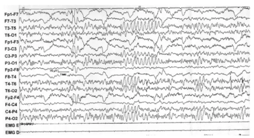
Fig 2. EEG showing OIRDA during hyperv e n t i l a -tion.
The patients were followed up in the outpatient unit for a period ranging from two to four years and thre e months (mean 40 months).
RESULTS
Pyknolepsy was re f e rred to in 13 patients (92.8 5 % ) . TA was the only seizure type in 13 children (92.85%) and in three (21.42%) there were myoclonic jerks du-ring the absences. One patient (7.14%) had also gene-ralized tonic-clonic seizures (GTCS).
A c c o rding to ILAE's syndromic classification ( 19 8 9 )1 2,
nine patients (64.18%) could be classified as having childhood absence epilepsy and the remaining ones did not present electroclinical characteristics that allo-wed syndromic classification1 3. When we used
Pana-yiotopoulos’ criteria1 4for absence seizures, nine
pati-ents (64.18%) could be classified with the childhood type.
In 13 patients (92.85%) the seizures could be con-t rolled: 11 in monocon-therapy wicon-th valproacon-te (VPA), m e a n dose of 22.7 mg/kg/d; one with divalproate (diVPA ) , another one with the association of VPA and etho-suximide (ESM) and in one (7.14%), the seizures per-sisted despite the use of VPA (34 mg/kg/d). This was probably due to no compliance to treatment.
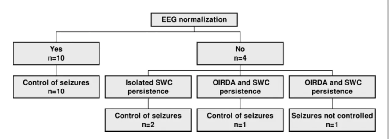
In 10 patients (71.42%) EEG became normal after c o n t rol of seizures. In nine of them OIRDA disappea-red simultaneously with SWC in mean time of 11 months (range 0-31) after treatment and in one pa-tient, 8 months before them. In three, there were persistent discharges re g a rding control of the seizure s as described by the family: in two of these isolated SWC and in one, OIRDA and SWC. In one patient sei-z u res were not controlled and SWC, as well as OIR-DA, still persisted. Diagram 1 shows EEG evolution in these patients. Figures 1 and 2 illustrate 2 cases.
DISCUSSION
D i ff e rently from FIRDA, posterior EEG slowing may be present either in physiological or patholog-ical conditions. Physiologpatholog-ical conditions are age re l a t-ed and have diff e rent morphological characteristics, such as slow alpha variant group and slow posterior waves of youth5. Among the pathological conditions,
basilar migraine may show signs of dysfunction of brain areas supplied by the vert e b robasilar art e r i e s during an attack with resolution during follow-up1 5.
Posterior fossa tumors, specially those of the 4t hv e
n-tricle, may show EEG abnormalities, such as posteri-or arrhythmic slow waves and transmitted rhythms, which are probably correlated with acute or subacu-te dilatation of the 3rdventricle16. Head trauma was
described as also associated to OIRDA in the past5 , 1 7.
Since the first studies in the 30s1 8 , 1 9a b n o rmal slow
waves were seen after closing the eyes in patients with “petit mal seizures”. As those authors described “these slow waves may continue for a short time, and then develop a rapid alternation between fast and slow waves which is characteristic of petit mal (that is, the 3 per second wave and spike), and a clin-ically characteristic petit mal seizure may occur”.
None of our patients had any other neuro l o g i c a l symptom besides seizures. All our patients were chil-d ren anchil-d 13 out of 14 hachil-d absences as the only seizu-re type, with mean age of seizuseizu-re onset 5.57 yrs. (ran-ge 1-10 yrs.) and control of seizures in 13 of them (92.85%). Cobb et al. described OIRDA in patients that had an early age of onset of absences, cessation at age 10-12 and a lesser tendency to develop grand mal7.
Although pyknolepsy was described in the ILAE’s c l a s s i f i c a t i o n1 2as a characteristic of childhood absence
The majority of patients who had their seizure s c o n t rolled (10/13) had normalization of the EEG and only three had their seizures controlled with persist-ence of SWC, one with associated OIRDA. The fact that one patient whose seizures were not contro l l e d p resented OIRDA as well as SWC may indicate that these two types of abnormalities are linked. Actually, one of our patients presented one episode of absence just after bursts of OIRDA. Aird and Gastaut described one patient with “petit mal” who showed marked occipital rhythmic slowing before a typical spike-wave burst (called larval or subclinical)5.
Cobb described two epileptic patients with abnor-mal activity distributed over the occipital re g i o n s , one with spike-wave activity and the other with a continuous delta rhythm which showed reaction to visual stimuli4. This author described the distribution
of grading among “single spikes, single spike-and-wave, bursts of spike-and-wave; bursts in which the spikes were barely discernible, bursts of ‘sine’ waves, and finally, rhythmic waves which were continuous or changed in amplitude only slowly…”. The mech-anisms involved in this activity have been discussed by several authors. Experimental stimulation of thal-amus promoted cortical discharges similar to those seen in petit mal2 1 - 2 3. The cortical theory by Gibbs and
Gibbs that proclaimed the cortex as the main site of the abnormalities in generalized epilepsies contra-dicted this concept18,19.
The theory of a “cerebral pacemaker” sustained that a system of subcortical pacemaker would con-t rol normal corcon-tical rhycon-thms like alpha and becon-ta fre-quencies and also abnormal activities (slow waves, wave and spikes, diffuse spikes)2 4. Hence,
dysfunc-tion either in this system, probably located in the brainstem and diencephalon, or in its nonspecific dif-fuse cortical projections would explain the coexis-tence of spike-wave activity and OIRDA in patients with typical absence seizures. The principle of inter-action between thalamus and cortex was studied
lat-Some authors consider OIRDA as a good pro g n o-sis factor in patients with absences7 , 8. This finding
p robably implies a clue for the diagnosis of the child-hood type of absence epilepsy in their papers. The fact that the thalamocortical dysfunction is age de-pendent was raised in 1995 by Noebels and Tharp who discussed the interactions of neuro b i o l o g i c a l , genetic and developmental aspects of absence seizu-re s2 9. A “seizure - p rone” cortex that undergoes
oscil-l a t o ry thaoscil-lamic infoscil-luence was aoscil-lso described in exper-imental work on spike-wave activity3 0. Although we
could not compare patients with TA without OIRDA with our patients, most of them had control of the s e i z u res and only one was re f r a c t o ry to AED, pro b a-bly due to no compliance to treatment.
In conclusion, in this series of typical absences and OIRDA we observed mean age of seizure onset befo-re 10, a common finding in pyknolepsy (92.85%) and also low incidence of GTCS (7.14%). Seizures could be controlled in 92.85% of the patients and from this g roup, in 71.42% there was EEG normalization. The-re f o The-re, in this series, OIRDA could be consideThe-red a good prognostic factor in TA being associated with the presence of generalized SWC. We propose that in patients with TA, OIRDA should be considered an e p i l e p t i f o rm EEG pattern that would lead to individ-ual appropriate investigation.
REFERENCES
1. Jasper HH, Solomon P, Bradley C. Electroencefalographic analyses of behavior problem children. Am J Psychiatry 1938;95:641-658. 2. Kellaway P. An orderly approach to visual analysis characteristics of
the normal EEG of adults and children. In Daly DD, Pedley TA (eds). C u r rent practice of clinical electro e n c e p h a l o g r a p h y. 2.Ed. New Yo r k : Raven Press, 1990:139-199.
3. Lindlsey DB, Cutts KK. Electroencephalograms of constitutionally infe-rior and behavior problem children: comparison with those of normal children and adults. Arch Neurol Psychiatry 1940;44:1199-1212. 4. Cobb WA. Rhythmic slow discharges in the electroencephalogram. J
Neurol Neurosurg Psychiatry 1945;8:65-78.
5. A i rd RB, Gastaut Y. Occipital and posterior electro e n c e p h a l o g r a p h i c rhythms. Electroenceph Clin Neurophysiol 1959;11:637-656. 6. Gullapalli D, Fountain NB. Clinical correlation of occipital intermittent
7. Cobb WA, Gordon N, Matthews C, Nieman EA. The occipital delta rhythm in petit mal. Electroenceph Clin Neurophysiol 1961;13:142-143. 8. Loiseau P, Pestre M, Dartigues JF, Commenges D, Barberg e r-Gateau C, Cohadon S. Long-term prognosis in two forms of childhood epilepsy: typical absence seizures and epilepsy with rolandic (centro t e m p o r a l ) EEG foci. Ann Neurol 1983;13:642-648.
9. Guilhoto LMFF. Caracterização clínica e eletrencefalográfica de crises de ausência típica. Tese. São Paulo, 1999.
1 0 . Commission on classification and terminology of the International Lea-gue against Epilepsy. Proposal for revised clinical and electro e n c e p h a l o-graphic classification of epileptic seizures. Epilepsia 1981;22:489-501. 11. Panayiotopoulos CP, Baker A, Grünewald R, Rowlinson S, Walsh P.
Breath counting during 3 Hz generalized spike and wave discharges. J Electrophysiol Technol 1993;19:15-23.
12. Commission on classification and terminology of the International League against Epilepsy. Proposal for revised classification of epilep-sies and epileptic syndromes. Epilepsia 1989;30:389-399.
13. Guilhoto LMFF, Manreza MLG, Yacubian EMT. Syndromic classifica-tion of patients with typical absence seizures. A rq Neuro p s i q u i a t r 2003;61:580-587.
14. Panayiotopoulos CP. Absence epilepsies. In Engel J, Jr, Pedley TA ( e d s ) . Epilepsy: a comprehensive textbook. Philadelphia: Lippincott-Raven, 1997:2327-2346.
15. Ramelli GP, Sturzewnegger M, Donatti F, Karbowski K. EEG findings during basilar migraine attacks in children. Electroenceph Clin Neurophysiol 1998;107:374-378.
16. Martinius J, Mattes A, Lombroso CT. Electroencephalographic feature s in posterior fossa tumors in children. Electroenceph Clin Neuro p h y s i o l 1968;25:128-139.
17. Pitot M, Gastaut Y. Aspects EEGraphiques inhabituels des séquelles des traumatismes crâniens avec rythmes postérieurs à quatre cycles-seconde: notions sur les caractères évolutifs. Quelques réflexions à pro-pos de l’éxpertise. Rev Neurol (Paris) 1957;96:551-552.
18. Gibbs FA, Gibbs EL, Lennox WG. Epilepsy: a paroxysmal cerebral dys-rhythmia. Brain 1937;60:377-314.
19. Gibbs FA, Gibbs EL, Lennox WG. The cerebral dysrhythmias of epilep-sy: measures for their control. A rch Neurol Psychiat (Chicago) 1938;39:298-314.
20. Tassinari CA, Lyagoubi S, Santos V, et al. Étude des décharges de pointe-ondes chez l’homme: II. Les aspects cliniques et électro e n c é p h a l o-graphiques des absences myocloniques. Rev Neurol (Paris) 1969;121: 379-383.
21. Morison RS, Dempsey EW. A study of thalamocortical relations. Am J Physiol 1942;135:281-292.
22. Hunter J, Jasper HH. Effects of thalamic stimulation in unanaesthetized animals. Electroenceph Clin Neurophysiol 1949;1:305-324.
23. Penfield W. Epileptic automatisms and the centrencephalic integrat-ing system. Res Publ Assoc Res Nerv Ment Dis 1950;30:513-528. 24. A i rd RB, Garoutte B. Studies on the cerebral pacemaker. Neuro l o g y
1958;8:581-589.
25. Gloor P, Quesney LF, Zumstein H. Pathophysiology of generalized penicillin epilepsy in the cat: the role of cortical and subcortical struc-tures. II. Topical application of penicillin to the cerebral cortex and to subcortical stru c t u res. Electroenceph Clin Neurophysiol 1977;43:79-94. 26. Steriade M, Gloor P, Llinás RR, Lopes da Silva, Mesulam MM. Basic mechanisms of cerebral rhythmic activities. Electroenceph Clin Neu-rophysiol 1990;76:481-508.
27. Snead OC III. Basic mechanisms of generalized absence seizures. A n n Neurol 1995;37:146-157.
28. Danober L, Deransart C, Depaulis M, Ve rgnes M, Marescaux C. Patho-physiological mechanisms of genetic absence epilepsy in the rat. Pro g Neurobiol 1998;55:27-57.
29. Noebels JL, Tharpe BR. Absence seizures in developing brain. In: Brain development and epilepsy. New York: Oxford, 1995;66-93.