D e p a rtment of Neuro l o g y, School of Medicine at Ribeirão Preto, University of São Paulo, Ribeirão Preto SP, Brazil: 1PhD Student, 3Associate Professor; Department of Neuro l o g y, School of Medicine, Federal University of Ceara, Fortaleza CE, Brazil; 2A s s i s t a n t
P ro f e s s o r. Support: Brazilian Council for Scientific and Technological Development (CNPq) and FA E PA (Fundação de Apoio ao Ensino, Pesquisa e Assistência do Hospital das Clínicas da Faculdade de Medicina de Ribeirão Preto).
Received 12 September 2005, received in final form 23 November 2005. Accepted 19 January 2006.
D r. Wilson Marques Jr Departamento de Neurologia da Faculdade de Medicina de Ribeirão Preto / Campus Universitário USP -14048-900 Ribeirão Preto SP - Brasil. E-mail: [email protected]
SPINOCEREBELLAR ATAXIA TYPE 7 (SCA7)
Family princeps’ history, genealogy
and geographical distribution
Salomão da Cunha Linhares
1, Wagner Goes Horta
2, Wilson Marques Júnior
3ABSTRACT - We conducted a 320 year re t rospective survey of the history and genealogy of a large Brazilian family with SCA7. The ancestral couple was from the State of Ceará, Brazil, and the genealogical tree was composed of 577 individuals, including 217 males (37.6%), 255 females (44.1%) and 105 individuals of unknown sex (18.1%). Based on collected information, the 118 individuals consistently affected were dis-tributed in generations IV (n=2), V (n=28), VI (n=57), VII (n=25) and VIII (n=6) of the genealogical tree. Sixty a ffected members are alive, 37 of them (61.6%) live in the Northeast region, 12 (20%) in the Southeast, 9 (15%) in the Center-West and 2 (3.3%) in the North. This genealogical survey was based only on 4 of the 10 children of the ancestral couple since the destiny of the remaining 6 is unknown. We propose that oth-er Brazilian families with SCA7 may have the same genetic origin.
KEY WORDS: autosomal dominant cerebellar ataxia (ADCA), spinocerebellar ataxia type 7 (SCA7), neu-rodegenerative disease, trinucleotide repeat expansion.
Ataxia espinocerebelar do tipo 7 (AEC7): história, genealogia e distribuição geográfica da família princeps
RESUMO - Avaliamos re t rospectivamente 320 anos da história e da genealogia de uma família brasileira portadora de ataxia espinocerebelar do tipo 7 (AEC7). O casal ancestral é oriundo do Estado do Ceará e a á rv o re genealógica foi composta de 577 indivíduos, sendo 217 do sexo masculino (37,6%), 255 do sexo feminino (44,1%) e 105 de sexo ignorado (18,1%). Até o presente momento, 118 indivíduos foram acometi-dos, distribuídos nas gerações IV (n=2), V (n=28), VI (n=57), VII (n=25) e VIII (n=6) da árv o re genealógica. E n t re os doentes atualmente vivos (n=60), 37 deles (61,6%) encontram-se na região Nordeste, 12 (20%) na região Sudeste, 9 (15%) na região Centro-Oeste e 2 (3,3%) na região Norte. Uma vez que a re c o n s t i t u-ição da árvore genealógica foi baseada em apenas 4 dos 10 filhos do casal ancestral devido ao desconhe-cimento do destino dos outros 6, levantamos a hipótese de que outras famílias brasileiras com AEC7 pos-sam ter a mesma origem genética.
PA L AV R A S - C H AVE: ataxia cerebelar autossômica dominante (ACAD), ataxia espinocerebelar tipo 7 (AEC7), doença neurodegenerativa, expansão de trinucleotídeos CAG.
S p i n o c e rebellar ataxia type 7 (SCA7) is an autoso-mal dominant neurodegenerative disorder mapped
on chromosome 3p12-p131 - 6whose mutation was
identified as being an abnormal CAG expansion in
the ataxia 7 gene7 , 8, with the contribution of the
ge-netic data of a large Brazilian family, first pre s e n t e d by Professor Wagner Horta in an informal case pre s-entation to the late Professor Anita E. Harding in the
1 6t hBrazilian Congress of Neurology happened in
F o rtaleza in 1994 (personal communication). The mutated protein results in neuronal loss aff e c t i n g mainly cells of the cerebellum, regions of the
brain-stem, inferior olivary complex and re t i n a9. Clinically,
the most important manifestations are pro g re s s i v e
cerebellar ataxia and visual loss10-13.
METHOD
The history and the genealogy of this family were inves-tigated through surveys of registries and registrations of c h u rches, interviews with family members belonging to dif-f e rent generations and data present in the dif-files odif-f the Be-neficent Association for help to the Carriers of Ataxia type II (ABPAT), maintained by members of the family. The “ancestral couple” was defined as the ancient couple fro m which all the family branches originated. Although their direct descendents were all normal, several of them origi-nated the first clearly affected cases. We excluded from the p resent study the characteristics of three branches of the family whose members were persistently normal thro u g h-out the generations.
The following parameters were analyzed: family histo-ry, geographic localization, consanguinity and gender. We considered a member to be affected when (1) abnormali-ties were clinically evident according to an objective neu-rological evaluation, perfomed always by the same author (SCL), or (2) if there was a clear report of a higly suggesti-ve manifestation such as walking or speaking like a dru n k or a history of pro g r essive visual loss. These re p o rts were c o n s i d e red only if the descendents or siblings of the re p o rt-ing person had a similar condition.
For the available members and after informed consent, DNA was extracted from peripheral blood cells accord i n g to routine methods. The analysis of the CAGn ex pansion in the ataxin 7 gene w as perfomed the polymerase chain reaction (PCR) according to David et al.7. The segment of i n t e rest was amplified with the forw a rd primer labeled with a fluoresc ent dye. The fluoresc ent PCR product was analyzed in a 377 ABI automatic Sequencer (Perkin Elmers-ABI) with the GENESCAN (Elmers-ABI) software.
The present study was approved by the Research Ethics Committee of our hospital.
RESULTS
The historical data gathered revealed that the an-cestral couple of this large Brazilian family with SCA7 originated from two clans, one descendant from Por-tugal and another from the State of Pernambuco, in the northeast of Brazil. Their migration to the State of Ceará happened at diff e rent times. The patriarc h of the Portuguese clan arrived in Brazil in the 1680s and established himself initially along the bank of San Francisco river, in a region that today is part of the State of Alagoas. His mission was to colonize the lands of the Brazilian northeast. To increase his ter-ritorial domains, he moved to Inhamuns, in the State of Ceará. His past history is unknown as is the fami-ly history of his wife. The patriarch of the Pern a m b u-co clan arrived in Ceará around 1800, and also set-tled at Inhamuns. Nothing is known about his wife and their previous life. The ancestral couple of this l a rge Brazilian family with SCA7 married in the 1820s, with the husband the one of Portuguese descent.
Seven females and 3 males were born from this union. We were unable to follow the destiny of 6 of them. From the known data, the third generation was composed of 27 individuals, including 15 males (55.5%) and 12 females (44.4%). In this generation, 8 consanguineous marriages were re g i s t e red and 89 individuals affected with the disease appeared in sub-sequent generations. In the fourth generation, 38 in-dividuals were identified, 22 of them males (57.8%) and 16 females (42.1%). Another consanguineous union occurred in this generation, from which 29 ad-ditional affected cases were identified, corre s p o n-ding to 118 known affected persons in the known branches of the family.
The consanguineous marriages among descen-dents the first cases of the disease registered in the family occurred in the third and fourth generations, being labeled for operational purposes as subfami-lies I to V.
Subfamily I, originated from a consanguineous m a rriage, is composed of 83 individuals, distributed into 4 generations, including 31 males (37%), 22 fe-males (25.9%) and 30 of unknown gender (37%). Twenty six of them are known to be affected, 13 of each gender. In the sixth generation of this subfam-i l y, 12 (66.6%) of the affected members dsubfam-id not form a family, consequently reducing the number of aff e c t-ed in the following generation (n=2). Eleven of the a ffected patients are still alive and 4 of them part i c-ipated in the clinical phase of this study (Fig 1).
Subfamily II, that also originated from a consan-guineous marriage, is composed of 93 individuals dis-tributed into 5 generations, with 27 being males (30.1%), 47 females (50.5%) and 18 of unknown sex (19.3%). This subfamily originated from subfamilies I and III. In this subfamily there were 29 cases of SCA7, 13 males (44.8%) and 16 females (55.1%). Tw e n t y -one of the affected patients are still alive, and 11 of them participated in this study.
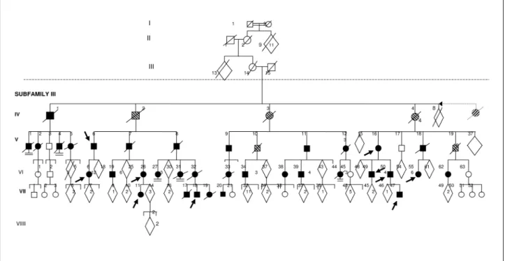
Subfamily III is composed of 162 individuals distri-buted into 6 generations, including 55 males (33.9%), 67 females (41.3%) and 40 subjects of unknown gen-der (24.6%). This subfamily also originated from a consanguineous marriage and 34 members are aff e c t-ed, including 18 males (52.9%) and 16 females (47%). In this subfamily, the first case of SCA7 appeared in the fourth generation. There are 19 affected alive and 10 of them agreed to participate in this study (Fig 2).
f rom a consanguineous marriage in the third gener-ation of the family and possesses 11 affected individ-uals, 8 of them males (72.7%) and 3 females (27.2%). Two of them are alive, but none of them was includ-ed in this study.
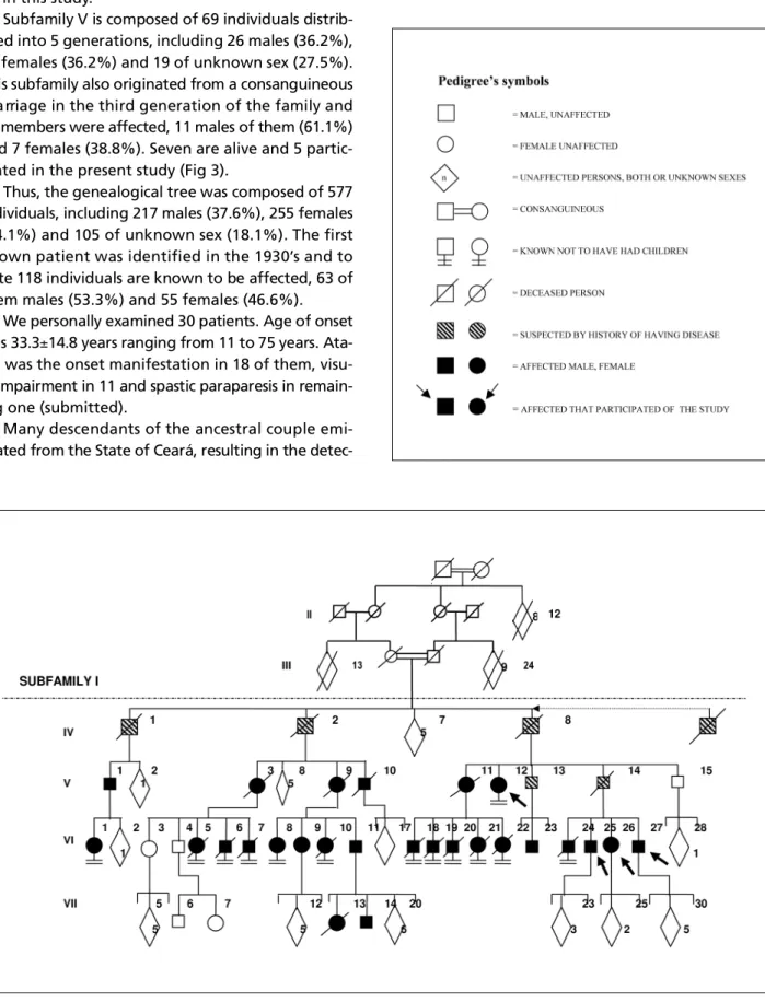
Subfamily V is composed of 69 individuals distrib-uted into 5 generations, including 26 males (36.2%), 25 females (36.2%) and 19 of unknown sex (27.5%). This subfamily also originated from a consanguineous m a rriage in the third generation of the family and 18 members were affected, 11 males of them (61.1%) and 7 females (38.8%). Seven are alive and 5 partic-ipated in the present study (Fig 3).
Thus, the genealogical tree was composed of 577 individuals, including 217 males (37.6%), 255 females (44.1%) and 105 of unknown sex (18.1%). The first known patient was identified in the 1930’s and to date 118 individuals are known to be affected, 63 of them males (53.3%) and 55 females (46.6%).
We personally examined 30 patients. Age of onset
was 33.3±14.8 years ranging from 11 to 75 years.
Ata-xia was the onset manifestation in 18 of them, visu-al impairment in 11 and spastic paraparesis in re m a i n-ing one (submitted).
Many descendants of the ancestral couple emi-grated from the State of Ceará, resulting in the
detec-tion of cases of SCA7 of the same genealogical ori-gin in other areas of the country. Considering only the 60 alive patients that we were able to identify, 37 of them (61.6%) still live in the Northeast region
Fig 2. Pedigree of subfamily III with 162 persons in 5 generations and 34 affected.
Fig 3. Pedigree of subfamily V with 69 persons in 5 generations and 18 affected.
of the country (26 in Ceará, 4 in Piauí, 3 in Maranhão, 2 in Bahia and 2 in Pernambuco), 12 (20%) in the Sou-theast region (6 in Rio de Janeiro, 5 in Minas Gerais
DISCUSSION
The cerebellar ataxias are neurodegenerative dis-o rders characterized by degeneratidis-on dis-of the cere b e l-lum and/or the cerebellar connections, either isolat-ed, or associated with involvement of other stru c t u-res such as the visual and auditory systems, the cor-tico-spinal pathways, the lower motor neurons, the
basal ganglia and the peripheral nerv e s7. The
inher-itance may be dominant, recessive or X-linked. At least 26 diff e rent types of autosomal dominant cere-bellar ataxias (ADCA) have been described thus far and at least 11 of them may be diagnosed based on molecular testing: SCAs 1, 2, 3, 6, 7, 8, 10 ,12, 14, 17 and DRPLA.
The association of spinocerebellar manifestations with retinal degeneration in a family with autoso-mal dominant inheritance and evidence of
anticipa-tion is highly suggestive of SCA79 , 1 4. The clinical
fea-t u res of fea-the esfea-tablished cases of fea-this Brazilian fami-ly with SCA7 included ataxic gait, visual loss, dysar-tria, nystagmus, ophthalmoplegia, dysphagia, dysto-nia, pyramidal and extrapyramidal signs, being
sim-ilar to those already described in the literature5.
Contrasting with this similarity, the Brazilian patients
with SCA10 described by Teive et al.1 5did not have
epilepsy, that was present in the Mexican patients16
and was supposed to be an essential clinical charac-teristic of the disease. Diff e rent phenotypes of the same disease in diff e rent populations have impor-tant clinical implications, and its origin still have to be determined.
Although generally considered rare, the pre s e n c e of SCA7 has already been confirmed in several re g i o n s of the world. It seems to be prevalent in United States
and France1 7, and is the most frequent identified form
of ADCA in Sweden and Finland18. In Spain, the
fre-quency of SCA7 among 72 patients with ADCA was 3%, being lower than the frequency of SCA3/MJD
(15%), SCA2 (15%) and SCA1 (6%)1 9. SCA7 also seems
to be uncommon in Brazil, with re p o rted fre q u e
n-cies ranging from 2%2 0to 4.4%1 5, much less fre q u e n t
than SCA3 or Machado-Joseph disease that re p
re-sents about 40% of the ADCAs21.
Although SCA7 is a dominant condition, several consanguineous marriages have been detected in the first generations of this family, apparently pre c e d i n g the existence of the disease. In spite of the high con-sanguinity rate and of the autosomal dominant char-acter of the disease, it was impossible to detect any patient with expansion of the two alleles, a rare o c c u rrence that, according to Palau and Sevilla2 2c o u l d
have a dosage effect, resulting in a more severe dis-ease.
In this Brazilian family, the first cases of the disea-se appeared in the fourth generation, but this obdisea-ser- obser-vation does not exclude the possibility that members of the previous generations carried abnormal alle-les. If this was the case, these individuals may not have been identified because they did not surv i v e long enough to manifest symptoms of the disease, since of death was very early at that time, or because the disease was very mild and late due to the small numbers of CAG repetitions. It is virtually impossible to test these hypotheses and, in addition, a new explanation that has become available is the
occur-rence of de novo mutation, that has been shown to
be the case in some SCA7 families23,24.
Most of those affected in the sixth generation of subfamily I did not constitute family. We are unable to state if this fact was a deliberate decision or a bio-logical limitation. Due to the phenomena of antici-pation, the disease may move towards extinction, but the presence of interm e d i a ry alleles may re s u l t
in the beginning of a new cycle of the disease23,24.
The ancestral couple we described had 10 descen-dents, but only 4 of them constituted the basis of this genealogical tree that we were able to constru c t . Once the destiny of the other 6 sibs and their descen-dants is currently unknown, it is highly probable that they have emigrated from the State of Ceará, with the possibility that other cases of SCA7 in other re-gions of Brazil might have the same family origin.
Acknowledgements - We are thankful to Mrs. Sandra
E.M. Nemoto for the excellent assistance with the labora-tory work.
REFERENCES
1. Benomar A, Le Guern, Durr A, et al. Autosomal-dominant cere b e l l a r ataxia with retinal degeneration (ADCA type II) is genetically diff e r-ent from ADCA type I. Ann Neurol 1994;35:439-444.
2. Benomar A, Stevanin G, Cancel G, et al. The gene for autosomal dom-inant ataxia with pigmentary dystrophy maps to chromosome 3p12-p21.1. Nat Genet 1995;10:84-88.
3. Gouw L, Kaplan C, Haines J, et al. Retinal degeneration characterizes a spinocerebellar ataxia mapping to chromosome 3p. Nat Genet 1995;10:89-93.
4. H o l m b e rg M, Johansson J, Forsgren J, Heijbel J, Sandgren O, Holmgre n G. Localization of autosomal dominant cerebellar ataxia associated with retinal degeneration and antecipation to chromosome 3p12-p21.1. Am J Hum Genet 1995;4:1441-45.
5. David G, Giunti P, Abbas N, et al. The gene for autosomal dominant ataxia Type II is located in a 5-cM in 3p12-13: genetic and physical map-ping of the SCA7 locus. Am J Hum Genet 1996;59:1328-1336. 6. K rols L, Martin JJ, David G, et al. Refinement of the locus for
autoso-mal dominant cerebellar ataxia type II to chromosome 3p21.1-14.1. Hum Genet 1997;99:225-232.
8. Del-Favero , Krols L, Michalick A, et al. Molecular genetic analysis of autosomal dominant cerebellar ataxia with retinal degeneration (ADCA type II) caused by CAG triplet repeat expansion. Hum Molec Genet 1998;7:177-186.
9. David G, Dürr A, Stevanin G, et al. Molecular and clinical corre l a t i o n s : c e rebellar ataxia with pro g ressive macular dystrophy (SCA7). Hum Molec Genet 1998;7:165-170.
10. Havener WH. Cere b e l l a r-macular abiotro p h y. A rch Ophthalmol 1951; 25:40-43.
11. Boudin G, Barbizet J, Le Hénaff MY. Hérédo-ataxie cérébelleuse avec amblyopie et paralysie de la verticalité du regar chez la mère et l’en-fant. Rev Neurol 1952;87:330-335.
12. Cooles P, Michaud R, Best PV. A dominantly inherited pro g ressive dis-ease in a black family characterized by cerebellar and retinal degener-ation, external ophthalmolplegia and abnormal mitochondria. J Neuro l Sci 1988;87:275-288.
13. Jampel RS, Okazaki H, Bersntein H. Ophthalmoplegia and re t i n a l degeneration associated with spinocerebellar ataxia. A rch Ophthalmol 1961;66:247-249.
14. Enovoldson TP, Sanders MD, Harding AE. Autosomal dominant cere-bellar ataxia with pigmentary macular dystrophy: a clinical and genet-ic study of eigth families. Brain 1994;117:445-460.
15. Teive HAG, Iwamoto FM, Camargo CH, Lopes-Cendes I, Werneck LC. Clinical phenotype of Brazilian families with spinocerebellar ataxia 10. Neurology 2004;63:1509-1512.
16. Zu L, Figueroa KP, Grewal R, Pulst SM. Mapping of a new autosomal dominant spinocerebellar ataxia to chromosome 22. Am J Hum Genet 1999;64:594-599.
17. Tang B, Liu C, Shen L, Dai H, et al. Frequency of SCA1, SCA2, SCA3/ MJD, SCA6, SCA7, and DRPLA CAG trinucleotide repeat expansion in patients with hereditary spinocerebellar ataxia from Chinese kin-dreds. Arch Neurol 2000;57:540-544.
18. Jonasson J. Evidence for a common spinocerebellar ataxia type 7 (SCA7) founder mutation in Scandinavia. Eur Hum Genet 2000;8:918-922. 19. Pujana MA, Corral J, Gratacós M, et al. Spinocerebellar ataxias in
Spa-nish patients: genetic analysis of familial and sporadic cases. Hum Genet 1999;104:516-522.
20. J a rdim LB, Silveira I, Pereira ML, et al. A survey of spinocerebellar ata-xia in South Brazil: 66 new cases with Machado Joseph disease, SCA 7, SCA 8 or unidentified disease causing mutations. J Neurol 2001; 248:870-876.
21. Lopes-Cendes I, Teive LGA, Calcagnoto ME, et al. Frequency of the d i ff e rent mutations causing spinocerebellar ataxia (SCA1, SCA2, MJD/SCA3, and DRPLA) in a large group of brazilian patients. A rq . Neuropsiquiatr 1997;55:519-529.
22. Palau F, Sevilla T. Genética de las neuropatias periféricas y las ataxias hereditarias. Neurologia 1995;10:32-43.
2 3 . Stevanin G, Giunti P, David G, et al. De novo expansion of intermediate
alleles in spinocerebellar ataxia 7. Hum Molec Genet 1998;7:1809-1813. 24. Giunti P, Stevanin G, Worth PF, David G, Brice A, Wood NW. Molecular and clinical study of 18 families with A D C A type II: evidence for genet-ic heterogeneity and de novo mutation. Am J Hum Genet 1999;64: