PB 203
Response of osteoblastic cells to titanium submitted
to three different surface treatments
Resposta de células osteoblásticas ao titânio submetido
a três diferentes tratamentos de superfície
Adriana Soares Santiago* Euler Araújo dos Santos** Márcia Soares Sader*** Marcelo Felippe Santiago**** Gloria de Almeida Soares*****
ABSTRACT: In the complex process of bone formation at the implant-tissue interface, surface properties are rel-evant factors modulating osteoblastic function. In this study, commercially pure titanium (cp Ti) samples were prepared with different surface characteristics using chemical attack with a sulfuric acid/hydrochloric acid based solution (treatment A); chemical attack plus anodic oxidation using phosphoric acid (treatment B); and chemical attack plus thermal oxidation followed by immersion in a sodium fluoride solution (treatment C). The samples were characterized by scanning electron microscopy (SEM), contact profilometry and contact angle. The biological performance of the prepared surfaces was evaluated using mice osteoblastic cell cultures for up to 21 days. Cells seeded on the different titanium samples showed similar behavior during cell attachment and spreading. However, cellular proliferation and differentiation were higher for samples submitted to treatments A and C (p ≤ 0.05; n = 3), which were less rough and showed surface free energy with smaller polar components.
DESCRIPTORS: Titanium; Surface properties; Osteoblasts; Biocompatible materials.
RESUMO: No complexo processo de formação óssea na interface implante-osso, as propriedades de superfície são um importante fator modulador da função osteoblástica. No presente estudo, foram preparadas amostras de titânio comercialmente puro (cp Ti) com diferentes propriedades de superfície por meio de ataque químico com solução à base de ácido sulfúrico/clorídrico (tratamento A); ataque químico seguido de oxidação anódica com ácido fosfórico (tratamento B); e ataque químico seguido de oxidação térmica e imersão em fluoreto de sódio (tratamento C). As chapas foram caracterizadas por meio de microscopia eletrônica de varredura (MEV), perfilometria e ângulo de contato. O comportamento de células osteoblásticas de camundongo foi acompanhado por três semanas. As células cultivadas sobre os diferentes substratos de titânio apresentaram um modelo de comportamento similar durante as etapas de adesão e espalhamento. No entanto, a proliferação e a diferenciação celulares foram maiores nas amostras submetidas aos tratamentos A e C, que se apresentaram menos rugosas e com energia livre de su-perfície com menores componentes polares.
DESCRITORES: Titânio; Propriedades de superfície; Osteoblastos; Materiais biocompatíveis.
INTRODUCTION
Titanium is a material widely used for implants because of its biocompatibility5. This material has
high corrosion resistance, suitable mechanical properties and can be easily produced in many different shapes and textures. A key question in most applications of titanium is how the mate-rial influences, and is influenced by the biological response that results from the contact between biomaterial and biological systems.
Titanium is a reactive metal that forms, spon-taneously, in the air, water or any other electrolyte, a thin native oxide film, which is responsible for titanium biocompatibility. Surface properties of an implant play a critical role in this process as bone cells can recognize and respond to surfaces6.
Surface modification aims to accelerate osseoin-tegration and includes hydroxyapatite (HA) coat-ings, mechanical blasting (using either Al2O3 or
204 205
204 205
TiO2 particles), anodic oxidation and
acid-etch-ing processes (different concentrations of H2SO4,
HF, HNO3 and HCl). Recently, some authors have
reported on significantly improved bone tissue re-actions by modification of surface oxide properties of titanium implants1,19 or ion release2. Chemical
modification of titanium implant surface is of par-ticular interest because it may enhance osseointe-gration without embedding surface contaminants, such as grit particles21.
The biological response of osteoblastic cells includes cell attachment, cell growth and func-tional activity7. Concerning osteoblastic
differen-tiation and metabolism, the results reported in the literature are somewhat controversial. While some papers show that increasing surface rough-ness enhances in vitro osteoblastic differentiation and inhibits cell proliferation3,11,12, others1,6,13,15,18,19
indicate that proliferation can be improved on specimens submitted to higher surface roughness. These results suggest that there are other aspects that also modulate proliferation, differentiation and extracellular matrix production of osteoblastic cells in vitro.
In this study, the behavior of mice osteoblastic cells seeded on titanium submitted to three differ-ent surface treatmdiffer-ents was evaluated in terms of cell attachment, cell proliferation (MTT assay) and cell differentiation (alkaline phosphatase – ALP ac-tivity). Additionally, a correlation between cell re-sponse and surface properties was established.
MATERIALS AND METHODS
Sample preparation
A commercially pure titanium (ASTM grade 2) (Titanews, São Paulo, Brazil) sheet with 1 mm in thickness was cut in 10 mm x 10 mm squares (for cell culture) and 20 mm x 10 mmrectangles (for samplecharacterization). According to the surface treatment employed, three groups of samples were obtained, as shown in Table 1. The aim of treat-ment A was to create surface roughness, while samples B and C had an additional treatment to increase oxide thickness. Anodization and thermal oxidation samples were based on the findings of Sena et al.18 (2003) and Vanzillotta20 (2003),
re-spectively. C samples were additionally immersed in a NaF solution to allow fluoride incorporation, which seems to be beneficial to bone formation8,9.
After surface treatments, the samples were cleaned using acetone (Merck, São Paulo, Brazil) and dis-tilled water (Quimis Q-341, Diadema, Brazil) in an ultrasonic cleaner and then sterilized by au-toclaving.
Scanning electron microscopy (Zeiss DSM 940A, Oberkochen, Germany), operating at 15 kV was employed for qualitative evaluation of the ti-tanium topography. Surface roughness was meas-ured with a contact stylus profilometer (Perthom-eter, Perthen, Gottingen, Germany) over a 250 µm2
area, using a Gaussian filter (80 µm) to exclude form and waviness characteristics from the rough-ness measurements, according to the DIN 4768 standard. For each group, three specimens were employed, and at least 10 measurements were made on different regions of each sample. Three parameters were used: Ra (the average surface roughness, or average deviation, of all points from a plane fit to the test part of the surface); Rq (the square-root of the average of the measured height deviations) and Rz (the average absolute value of the five highest peaks and the five lowest valleys over the evaluation length).
Contact angle measurements were used to cal-culate the surface free energy (SFE) of the titanium samples. Contact angles (θ) on the sterilized tita-nium surfaces were measured with a goniometer (Ramé-Hart Instrument Co., Netcong, NJ, USA) by the captive air bubble method. SFE components were obtained by the equation14:
(1 + cos θ) γl = 2[(γsdγld)1/2 + (γspγlp)1/2]
where: θ is the contact angle between the liquid and the captive air bubble; subscript s and l are the solid and
liquid surfaces, respectively;
γd stands for the dispersion compo-
nent of the total surface energy (γ);
γp is the polar component.
Water (γl = 72.8 mJ/m2; γld = 21.9 mJ/m2;
γlp = 51.0 mJ/m2) and glycerol (γl = 64.0 mJ/m2;
TABLE 1 - Surface treatments employed.
Sample Treatment
A 18% HCl + 48% H2SO4 (55°C – 60 s)
B
First step: 18% HCl + 48% H2SO4 (55°C –
60 s); Second step: anodic oxidation (IPRJ, Nova Friburgo, Brazil) with 8% H3PO4 in ethanol solution (20 V – 10 min)
C
First step: 18% HCl + 48% H2SO4
(55°C – 60 s); Second step: thermal oxidation (Quimis, Diadema, Brazil) (450°C – 60 min); Third step: immersion in 4% NaF (40 min)
204 205
204 205
γld = 34.0 mJ/m2; γlp = 30.0 mJ/m2) were used as
the standard liquids.
Cell culture
Mice were obtained from our breeding colony. All experiments were performed in accordance with the National Institute of Health Guide for the Care and Use of Laboratory Animals, and the experi-mental protocols were approved by the Commit-tee for the Use of Experimental Animals of our Institution.
Mice osteoblastic cells were cultured in Dul-becco’s Modified Eagle Medium (DMEM, Gibco BRL, Gaithersburg, MD, USA) containing 10% of fetal bovine serum. Primary cultures were main-tained until near confluence and, at the 6th
pas-sage, adherent cells were enzymatically released (0.04% trypsin – Sigma, St. Louis, MD, USA – and 0.025% collagenase – Sigma, St. Louis, MD, USA) and seeded in 24-well plates (Sigma, St. Louis, MD, USA) at a density of 104 cells/cm2. Incubation
was carried out in a humidified atmosphere of 95% air and 5% CO2 at 37°C, and culture medium was
changed twice a week. Cells were cultured on both control (glass – Elzividros, Rio de Janeiro, Brazil) and titanium samples for up to 21 days.
Cell attachment was observed by fluorescence microscopy 5 and 24 hours after incubation. Tita-nium substrates (n = 3 for each treatment) were stained with 4-6-diamidino-2-phenylindole (DAPI, Sigma, St. Louis, MO, USA), and the number of adherent cells was determined for each of the three specimens.
As for cell viability/proliferation evaluation, the cells were cultured for 7, 14 and 21 days on titanium specimens and analyzed using the MTT (Sigma, St. Louis, MD, USA) assay. This method is
based on the reduction of 3-(4,5-dimethylthiazol-2-yl)-2,5-diphenyltetrazolium bromide by viable cells to a purple formazan salt. The cells were incubated with 50 µl of MTT for 4 hours at 37°C. The dark blue formazan crystals were dissolved with acid sodium dodecyl sulfate (0.1 N HCl SDS, Merck, Darmstadt, Germany) and then they were kept in the acid solution overnight to ensure complete dissolution of crystals. The absorbance was de-termined at 595 nm in an ELISA reader (Bio-Rad, Hercules, USA).
Alkaline phosphatase (ALP) activity was as-sayed by the hydrolysis of p-nitrophenyl phosphate (Sigma, St. Louis, MD, USA) in alkaline buffer solution (pH 10.3) (Sigma, St. Louis, MD, USA), and colorimetric determination of the product (p -nitrophenol) was carried out at 405 nm (ELISA reader, Bio-Rad, Hercules, USA). ALP activity was calculated from a standard curve and results were expressed in nanomoles of p-nitrophenol produced per minute.
Statistical analysis
For cell culture analysis, data are presented from one of two replicate experiments, both of which yielded comparable results. For any giv-en experimgiv-ent, each data point represgiv-ents the mean ± standard deviation of three replicates. Statistical analysis was done by one-way analysis of variance (ANOVA), and statistical differences between the three samples were determined by Tukey’s multiple comparison post hoc test. Only p values ≤ 0.05 were considered significant.
RESULTS
SEM micrographs (Figure 1) show a similar rough topography for the three samples (A, B, C) FIGURE 1 - SEM micrographs (1,000 X) of the titanium samples submitted to: A) Treatment A; B) Treatment B; C) Treatment C.
206 207
206 207
Alkaline phosphatase (ALP), which is produced during osteoblastic differentiation, diffuses into circulation and is used as a serum biochemical marker of bone formation. Until day 14, ALP activ-ity was reduced in all samples, however, samples A and C exhibited a greater activity, which remained at higher levels at day 21 when compared to control and sample B surfaces (p ≤ 0.05; n = 3) indicating an osteoblastic differentiation stimulus.
DISCUSSION
The present work showed that different chemi-cal treatments may produce similar topographies of cp Ti surfaces (Figure 1). This result was quite expected as oxide films are very thin (from 30 to 80 nm)20 and may not influence topography on a
micrometer scale.
Surfaces submitted to treatments A and C exhibited quite similar and smaller polar compo-nents. Hallab et al.10 (2001) demonstrated that
SFE is a more relevant surface characteristic than surface roughness for cellular adhesion strength and proliferation, and that surface energy compo-nents of the various tested materials proved to be related to cellular adhesion strength: poor correla-tion was observed between the dispersive compo-nent of SFE and adhesion strength when compared to the polar component of SFE. Similar correla-tions were observed by Ponsonnet et al.16 (2003).
The biological performance of the titanium samples was evaluated in terms of their ability to allow cell attachment, proliferation and differen-tiation. Cell adhesion is one of the initial events essential to subsequent proliferation and differen-tiation of bone cells before bone tissue formation. Consequently, many in vitro evaluations of cell adhesion on substrates with various roughness levels have been performed in order to identify the main surface properties influencing cell response to implant surface5. Cell adhesion is a very specific
parameter and describes the relative adherence of TABLE 3 - Polar (γsp) and dispersive (γsd) components of
the surface free energy (SFE) calculated by Owens-Wendt’s method.
Sample γ (mJ/m2) γ
sp (mJ/m2) γsd (mJ/m2)
A 56 8 48
B 43 14 29
C 42 9 33
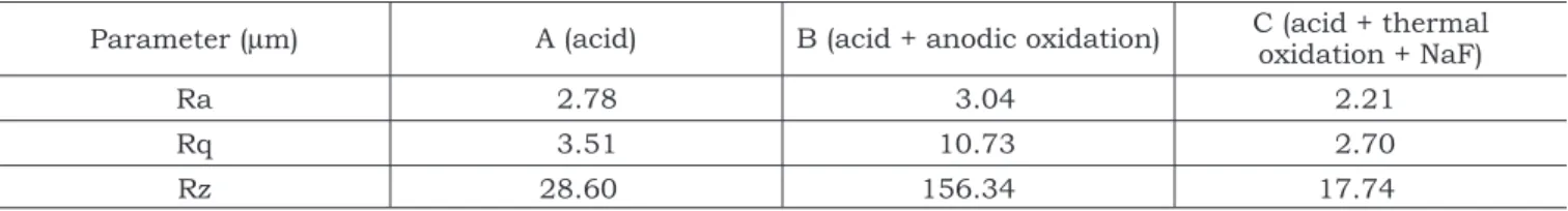
studied. Table 2 shows the roughness parameters (Ra, Rq and Rz) for the titanium samples and Ta-ble 3 shows surface free energy (γs) and their polar
(γsp) and dispersive (γsd) components calculated by
solving simultaneous equations (Owens-Wendt ap-proach14).
Ra was not significantly affected by the treat-ments used for surface preparation. However, the two other roughness parameters (Rq and Rz) were significantly different when samples A and C were compared to sample B (p < 0.05), with A and C showing smaller roughness values. Surfaces submitted to the treatments of samples A and C showed quite similar and smaller polar compo-nents.
Data on cell attachment are presented in Graph 1, which shows that attachment was not affected by surface topography either after 5 h or after 24 h. However, cell attachment was higher on control (glass) substrates (p ≤ 0.05; n = 3) than on Ti samples. The results of cell viability/prolifera-tion and of ALP activity are presented in Graph 2 and Graph 3, respectively. Results concerning cell viability/proliferation evaluated by MTT assay showed that cell growth was not affected by surface treatment after 7 days in culture, where osteoblas-tic cells proliferated very well on all substrates. The behavior in control cultures showed cell growth during two weeks, decreasing after that (p ≤ 0.001; n = 3). Treatment B resulted in a high decrease in the cell growth rate after 14 days and mainte-nance of this rate after 3 weeks (p ≤ 0.05; n = 3). TABLE 2 - Roughness parameters for titanium samples.
Parameter (µm) A (acid) B (acid + anodic oxidation) C (acid + thermal oxidation + NaF)
Ra 2.78 3.04 2.21
Rq 3.51 10.73 2.70
Rz 28.60 156.34 17.74
206 207
206 207
a cell to its substrate, generally at an early stage of culture when cells are directly in contact with the material surface4.
The similarity observed in all samples after 5 or 24-hour inoculation can have two explana-tions: 1) it is possible that, after the initial hours, osteoblastic cells can easily attach, spread over the entire surface, and then start to die because of growth limitation (in this case, after 24 hours, cell number could be reduced to the same value observed after 5 hours); or 2) osteoblastic cell at-tachment could be the same after both studied times. An in vitro evaluation of cell adhesion 24 hours after seeding is not sufficient to anticipate the future integration of a material several weeks after implantation.
Analyzing data of the ALP assay, the most rel-evant aspect was that, after 14 days, osteoblastic differentiation showed to be affected by surface treatment, and the best behaviors were observed in samples A and C. These observations support the results concerning cell viability/proliferation evaluated by the MTT assay, and indicate a bet-ter behavior of osteoblasts seeded on substrates A and C.
Findings of the present work show that pro-liferation is unfavorably affected by increasing surface roughness, in agreement with other stud-ies3,12,17 that observed better cell responses on more
organized surfaces. This behavior was probably due to the slightly smoother surface and to the sur-face free energy with smaller polar components.
CONCLUSIONS
Cell differentiation/viability/proliferation was higher for Ti samples submitted to treatments A (chemical attack with a sulfuric acid/hydrochloric acid based solution) and C (chemical attack plus thermal oxidation followed by immersion in so-dium fluoride solution). Therefore, these treated surfaces seem to provide a better environment for mice osteoblastic cell integration. These results suggest that the treatments used in the present study may support favorable biological responses in vivo.
400 350 300 250 200 150 100 50 0 Control C el ls /mm 2
A B C
5 hours 24 hours 33 9. 3 32 4. 0 24 7. 3 25 4. 7 24 8. 0 23 0. 7 26 6. 0 24 0. 7
GRAPH 1 - Number of adherent osteoblast cells after 5 and 24 hours in culture on treated Ti and control surfaces. All data are reported as mean ± standard de-viation (n = 3).
0.12 0.11 0.10 0.09 0.08 0.07 0.06 0.05 0.04 0.03 0.02 0.01 0.00 7 O pt ica l d en si ty 14 Days 21 Control A B C 0. 01 4 0. 05 8 0. 01 4 0. 02 2 0. 07 1 0. 06 8 0. 06 3 0. 07 6 0. 04 8 0. 04 8 0. 05 6 0. 05 6
GRAPH 2 - Viability/proliferation (MTT assay) of mice osteoblastic cells on control and treated surfac-es after 7, 14 and 21 days. All data are reported as mean ± standard deviation (n = 3).
12 8 10 6 4 2 0 7 10
-7 n
mo l PN P/ mi n/ mg p ro te in 14 Days 21 3.
24 3.74
2. 34 2. 43 4. 61 4. 95 4. 67 2. 38 9. 99 9. 89 8. 71 8. 86 Control A B C
208 PB
ACKNOWLEDGMENTS
The authors acknowledge the financial sup-port given by the José Bonifácio University Foun-dation (FUJB) and by the Molecular and Interfaces
Nanotechnology Research Network/CNPq. Marcelo Felippe Santiago is supported by FAPERJ and a grant from the Ministry of Science and Technol-ogy (MCT) to the Millennium Institute for Tissue Bioengineering, Brazil.
REFERENCES
1. Albrektsson T, Johansson C, Lundgren A, Sul YT. Experi-mental studies on oxidized implants. A histomorphomet-rical and biomechanical analysis. Appl Osseointegr Res 2000;1:21-4.
2. Anderson PA, Copenhaver JC, Tencer AB, Clark JM. Re-sponse of cortical bone to local controlled release of sodium fluoride: the effect of implant insertion site. J Orthop Res 1991;9:890-901.
3. Anselme K, Bigerelle M, Noel B, Dufresne E, Judas D, Iost A, et al. Qualitative and quantitative study of human os-teoblast adhesion on materials with various surface rough-nesses. J Biomed Mater Res 2000;49:155-66.
4. Bigerelle M, Anselme K, Noel B, Ruderman I, Hardouin P, Iost A. Improvement in the morphology of titanium-based surfaces: a new process to increase in vitro human osteo-blast response. Biomaterials 2002;23:1563-77.
5. Branemark PI, Adell R, Breine U, Hansson BO, Lindstrom J, Ohlsson A. Intra-osseous anchorage of dental prosthe-ses. I. Experimental studies. Scand J Plast Reconstr Surg 1969;3(2):81-100.
6. Chehroudi B, Brunette DM. Subcutaneous microfabri-cated surfaces inhibit epithelial recession and promote long-term survival of percutaneous implants. Biomaterials 2002;23:229-37.
7. Diniz MG, Soares GA, Coelho MJ, Fernandes MH. Surface topography modulates the osteogenesis in human bone marrow cell cultures grown on titanium samples prepared by a combination of mechanical and acid treatments. J Mater Sci Mater Med 2002;13:421-32.
8. Ecki P. Evaluation of the repair process in mechanically injured rat bone stimulated by sodium fluoride with non-toxic doses. Ann Acad Med Stetin 1999;45:195-209. 9. Ellingssen JE. Surface configuration of dental implants.
Periodontol 2000 1995;17:36-46.
10. Hallab N, Bundy K, O’Connor RL, Jacobs JJ. Evalu-ation of metallic and polymeric biomaterial surface energy and surface roughness characteristics for directed cell ad-hesion. Tissue Eng 2001;71:55-71.
11. Lange R, Luthen F, Beck U, Rychyl J, Baumann A, Nebe B. Cell-extracellular matrix interaction and physico-chemical characteristics of titanium surfaces depend on the roughness of the material. Biomol Eng 2002;19:255-61.
12. Martin JY, Schwartz Z, Hummert TW. Effect of titani-um surface roughness on proliferation, differentiation, and protein synthesis of human osteoblast-like cells (MG63). J Biomed Mater Res 1995;29:389-401.
13. Owen TA, Aronow M, Shalhoub V. Progressive develop-ment of the rat osteoblast phenotype in vitro: reciprocal rela-tionships in expression of genes associated with osteoblast proliferation and differentiation during formation of the bone extracellular matrix. J Cell Physiol 1990;143:420-30. 14. Owens D, Wendt RC. Estimation of the surface free
energy of polymers. J Appl Polym Sci 1969;13:1741-7. 15. Perizzolo D, Lacefield WR, Brunette DM.
Interac-tions between topography and coating in the formation of bone nodules in culture for hydroxyapatite- and tita-nium-coated micromachined surfaces. J Biomed Mater Res 2001;56:494-503.
16. Ponsonnet L, Reybier K, Jaffezic N, Comte V, Lag-neau C, Lissac M. Relationship between surface properties (roughness, wettability) of titanium and titanium alloys and cell behavior. Mater Sci Eng 2003;23:551-60.
17. Rosa AL, Beloti MM, Noort RV. Osteoblastic differentiation of cultured rat bone marrow cells on hydroxyapatite with different surface topography. Dent Mater 2003;19:768-72. 18. Sena LA, Rocha NCC, Andrade MC, Soares GA.
Bioactivity assessment of titanium sheets electrochemi-cally coated with thick oxide film. Surface Coat Technol 2003;166:254-8.
19. Sul YT, Johansson CB, Roser K, Albrektsson T. Quali-tative and quantiQuali-tative observations of bone tissue reactions to anodized implants. Biomaterials 2002;23:1809-17. 20. Vanzillotta PS. Avaliação in vitro de chapas de titânio
com diferentes tratamentos de superfície em solução simu-ladora de plasma humano [Dissertação de Mestrado]. Rio de Janeiro: Instituto Alberto Luiz Coimbra de Pós-Gradu-ação e Pesquisa de Engenharia da UFRJ; 2003.
21. Xavier SP, Carvalho PSP, Beloti MM, Rosa AL. Re-sponse of rat bone marrow cells to commercially pure ti-tanium submitted to different surface treatments. J Dent 2003;31:173-80.