Membrane-To-Nucleus Signaling Links Insulin-Like
Growth Factor-1- and Stem Cell Factor-Activated
Pathways
Yujiro Hayashi
1,2, David T. Asuzu
1,2, Simon J. Gibbons
1, Kirsten H. Aarsvold
1,2, Michael R. Bardsley
1,2,
Gwen A. Lomberk
2, Angela J. Mathison
2, Michael L. Kendrick
3, K. Robert Shen
3, Takahiro Taguchi
4, Anu
Gupta
5, Brian P. Rubin
5, Jonathan A. Fletcher
6, Gianrico Farrugia
1,7, Raul A. Urrutia
2,7, Tamas Ordog
1,2,7* 1 Enteric Neuroscience Program, Department of Physiology and Biomedical Engineering, Mayo Clinic, Rochester, Minnesota, United States of America, 2 Gastroenterology Research Unit, Division of Gastroenterology and Hepatology, Mayo Clinic, Rochester, Minnesota, United States of America, 3 Department of Surgery, Mayo Clinic, Rochester, Minnesota, United States of America, 4 Division of Human Health and Medical Science, Graduate School of Kuroshio Science, Kochi University, Nankoku, Kochi, Japan, 5 Departments of Anatomic Pathology and Molecular Genetics, Lerner Research Institute and Taussig Cancer Center, Cleveland Clinic, Cleveland, Ohio, United States of America, 6 Department of Pathology, Brigham and Women’s Hospital and Harvard Medical School, Boston, Massachusetts, United States of America, 7 Center for Individualized Medicine, Mayo Clinic, Rochester, Minnesota, United States of AmericaAbstract
Stem cell factor (mouse: Kitl, human: KITLG) and insulin-like growth factor-1 (IGF1), acting via KIT and IGF1 receptor (IGF1R), respectively, are critical for the development and integrity of several tissues. Autocrine/paracrine KITLG-KIT and IGF1-IGF1R signaling are also activated in several cancers including gastrointestinal stromal tumors (GIST), the most common sarcoma. In murine gastric muscles, IGF1 promotes Kitl-dependent development of interstitial cells of Cajal (ICC), the non-neoplastic counterpart of GIST, suggesting cooperation between these pathways. Here, we report a novel mechanism linking IGF1-IGF1R and KITLG-KIT signaling in both normal and neoplastic cells. In murine gastric muscles, the microenvironment for ICC and GIST, human hepatic stellate cells (LX-β), a model for cancer niches, and GIST cells, IGF1 stimulated Kitl/KITLG protein and mRNA expression and promoter activity by activating several signaling pathways including AKT-mediated glycogen synthase kinase-γ inhibition (GSKγi). GSKγi alone also stimulated Kitl/KITLG expression without activating mitogenic pathways. Both IGF1 and GSKγi induced chromatin-level changes favoring transcriptional activation at the Kitl promoter including increased histone Hγ/H4 acetylation and Hγ lysine (K) 4 methylation, reduced HγK9 and HγKβ7 methylation and reduced occupancy by the HγKβ7 methyltransferase EZHβ. By pharmacological or RNA interference-mediated inhibition of chromatin modifiers we demonstrated that these changes have the predicted impact on KITLG expression. KITLG knock-down and immunoneutralization inhibited the proliferation of GIST cells expressing wild-type KIT, signifying oncogenic autocrine/paracrine KITLG-KIT signaling. We conclude that membrane-to-nucleus signaling involving GSKγi establishes a previously unrecognized link between the IGF1-IGF1R and KITLG-KIT pathways, which is active in both physiologic and oncogenic contexts and can be exploited for therapeutic purposes.
Citation: Hayashi Y, Asuzu DT, Gibbons SJ, Aarsvold KH, Bardsley MR, et al. (β01γ) Membrane-To-Nucleus Signaling Links Insulin-Like Growth Factor-1-and Stem Cell Factor-Activated Pathways. PLoS ONE 8(10): e768ββ. doi:10.1γ71/journal.pone.00768ββ
Editor: Anette Duensing, University of Pittsburgh Cancer Institute, United States of America Received May 7, β01γ; Accepted August β9, β01γ; Published October 7, β01γ
Copyright: © β01γ Hayashi et al. This is an open-access article distributed under the terms of the Creative Commons Attribution License, which permits unrestricted use, distribution, and reproduction in any medium, provided the original author and source are credited.
Funding: This work was supported, in part, by National Institutes of Health (http://www.nih.gov/) grants R01 DK058185, P01 DK068055 (Project β), Pγ0 DK084567, Pγ0 CA01508γ, P50 CA1β700γ, as well as by the GIST Cancer Research Fund (http://www.gistinfo.org/) and the Mayo Clinic Center for Individualized Medicine http://mayoresearch.mayo.edu/center-for-individualized-medicine/). The funders had no role in study design, data collection and analysis, decision to publish, or preparation of the manuscript.
Competing interests: The authors have declared that no competing interests exist. * E-mail: [email protected]
Introduction
Stem cell factor (mouse: Kitl; human: KITLG) is the natural ligand of the type γ receptor tyrosine kinase (RTK) KIT. Kitl/ KITLG is widely expressed in stromal tissues and is critical for the differentiation, proliferation, migration, survival and functional activation of germinal, erythroid and mast cells and
Kitlβ48/KITLGβ48) at a site encoded within exon 6. A
ββ0-amino-acid isoform, which only generates secreted peptide at a slow rate, is produced from an alternatively spliced transcript lacking exon 6 (“membrane-bound” isoform; Kitlββ0/KITLGββ0) [γ].
Autocrine/paracrine activation of KIT signaling by KITLG plays a role in several tumors and hematologic malignancies [1]. In other cancers including the majority (75-80%) of gastrointestinal stromal tumors (GIST), which originate from cells of the ICC lineage [4,5], KIT signaling is constitutively active due to oncogenic mutations [6]. GIST lacking mutated
KIT may harbor activating mutations in PDGF receptor α (PDGFRA) [7] or have neither KIT nor PDGFRA mutations (“wild-type (WT)” GIST) [6]. KIT/PDGFRA inhibitors such as imatinib mesylate are the mainstay of medical treatment for advanced GIST but they are not curative due in part to secondary mutations interfering with drug action [6] or lack of dependence of cancer-initiating cells on KIT/PDGFRA signaling [8]. Since imatinib preferentially targets mutant receptors [6], reduced drug responsiveness [9,10] and aggressive GIST behavior [11] may also reflect activation of WT KIT expressed in the majority of GIST by KITLG originating from the circulation, the tumor cells, or their niche [9,11-1γ]. However, direct evidence of KITLG-driven GIST cell proliferation is lacking.
Similarly to KIT, PDGFRA and their ligands, insulin-like growth factor (IGF)-1 receptor (IGF1R), a type β RTK, and its ligands IGF1 and IGFβ play critical roles in normal growth and development, as well as in cellular stress, aging and cancer by stimulating protein synthesis and the cell cycle [14,15]. IGF1R is expressed and activated in some GIST [16] and is overexpressed in WT GIST [16,17]. Via an autocrine loop, IGF1 stimulates the growth and survival of gastrointestinal smooth muscle cells [18-β0], and, thereby, promotes the differentiation of ICC [β1] by increasing Kitl availability in their microenvironment [ββ]. IGF1 also activates gene transcription e.g. via p44/p4β mitogen-activated protein kinase (ERK1/β MAPK) signaling [15] or by promoting the nuclear translocation and binding to the chromatin of IGF1R [βγ]. Together, these observations suggest that there may be cooperation between the IGF1-IGF1R and KITLG–KIT signaling pathways both in normal tissues and certain cancers including GIST; these interactions may be mediated by epigenetic control of gene transcription; and increased Kitl/KITLG expression may result in autocrine/paracrine stimulation of proliferation in cells expressing WT KIT. Here, we tested these hypotheses by investigating the effects of IGF1 on endogenous Kitl/KITLG
expression and the underlying epigenetic mechanisms and signaling pathways in IGF1R-expressing cells and tissues including gastric smooth muscles [18-β0,ββ], the natural microenvironment for ICC and GIST; in human GIST cells [10,16], and in LX-β human hepatic stellate cells [β4], a model for cancer niches [β5]. Our findings indicate that IGF1 stimulates KITLG transcription by inducing coordinated chromatin modifications in part via glycogen synthase kinase (GSK)-γ inhibition. We also provide evidence supporting KITLG-mediated autocrine/paracrine stimulation of cell proliferation in GIST cells expressing WT KIT.
Materials and Methods
Ethics statements
Mice were maintained and the experiments were performed in accordance with the National Institutes of Health Guide for the Care and Use of Laboratory Animals. The protocol was approved by the Institutional Animal Care and Use Committee of the Mayo Clinic (A6481β). De-identified human gastric tissues used for the preparation of primary cell cultures were obtained as surgical waste tissue from patients undergoing bariatric surgery with the approval of the Mayo Clinic Institutional Review Board (07-00γγ71). The Mayo Clinic Institutional Review Board waived the need for written informed consent from the participants.
Tissue preparation
BALB/c mice aged 14-16 days were obtained from breeder pairs purchased from Harlan Laboratories (Madison, WI). LONG Rγ-recombinant human IGF1 (LRγ-rhIGF1; Research
Peptides, Orlando, FL) was administered by a single i.p. injection. Mice were anesthetized with isoflurane (AErrane; Baxter Healthcare, Deerfield, IL) inhalation and killed by decapitation. Intact gastric corpus and antrum tunica muscularis tissues were dissected as described [ββ] and used either as organotypic cultures [ββ] or primary cell cultures [β6]. Primary cell cultures were prepared from human tunica muscularis as described for murine tissues [β6].
Tissue culturing and drug treatment
Gastric tunica muscularis organotypic cultures were maintained for up to β4 h. Pharmacological agents were applied as indicated in the Results and Figures. In some experiments tissues were preincubated for γ h with specific, cell-permeable inhibitors or dimethyl sulfoxide (DMSO) vehicle before exposure to rhIGF1 for 18 h in the continuing presence of the inhibitors. Pharmacological agents included rhIGF1, the ATP-competitive GSKγα/ inhibitors SB415β86 and SBβ1676γ [β7], the non-ATP-competitive GSKγα/ inhibitor TDZD-8 [β8], adenosine dialdehyde (Adox) [β9], an inhibitor of adenosylhomocysteine hydrolase and indirect inhibitor of
S-adenosyl-methionine-dependent methylation reactions
including trimethylation of lysine β7 of histone γ (HγKβ7meγ) by the polycomb repressive complex β (PRCβ) member enhancer of zeste homolog β (EZHβ) (Sigma-Aldrich, St. Louis, MO); Tyrphostin AG10β4, a specific inhibitor of IGF1 and insulin RTK activity [γ0]; AKT Inhibitor X (10-(4'-(N-diethylamino)butyl)-β-chlorophenoxazine, HCl), an inhibitor of AKT phosphorylation, in-vitro kinase activity and IGF1-induced nuclear translocation [γ1]; rapamycin, inhibitor of mechanistic target of rapamycin (MTOR) complex 1 and ribosomal p70 S6 kinase (p70S6K) phosphorylation [β0]; and PD98059, inhibitor of MEK1/β ERK MAPK kinases [γβ] (EMD Chemicals, Inc., Gibbstown, NJ).
Cell cultures and antibody/drug treatment
[β6]. Stromal cells derived from the fetal hematopoietic microenvironment of Kitl-deficient Sl/Sl4 mice, Sl/Sl4 stromal
cells genetically modified to express full-length murine Kitl (Sl/Sl4-Kitlβ48) (generously donated by Dr. David Williams,
Indiana University School of Medicine, Indianapolis, IN) [γ], human LX-β spontaneously immortalized hepatic stellate cells (kindly provided by Dr. Scott Freeman, Mount Sinai School of Medicine, New York, NY) [γγ], and human GIST-T1 cells derived from metastatic pleural tumor from a gastric GIST containing a heterozygous deletion of 57 bases in exon 11 juxtamembrane domain of KIT (contributed by Dr. Takahiro Taguchi) [γ4] were cultured in high-glucose Dulbecco’s modified Eagle’s medium (DMEM; Gibco, Life Technologies, Carlsbad, CA) containing 10% fetal bovine serum (FBS) and 1% antibiotic-antimycotic (Gibco) at γ7 °C in the presence of 5% COβ. GIST-T1-5R cells derived from GIST-T1 cells by
prolonged in vitro exposure to imatinib and carrying a secondary, imatinib-resistant T670I mutation in exon 14 (contributed by Dr. Anu Gupta and Dr. Brian P. Rubin) were propagated in the presence of 1 µM imatinib mesylate (LC Laboratories, Woburn, MA). Imatinib was removed from the culture media 4 days prior to the experiments. GIST88β human cells from a primary GIST containing a homozygous KIT exon 1γ missense mutation leading to K64βE substitution in the first part of the split tyrosine kinase domain (contributed by Dr. Jonathan Fletcher) [γ5] were also cultured in high-glucose DMEM containing 10% FBS and 1% antibiotic-antimycotic but were maintained in a milieu of 4% Oβ, 5% COβ and 91% Nβ.
GIST48B KITlow/– human cells derived from GIST48 cells
containing KIT exon 11 (homozygous V560D: imatinib-sensitive) and exon 17 phosphotransferase domain (heterozygous D8β0A: imatinib-resistant) mutations by prolonged heat shock protein 90 inhibition (contributed by Dr. Jonathan Fletcher) [γ6] were maintained with Iscove’s DMEM (high glucose) containing 15% FBS, 1% L-glutamine and 1%
antibiotic-antimycotic (Gibco) at γ7 °C in the presence of 5% COβ. LX-β, GIST-T1 and GIST88β cells have previously been
demonstrated to express IGF1R [16,β4]; results showing IGF1R α and chain expression in GIST48B cells and IGF1 secretion by LX-β, GIST-T1, GIST88β and GIST48B cells are shown in Figure S1F-G. The role of endogenous KITLG in the proliferation of GIST-T1, GIST-T1-5R, GIST88β, GIST48B and LX-β cells was tested by culturing in the above media in the presence of purified, azide-free goat polyclonal anti-human KITLG antibody (AB-β55-NA, R&D Systems, Minneapolis, MN; applied for 4 days at concentrations indicated in the Results), which has been shown to neutralize KITLG-induced proliferation in the TF1 human erythroleukemic cell line [γ7]. The specificity of KITLG immunoneutralization was verified by preabsorbing the anti-KITLG antibody with rhKITLG (R&D Systems) applied at 10:1 molar ratio overnight at 4 °C. Cell proliferation was assessed by the CellTiter 96® AQ
ueous
Non-Radioactive Cell Proliferation Assay (Promega, Madison, WI) according to the manufacturer’s protocol. To examine the effects of directly modifying the epigenetic status of chromatin on KITLG expression, LX-β and GIST-T1 cells were treated after β4-h serum starvation with the following specific, cell-permeable drugs or DMSO vehicle for β4-7β h at the
concentrations indicated in the text: Adox, BIX-01β94, a non-S-adenosyl-methionine analog-based inhibitor of the histone-lysine methyltransferases (HKMT) G9A (EHMTβ) and GLP (EHMT1) and the HγK9me1/β (histone Hγ lysine 9 mono/ dimethylation) modification they catalyze [γ8,γ9]; chaetocin, an S-adenosyl-methionine-competitive inhibitor of the SUVγ9 family of HγK9 HKMTs including G9A, GLP and SUVγ9H1 [γ9,40]; garcinol, an inhibitor of histone acetyltransferases (HAT) pγ00 (EPγ00) and PCAF (KATβB) (Sigma-Aldrich) [41]; and suberoylanilide hydroxamic acid (SAHA; vorinostat), a class I-II histone deacetylase (HDAC) inhibitor (Santa Cruz Biotechnology, Dallas, TX) [4β].
RNA interference (RNAi)
RNAi against KITLG and heterochromatin protein 1 (HP1) homolog α (CBX5) was performed using Dharmacon ON-TARGETplus® SMARTpool® small interfering RNA (siRNA) or corresponding scrambled sequences (β5 nM) and DharmaFECT 4 Transfection Reagent (Thermo, Fisher Scientific, Waltham, MA) according to the manufacturer’s protocol. HP1 (CBX1) and HP1 (CBXγ) were targeted with short hairpin RNAs (shRNAs) containing 19-mer antisense sequences (HP1 : GAAAGGGAGATGGGTAGCATC; HP1 : GCAAATCAAAGAAGAAAAG). The sense-loop-antisense-terminator shRNA template inserts were cloned in under the RNA polymerase III H1 promoter in a bicistronic plasmid assembled to express green fluorescent protein from a CMV promoter. Plasmids were transformed into DH5a competent cells (Invitrogen, Carlsbad, CA), expanded and purified using a Plasmid Maxi Kit (QIAGEN, Germantown, MD). Plasmids (γ0 µg) were electroporated γ times into LX-β cells. Transfection efficacy was estimated after β4 h by fluorescence microscopy; cells were harvested after 7β h. Off-target effects were controlled for by transfecting cells with empty vectors.
Reverse transcription-polymerase chain reaction (RT-PCR).
Kitl/KITLG transcription was monitored by real-time or traditional RT-PCR (see details including controls in ref [4γ].) using specific, intron-spanning primers published previously [ββ] or designed for this study (human KITLG exons 1-β:
forward: TGCGCTCGGGCTACCCAATG; reverse:
GCAGATCCCTTCAGTTTTGACGAGAG). Transcriptional
quantification was obtained by the ΔΔCT method on a Bio-Rad
Laboratories (Hercules, CA) CFX96 real-time PCR detector.
KITLG transcriptional activity
A human KITLG promoter-pGLγb luciferase construct was generated in a two-step process. First, a 145β-bp product of the 5' promoter region (-β1β0 bp to -669 bp) of KITLG was obtained by PCR amplification of human genomic DNA using specific primers. The 5' primer contained a KpnI restriction site at the 5' end for incorporation into the vector. The BglII site contained within the promoter region (-84β bp to -8γ7 bp) was utilized as the γ' restriction site and the 1β80-bp KpnI-BglII digestion product was ligated into the KpnI-BglII sites of the pGLγ basic vector (Promega). The 5' KITLG promoter sequence was confirmed and, subsequently, the vector was reopened at the BglII site. A 1β80-bp product of the γ' KITLG
utilizing human genomic DNA and specific primers with the γ' primer containing a BglII site at the 5' end for incorporation into the vector. Again, the BglII site contained within the promoter region was utilized this time as the 5' restriction site and the 1β45 bp BglII-BglII digestion product was ligated into the BglII site of the 5' KITLG promoter (-β1β0 bp to -8γ7 bp)-pGLγb construct. Orientation and the entire promoter sequence was confirmed by sequencing to obtain the full length KITLG
promoter (-β1β0 bp to +407 bp)-pGLγb luciferase construct. Transfection of LX-β and GIST-T1 cells, as well as primary cell cultures prepared from murine gastric smooth muscles was performed as described above. Cells were harvested, lysed and assayed for luciferase activity 48 h after transfection using the Promega Luciferase Assay System.
Western immunoblotting
Tissue and cell lysates were prepared and subjected to sodium dodecyl sulfate–polyacrylamide gel electrophoresis and immunoblotting as described previously [44] (see antibodies in Table S1). Bound antibodies were visualized using an Odyssey Infrared Imaging System (LI-COR Bioscience, Lincoln, NE) and Bio-Rad Quantity One 4.5.1 software. Protein and phosphoprotein bands of interest were expressed in densitometric units normalized to the loading control (glyceraldehyde-γ-phosphate dehydrogenase; Gapdh/GAPDH or -actin; Actb) and the corresponding total protein, respectively, detected simultaneously in the same sample.
Chromatin immunoprecipitation (ChIP)
DNA–protein complexes from juvenile murine gastric smooth muscle were cross-linked using fresh 1% formaldehyde (Thermo, Fisher) for 10 minutes, followed by glycine quenching. DNA-protein complexes were sheared on cold water using a Bioruptor sonicator (Diagenode, Denville, NJ) at settings optimized for obtaining DNA fragments ranging from ~150 to 600 bp. Histone and chromatin-binding proteins were purified and immunoprecipitated overnight at 4 °C along with bound DNA fragments using reagents and magnetic beads from a EZ-Magna ChIP™ G Chromatin Immunoprecipitation Kit (Millipore, Billerica, MA) and ChIP-grade antibodies against EZHβ, HγKβ7meγ, HγK4meβ (dimethylated lysine 4 of histone γ), HγK9meβ, HγK9meγ, HγK9ac (acetylated lysine 9 of histone γ) and H4ac (acetylated histone 4) (Table Sβ). The ability of these antibodies to enrich target DNA was verified by PCR and antibodies against RNA polymerase II (positive control), Gapdh (negative control) and “non-immune” mouse IgG (negative control). Bound DNA fragments were isolated by proteinase K digestion for β hours at 6β °C, and magnetic beads were dissociated by incubation at 95 °C for 10 minutes. Immunoprecipitated DNA fragments were analyzed by quantitative real-time PCR using primers designed to target the mouse Kitl promoter (-γ00 bp to -β14 bp): forward:
GCTGGTGAGCTTGCTGCGGA; reverse:
TGAGGCACCGGGAGTCTCGG. PCR results were quantified by the ΔΔCT method using input DNA as reference and the
vehicle (DMSO)-treated samples as control.
Statistical analyses
Each data point (n) represents one freshly isolated or cultured stomach or a biological replicate experiment in cultured cells. Data are expressed as mean ± standard error of the mean (SEM) or median and interquartile range. Student’s t
test or Mann-Whitney rank sum test were used for comparing two data sets. Three groups or more were compared by one-way analysis of variance (ANOVA) or ANOVA on ranks followed by multiple comparisons. A probability value of P<0.05 was used as a cut-off for statistical significance in all statistical procedures. IC50 values were obtained by nonlinear curve fitting
applied to dose-response data using the equation library in SigmaPlot 10.0 (Systat Software, Chicago, IL).
Results
Kitl/KITLG protein is expressed in IGF target cells and tissues
We investigated Kitl/KITLG protein expression in IGF1R-expressing gastrointestinal smooth muscles [18,β0,ββ], LX-β hepatic stellate cells [β4] and GIST cell lines (see ref [16]. and Figure S1F). In murine gastric muscles, Kitl protein could be readily detected by Western immunoblotting as a ~γ1-kDa band (Figure S1A) corresponding to uncleaved, cell-associated Kitlββ0 and secreted Kitl produced from the Kitlβ48 isoform [γ].
Occasionally, we also detected a 4γ-kDa band likely representing residual, uncleaved Kitlβ48 and a β1-kDa band,
which corresponds to secreted Kitl produced from Kitlββ0 [γ].
However, the low abundance of these minor peptides did not allow quantification. In GIST and LX-β cells, only the γ1-kDa KITLG band was detected (Figure S1B). We validated our method in Sl/Sl4 fibroblasts lacking full-length Kitl and in Sl/Sl4
cells engineered to stably express Kitlβ48 (Figure S1C) or Kitlββ0
(not shown) [γ]. KITLG expression was similar in KIT+ GIST-T1
cells expressing WT KIT, in KIT+ GIST88β and KITlow/–
GIST48B cells lacking a WT KIT allele, and in KIT- LX-β cells
(Figure 1D-E). These results demonstrate Kitl/KITLG expression in all our models.
Endogenous KITLG stimulates the proliferation of GIST cells expressing WT KIT
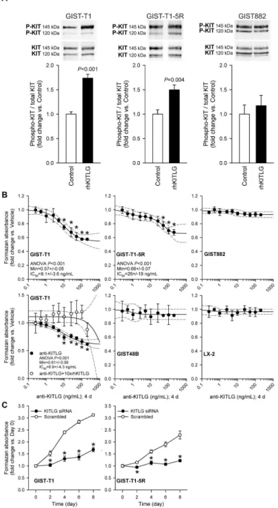
To investigate the role of autocrine/paracrine KITLG–KIT signaling in GIST proliferation, we examined the effect of KITLG immunoneutralization and RNAi-mediated knock-down in GIST cells expressing or lacking WT KIT. Control experiments verified the induction of KIT phosphorylation on Y7β1, the docking site for the p85 subunit of PIγK [1], by exogenous KITLG in GIST-T1 cells containing a heterozygous activating KIT mutation (Figure 1A; see reagent concentrations, exposure times, replicate numbers and other statistical details in the figures and their legends). KIT phosphorylation was also increased, albeit to a lesser degree, in GIST-T1-5R cells, derivatives of GIST-T1 cells containing an additional, imatinib-resistant KIT mutation. In contrast, no ligand-dependent KIT phosphorylation was detected in GIST88β cells, which are homozygous for the activating KITK64βE mutation. Culturing with
Figure 1. Contribution of KITLG-activated KIT signaling to baseline proliferation of GIST and LX-2 cells. A, KIT Y7β1 phosphorylation was activated by exogenous rhKITLG (100 ng/mL for 10 min following β h serum deprivation [1γ,γ5]) in GIST-T1 cells containing a heterozygous activating KIT mutation (n=4/group) and in GIST-T1-5R cells containing an additional, imatinib-resistant KIT mutation (n=6/group), but not in GIST88β cells lacking a WT KIT allele (n=5/group). B, Culturing with anti-human KITLG neutralizing antibody for 4 days inhibited baseline proliferation of GIST-T1 cells (P<0.001; n=γ; regression and 95% confidence band are shown as solid and dashed lines, respectively) and GIST-T1-5R cells (P<0.001; n=6). The effect of KITLG immunoneutralization on GIST-T1 cells was blocked by preabsorbing the antibody with 10-fold molar excess of rhKITLG (see open symbols in the left panel, second row; n=γ/group). KITLG immunoneutralization did not inhibit the proliferation of GIST88β or GIST48B cells lacking a WT KIT allele (note that GIST48B cells also express very low to undetectable levels of KIT protein, see Figure S1E) and of LX-β cells lacking KIT protein (Figure S1E) (n=γ/cell line). C, Inhibition of the proliferation of T1 and GIST-T1-5R cells by siRNA-mediated knock-down of KITLG (n=4/cell line/group; PGIST-T1: day β: 0.008, days 4, 6 and 8: <0.001; PGIST-T1-5R:
day β: <0.0β, day 4: 0.004, days 6 and 8: <0.001). doi: 10.1γ71/journal.pone.00768ββ.g001
ng/mL) and GIST-T1-5R cells by ~γ4% (IC50: ~β6 ng/mL)
(Figure 1B), whereas the same treatment had no effect on KIT+
GIST88β and KITlow/– GIST48B cells lacking WT KIT allele or on
KIT- LX-β cells. The effect of KITLG immunoneutralization on
GIST-T1 cells was prevented by preabsorbing the antibody with 10-fold molar excess of rhKITLG (Figure 1B). The proliferation of both GIST-T1 and GIST-T1-5R cells could also be inhibited by RNAi targeting KITLG (Figure 1C). These findings provide direct evidence that activation of KIT signaling by endogenous KITLG contributes to the proliferation of GIST cells expressing WT receptors.
IGF1 stimulates Kitl protein expression
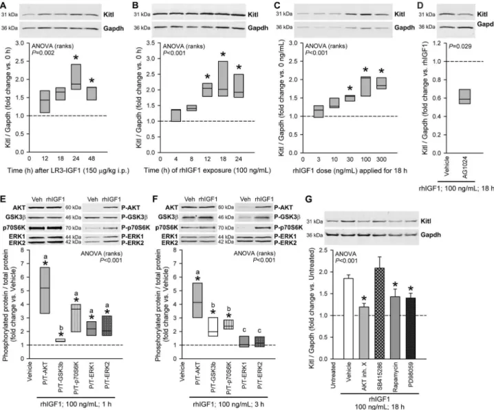
To investigate the direct effects of IGF1 on Kitl expression, we first administered 150 µg/kg LRγ-rhIGF1, a potent IGF1 analog with reduced affinity for IGF-binding proteins, to 14-16-day-old BALB/c mice in a single i.p. injection and measured Kitl protein in the gastric tunica muscularis by Western blotting. Kitl expression increased in a time-dependent manner, with maximum effect occurring at β4 h (Figure βA). In short-term cultures of intact gastric corpus+antrum muscles from 14-16-day-old BALB/c mice, 100 ng/mL rhIGF1 caused maximum stimulation of Kitl protein between 1β and β4 h (Figure βB). This effect was dose-dependent, plateauing between 100 and γ00 ng/mL (Figure βC). The upregulation of Kitl expression seen in response to 100 ng/mL rhIGF1 applied for 18 h was also verified using Actb as loading control (Figure Sβ). Tyrphostin AG10β4, a specific inhibitor of IGF1R and insulin RTK activity [γ0], reduced the rhIGF1 effect on Kitl (Figure βD) indicating that it was likely mediated by IGF1R or insulin receptor/IGF1R heterodimers [15].
Multiple signaling pathways mediate IGF1-induced Kitl protein expression
To investigate the mechanisms of IGF1-induced Kitl expression, we first detected the phosphorylation of key IGF1 signaling intermediates [19,β0] in murine gastric muscles stimulated by exogenous IGF1 (Figure βE-F). rhIGF1 elicited time-dependent increase in the phosphorylation of AKT, GSKγ , p70S6K and ERK1/β: After 1 h, we detected increased activating phosphorylation on S47γ/S474/S47β of AKT isoforms, p70S6K phosphorylation on Tγ89 [19], elevated ERK1 and ERKβ phosphorylation on Tβ0β/Yβ04 and T185/ Y187, respectively, and increased inhibitory phosphorylation on S9 of GSKγ [45] (Figure βE). After γ h, ERK1/β phosphorylation returned to baseline but phosphorylated AKT, p70S6K and GSKγ remained elevated (Figure βF). We then probed the contribution of these intermediates to IGF1-induced Kitl expression by using pathway-specific inhibitors (Figure βG): rhIGF1-stimulated Kitl expression was reduced by AKT Inhibitor X [γ1]; by rapamycin, which selectively inhibits the activation of p70S6K by MTOR complex 1 [19]; and by PD98059, a selective MEK inhibitor [γβ]. SB415β86, a selective competitive inhibitor of ATP binding to GSKγα/ and functional mimic of AKT-mediated GSK-γ phosphorylation and inactivation [β7], only minimally increased Kitl expression beyond the near-maximal stimulation caused by IGF1. These
results indicate cooperation among several major IGF1 intermediate pathways in stimulation of Kitl expression.
GSK-3 inhibition (GSK3i) stimulates Kitl expression without directly activating pathways involved in cell growth and proliferation
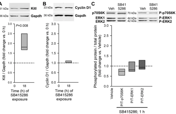
To better understand the role of GSKγi in the stimulation of Kitl expression, we evaluated the effects of SB415β86 in the absence of exogenous IGF1 in isolated murine gastric muscles. Under this condition, SB415β86 stimulated Kitl expression (Figure γA). Despite mimicking the effect of IGF1 on Kitl, SB415β86 did not stimulate the expression of cyclin D1, a key mediator of IGF1-induced cell cycle progression [15] (Figure γB), or the phosphorylation of p70S6K and ERK1/β (Figure γC) at time points when IGF1 effects on these parameters were prominent. We obtained similar results with SBβ1676γ, another ATP-competitive GSKγα/ inhibitor and TDZD-8, a non-ATP-competitive GSKγα/ inhibitor (Figures Sγ and S4). Thus, GSKγi alone specifically stimulates Kitl expression offering a pharmacological approach to increase Kitl/KITLG levels in gastrointestinal muscles without reproducing IGF1’s actions promoting cellular stress, aging and cancer [14,15].
IGF1 and GSK3i activate Kitl/KITLG transcription
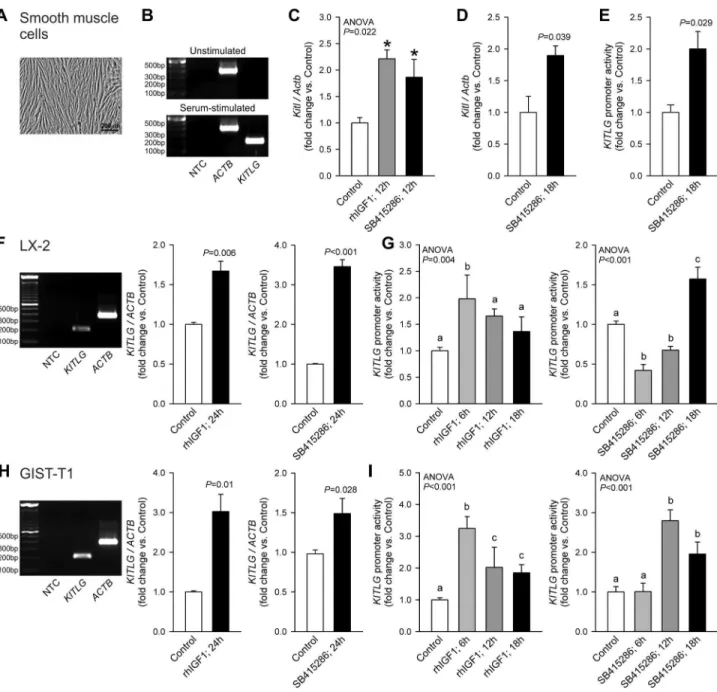
Next, we investigated the role of gene transcription in the IGF1 and GSKγi effects. In primary human smooth muscle cells (Figure 4A), KITLG mRNA was expressed in a serum-dependent fashion (Figure 4B). In intact murine smooth muscles, 1β-h exposure to rhIGF1 or SB415β86 upregulated
Kitl mRNA (Figure 4C). In primary murine smooth muscle cells, SB415β86 stimulated endogenous Kitl expression (Figure 4D) and activated transcription from an episomally expressed human KITLG promoter- luciferase construct (Figure 4E). In LX-β and GIST-T1 cells, both rhIGF1 and SB415β86 increased
KITLG mRNA (Figure 4F, H), although SB415β86 was more effective in LX-β cells and IGF1 had a greater effect in GIST-T1 cells. KITLG promoter activity was also increased by both IGF1 and SB415β86 (Figure 4G, I). In both cell lines, IGF1 displayed more rapid action on KITLG promoter activity than SB415β86. Nevertheless, these results indicate that the stimulation of Kitl protein expression by IGF1 and GSKγi primarily occurs at the transcriptional level and led us to investigate the nature of chromatin remodeling events that account for this effect.
Coordinated chromatin modifications underlie the activation of Kitl/KITLG transcription by IGF1 and GSK3i
Figure 2. Kitl protein expression is stimulated by IGF1 via multiple IGF1R-activated pathways. A, Time-dependent stimulation of Kitl expression in the gastric tunicamuscularis of 14-16-day-old BALB/c mice by the potent IGF1 analog LRγ-rhIGF1 administered in a single 150 µg/kg dose i.p.; n=5 mice/group. B, Time-dependent stimulation of Kitl expression by 100 ng/mL rhIGF1 in gastric corpus+antrum tunica muscularis organotypic cultures from 14-16-day-old BALB/c mice; n=4-5/group. C, Concentration-dependent stimulation of Kitl protein expression by 18-h rhIGF1 treatment; n=7 organotypic cultures/group. D, Blockade of the rhIGF1-induced Kitl expression in organotypic cultures by AG10β4 (1 µM), a specific inhibitor of IGF1/insulin receptor tyrosine kinase activity; n=4/group. E-F, Effects of 1-h (E) and γ-h (F) rhIGF1treatment on AKT (S47γ/S474/S47β), GSKγ (S9), p70S6K (Tγ89) and ERK1/β (Tβ0β/Yβ04 and T185/Y187) phosphorylation; n=5 organotypic cultures/group. G, Blockade of rhIGF1-induced Kitl expression in organotypic cultures by specific inhibitors of AKT (AKT Inhibitor X; 150 µM), MTOR–p70S6K (rapamycin; 1.5 nM) and MEK–ERK (PD98059; 50 µM); n=5-16/group. The GSK-γ inhibitor SB415β86 (γ0 µM), which is expected to mimic, rather than inhibit, the effect of the IGF1-induced inhibitory phosphorylation of GSK-γ, had no significant effect. Kitl and Gapdh or total (T) and phosphorylated proteins (P) were simultaneously detected in the same samples by two-color immunofluorescence (Figure S1). Representative immunoblots show identical areas of the blots imaged at different wavelengths. Box plots show medians and interquartile ranges; bar graphs indicate means±SEM. Data were normalized to the control groups indicated in the panels (dashed lines). Groups marked by asterisk are different from the control group, and groups not sharing the same superscript letter are different from each other (P<0.05 by post-hoc multiple comparisons). IGF1 stimulated Kitl expression in gastric smooth muscles in vivo and in vitro in a time- and concentration-dependent manner by activating IGFR1 and the AKT– GSKγ, MTOR–p70S6K and ERK MAPK pathways.
doi: 10.1γ71/journal.pone.00768ββ.g00β
pharmacological inhibitors and RNAi in LX-β and GIST-T1 cells using KITLG expression as readout. We first studied the role of histone acetylation, which is almost invariably associated with transcriptional activation [46] (Figure 5). In murine gastric smooth muscles, both rhIGF-I and SB415β86 increased the occupancy of the Kitl promoter by H4ac (which may include acetylated K5, K8, K1β and K18 [47]) (Figure 5A), whereas increased occupancy by HγK9ac was only detected in response to GSKγi (Figure 5B). Consistent with these findings, inhibition of HDAC classes I-II with SAHA led to a dose-dependent increase in KITLG mRNA in LX-β cells (Figure 5C). The role of increased HγK9 acetylation in the GSKγi effect was supported by the dose-dependent reduction of SB415β86-induced increase in KITLG mRNA by the pγ00/PCAF HAT inhibitor garcinol (Figure 5D). In GIST-T1 cells, the transcriptional effects of HDAC inhibition could not be determined due to rapid cell death likely reflecting the apoptotic effects of acetylation and consequent loss of function of the KIT chaperone heat shock protein 90 [48].
Next, we examined whether histone methylation, a
biochemical mechanism associated with long-term
transcriptional memory, also contributes to the regulation of
Kitl/KITLG expression. First we studied the activating HγK4meβ mark, which binds enhancer and promoter regions and gene bodies of actively transcribed or transcriptionally poised but repressed, tissue-specific genes [49]. Whereas rhIGF1 increased the level of HγK4meβ on the Kitl promoter ~β.8-fold, SB415β86 only had a modest effect (Figure 6A). Since transcriptional activation mediated by HγK4 methylation is often antagonized by PcG complexes [50,51], we also measured the association with the Kitl promoter of the PcG HKMT EZHβ and the repressive HγKβ7meγ modification it catalyzes [50]. These experiments showed that rhIGF1 and SB415β86 reduced the presence of both EZHβ and the HγKβ7meγ mark on the Kitl promoter (Figure 6B-C). To mechanistically explore the functional impact of the PcG-mediated repression on Kitl/KITLG expression, we inhibited EZHβ activity with the indirect HKMT inhibitor Adox [β9]. Adox increased KITLG mRNA in GIST-T1 cells ~β.9-fold (Figure 6F) but had more modest, albeit statistically significant, effects in murine gastric smooth muscles and LX-β cells (Figure 6D-E).
Figure 3. GSK3i stimulates Kitl expression without activating cyclin D1 expression and p70S6K and ERK1/2 phosphorylation. The GSKγα/ inhibitor SB415β86 was applied to organotypic cultures of gastric corpus+antrum muscles from of 14-16-day-old BALB/c mice at γ0 µM. A, Effect of 18-h application of SB415β86 on Kitl expression; n=5/group. B, Effect of the same treatment on cyclin D1 expression; n=γ/group. C, Effects of 1-h exposure to SB415β86 on p70S6K and ERK1/β phosphorylation; n=γ/group. See Figure β for further details.
Figure 4. IGF1 and GSK3i stimulate Kitl/KITLG transcription. Results obtained in murine and human smooth muscle cells (A
-E), human LX-β stellate cells (F-G) and human GIST-T1 cells (H-I) are shown. A, Hoffman modulation contrast image of primary human gastric smooth muscle cells. B, KITLG mRNA (total: soluble+membrane-bound) was readily detectable in primary human smooth muscle cells (passage γ) maintained with Smooth Muscle Growth Medium-β containing insulin, hFGF-B, hEGF and 5% FBS (Lonza) but not in β4-h growth factor- and serum-deficient basal medium. C, Both IGF1 (100 ng/mL) and the GSKγα/ inhibitor SB415β86 (γ0 µM) stimulated Kitl expression in murine gastric tunicamuscularis organotypic cultures (n=γ/group). D-E, SB415β86 stimulated endogenous Kitl expression in murine primary gastric smooth muscle cells (D; n=γ/group) and KITLG transcriptional activity in the same cell type transfected with a KITLG promoter (-β1β0 bp to +407 bp)-pGLγ luciferase construct (E; n=γ/group). IGF1 (100 ng/mL; n=γ/group) and SB415β86 (γ0 µM; n=6/group) also increased endogenous KITLG mRNA expression in LX-β (F) and GIST-T1 cells (H) and stimulated KITLG promoter activity in a time-dependent fashion (LX-β: n=6-9/group; G; GIST-T1: n=γ-11/ group; I). Groups marked by asterisk are different from the control group, and groups not sharing the same superscript letter are different from each other (P<0.05 by post-hoc multiple comparisons).
doi: 10.1γ71/journal.pone.00768ββ.g004
These results indicate that PcG inhibits Kitl/KITLG expression under basal conditions.
We also studied the involvement HγK9 methylation in the regulation of Kitl/KITLG transcription. The best understood function of HγK9meβ/γ is stable gene silencing through heterochromatin formation [5β]. This process requires the binding of HP1 proteins and consequent recruitment of the HγK9me1/β HKMTs G9A and GLP and the HγK9meγ HKMT SUVγ9H1, which reinforce the silencing of genes located within the constitutive heterochomatin and initiate the repression of genes previously embedded in euchromatin [5γ]. In murine gastric muscles, we detected both HγK9meβ and HγK9meγ on the Kitl promoter (Figure 7A-B), and both marks were reduced by rhIGF1. GSKγi had less pronounced effects. In LX-β cells,
KITLG mRNA increased in response to RNAi targeting HP1 (CBXγ) but was unaffected by HP1α (CBX5) or HP1 (CBX1) knockdown (Figure 7C-D). These findings are consistent with HP1 ’s role in euchromatic silencing [5γ]. In response to the G9A/GLP inhibitor BIX-01β94 we also detected a dose-dependent increase in KITLG mRNA in LX-β cells, and a ~100-fold increase in GIST-T1 cells (Figure 7E-F). In LX-β cells,
KITLG expression was also dose-dependently stimulated by chaetocin, a specific inhibitor of SUVγ9H1 (Figure 7G). The transcriptional effects of chaetocin could not be assessed in GIST-T1 cells due severe cytotoxicity. These results reveal an important role for HP1 and HγK9meβ/γ in IGF1-mediated Kitl/ KITLG expression.
In summary, our data support the model that IGF1 promotes autocrine/paracrine Kitl/KITLG–KIT signaling in part via AKT-mediated GSKγi and coordinated chromatin modifications favoring increased Kitl/KITLG transcription, and that these effects can be uncoupled from the direct mitogenic actions of
IGF1. To allow Kitl/KITLG expression, IGF target cells must increase the levels of activating H4ac, HγK9ac and HγK4meβ marks and decrease the levels of the repressive marks HγKβ7meγ and HγK9meβ/γ. While IGF1 and GSKγi appeared to exert their effects via the same general mechanisms, they also showed differences in their actions. The results presented herein outline a novel RTK cross-talk that regulates ligand-induced KIT activation in both physiologic/developmental and oncogenic contexts.
Discussion
Adequate stimulation of the KIT receptor by its ligand Kitl/ KITLG is required for diverse developmental and physiological
processes such as hematopoiesis, pigmentation,
gametogenesis, spatial learning, cardiomyocyte differentiation and repair, vasculogenesis and angiogenesis, lung function and gastrointestinal motility [1,β]. In contrast, ligand-independent activation of KIT due to oncogenic mutations is associated with several types of cancer such as GIST, seminomas, acute myeloid leukemia, melanomas and systemic mastocytosis [1,6]. However, abnormal ligand-dependent signaling occurring in the absence of mutations and reflecting altered expression of KIT and/or Kitl/KITLG contributes to the pathogenesis of not only non-neoplastic disorders such as gastrointestinal dysmotilities and allergies [1,β1,ββ] but also various cancers including subsets of GIST, acute myeloid leukemia, small-cell lung carcinoma, breast and colorectal cancer, ovarian cancer and neuroblastoma [1,9,11-1γ]. Therefore, understanding the factors and mechanisms regulating Kitl/KITLG expression will likely provide novel therapeutic tools for both functional disorders and cancers. We
Figure 5. Role of histone acetylation in the activation of Kitl/KITLG transcription by IGF1 and GSK3i. A, Increased occupancy of the Kitl core promoter region by H4ac in response to 6-h rhIGF1 (100 ng/mL) and SB415β86 (γ0 µM) treatment in murine gastric smooth muscles. Only SB415β86 increased occupancy by HγK9ac (B). Representatives of two independent experiments, each performed in triplicates, are shown. C, Dose-dependent stimulation of KITLG expression in LX-β cells by β4-h treatment with the class I-II HDAC inhibitor SAHA (n=γ/group). D, Dose-dependent inhibition of the SB415β86-induced stimulation of KITLG expression by the pγ00/PCAF HAT inhibitor garcinol (β4 h) in LX-β cells (n=γ/group). Drugs were applied following β4-h serum deprivation. Groups marked by asterisk are different from the control group, and groups not sharing the same superscript letter are different from each other (P<0.05 by post-hoc multiple comparisons).
have previously shown that long-term IGF1 treatment of intact murine gastric tunica muscularis increases Kitl availability in part by stimulating the growth and survival of smooth muscle cells, the primary source of Kitl in this tissue [ββ]. Here we demonstrate that IGF1 directly stimulates Kitl/KITLG expression in gastrointestinal smooth muscles, the natural microenvironment for ICC and GIST [8,β1,ββ], in GIST cells and in LX-β cells derived from hepatic stellate cells [γγ], the presumed niche for KIT+ hepatic progenitors [54], inflammatory
cells [55] and hepatocellular carcinoma [β5].
Our study outlines a transcriptionally mediated autocrine/ paracrine loop between two distinct RTK signaling systems, the Kitl/KITLG–KIT and IGF–IGF1R systems. Locally produced and circulating IGF1 affects almost all tissues and plays key roles in the regulation of body size, skeletal acquisition, muscle mass, reproduction, metabolism and life/health span [56]. There is
also a strong positive correlation between circulating IGF levels and cancer risk and prognosis [14,15]. IGF1R signaling facilitates cell cycle progression mainly at the G1-S transition
reflecting increased cyclin D1 transcription and translation stimulated via the ERK MAPK and AKT–MTOR–p70S6K pathways, respectively [15]. As our data show, increased IGF1R activation (in part from autocrine IGF1; Figure S1G) can, in turn, increase Kitl/KITLG expression and thus potentially amplify its oncogenic potential in KIT-expressing cells and tissues. For example, in WT GIST, an IGFβ-IGF1R autocrine/paracrine loop sustained by overexpression of both the receptor and its ligand in the same tumor microenvironment [5,16,17,57,58] may activate a secondary autocrine/paracrine loop formed by KITLG and WT KIT leading to increased KIT phosphorylation (refs. 9,10,1γ and present results) and cell proliferation. Indeed, our data in the heterozygous KIT mutant
Figure 6. Role of trithorax- and polycomb-mediated histone modifications in IGF1- and GSK3i-induced activation of Kitl/ KITLG transcription. A, Increased occupancy of the Kitl core promoter by the trithorax group-mediated, activating HγK4meβ histone modification in response to 6-h rhIGF1 (100 ng/mL) and SB415β86 (γ0 µM) treatment in murine gastric smooth muscles. B
-C, Reduced occupancy of the Kitl core promoter by the PRCβ-mediated, repressive HγKβ7meγ histone modification (B) and by the PRCβ histone methyltransferase EZHβ (C) in response to rhIGF1 and SB415β86 in the same tissues. D-F, Stimulation of KITLG
expression by the indirect histone methyltransferase inhibitor Adox in murine gastric smooth muscles (D), LX-β cells (E) and GIST-T1 cells (F) (n=γ/group). Adox was applied for 7β h following β4-h serum deprivation. See further details in the legend to Figure 5. doi: 10.1γ71/journal.pone.00768ββ.g006
GIST-T1 cells indicate that autocrine/paracrine KITLG-mediated KIT signaling may account for ~40% of baseline proliferation even in cells with a secondary, imatinib-resistant mutation and can be inhibited with antibodies or RNAi targeting KITLG. Since imatinib preferentially targets mutant receptors [6,9], inhibiting the IGF–IGF1R and KITLG–KIT coupled autocrine/paracrine loops may be beneficial in GIST, which are in most cases heterozygous for a given KIT mutation and continue expressing WT KIT protein [5]. However, the contribution of this pathway to GIST growth in vivo remains to be established. Interestingly, even in GIST lacking WT KIT, anti-KIT antibodies inhibited tumor growth with an efficacy similar to what we observed in GIST-T1 cells following KITLG immunoneutralization, although the former effect was attributed to increased phagocytosis and stimulation of KIT degradation
rather than prevention of KITLG binding [59]. In hepatic stellate cells, KITLG expression stimulated by IGF1R signaling may facilitate the KIT-dependent recruitment of inflammatory cells in injury and fibrosis [55] and progenitor cells in hepatic failure [54], and may thus be associated with both disease progression and tissue regeneration. In gastrointestinal neuromuscular tissues, stimulation of Kitl/KITLG–KIT signaling could prevent or counter ICC loss associated with several motility disorders and conditions including diabetic gastroparesis [ββ,60] and aging [4γ,61], where reduced Kitl/ KITLG expression is a pathogenetic factor [ββ,4γ]. Since ICC differentiation cannot be supported solely by supplying free Kitl/ KITLG [β,β1], pharmacological interventions will likely require stimulation of local production of both soluble and membrane-associated Kitl/KITLG, which, as our data show, could be
Figure 7. Role of reduced H3K9 methylation in the activation of Kitl/KITLG transcription by IGF1 and GSK3i. A-B, Reduced occupancy of the Kitl core promoter by the repressive HγK9meβ (A) and HγK9meγ (B) histone modifications in response to 6-h rhIGF1 (100 ng/mL) and SB415β86 (γ0 µM) treatment in murine gastric smooth muscles. C-D, Probing the role of HP1 isoforms in transcriptional repression of KITLG in LX-β cells by siRNA- (CBX5: HP1α; β5 nM, 7β h; C) or shRNA-mediated knock-down (CBX1: HP1 ; CBXγ: HP1 ; γ0 µg plasmid, 7β h; D). Note activation of KITLG expression by shRNA-mediated knock-down of HP1 (n=γ/ group). E-F, Stimulation of KITLG expression by the G9A/GLP HγK9me1/β HKMT inhibitor BIX-01β94 in LX-β cells (E) and GIST-T1 cells (F) (n=γ/group). G, Stimulation of KITLG expression by the HγK9 HKMT inhibitor chaetocin in LX-β cells (n=γ/group). Drugs were applied for β4 h following β4-h serum deprivation. See further details in the legend to Figure 5.
accomplished by IGF1 administration or GSKγi. In summary, the crosstalk between the IGF and the Kitl/KITLG pathways revealed by our results is potentially important for understanding and controlling the mechanisms of cell proliferation in both neoplastic and functional gastrointestinal diseases such as GIST, liver disease and gastroparesis. Therefore, it is also important to discuss the information we have obtained on the pathway mediating signal transduction from the IGF1R to the nucleus to regulate Kitl/KITLG expression.
Our initial studies focused on the intracellular kinases that transduce signals downstream of IGF1R. In our models, Kitl/ KITLG expression could also be stimulated by inhibiting GSK-γ activity with both ATP-competitive and non-ATP-competitive inhibitors. These results were unexpected because IGF1 effects on Kitl appeared to involve overlapping actions of several major pathways including the ERK MAPK, AKT and MTOR–p70S6K pathways, whereas GSKγi stimulated Kitl expression without increasing ERK1/β or p70S6K phosphorylation and cyclin D1 expression at time points when IGF1 effects on these parameters were prominent. The potential translational significance of these findings is that GSKγi may allow the stimulation of KITLG expression e.g. in patients with ICC loss without concomitant activation of the pathways that mediate IGF1’s actions promoting cellular stress, aging and cancer [14,15]. While GSKγi may affect ERK1/β and MTOR–p70S6K at other time points, our results agree with Kuemmerle’s finding that in gastrointestinal smooth muscle cells, the AKT–GSKγi-mediated and p70S6K- and ERK1/β-dependent mechanisms are functionally segregated, with AKT-dependent signaling being involved only in anti-apoptotic but not in the proliferation-stimulating actions of IGF1 [19,β0]. Indeed, despite mediating the -catenin-stabilizing effect of WNT signaling, long-term GSKγi has been associated with reduced, rather than increased, cancer risk possibly due to the concomitant activation of forkhead transcription factors [β8], and numerous GSK-γ inhibitors are under investigation for their beneficial effects in diabetes, inflammation, central nervous system injuries, bipolar disorder, Alzheimer’s disease and cancer [45].
Subsequently, we investigated how the IGF1- and GSKγi-induced signaling activates nuclear events to bring about increased Kitl/KITLG expression. By measuring Kitl/KITLG
transcript levels and KITLG promoter activity we found that Kitl/ KITLG protein expression induced by IGF1 or GSKγi indeed reflected increased gene transcription. However, our data also revealed differences in the time-courses of the IGF1 and GSKγi effects along with preferential stimulation by IGF1 and GSKγi in GIST-T1 and LX-β cells, respectively. Therefore, we studied the role of several repressive and activating histone modifications, as well as the enzymes and regulatory factors responsible for their establishment by ChIP and pharmacologic and RNAi-mediated interventions. In selecting our ChIP targets we considered that both the mouse and human Kitl/KITLG
promoter contain a CpG island but are unlikely to be DNA-methylated, given that we detected significant RNA polymerase II occupancy, baseline expression and transcriptional activity in our models. We found that activation of Kitl expression in
murine gastric smooth muscles by both IGF1 and GSKγi elicited coordinated changes in the chromatin involving reduced and increased occupancy of the Kitl promoter by repressive and activating histone marks, respectively. These events involved the activating histone marks H4ac, HγK9ac, HγK4meβ, the repressive marks HγK9meβ/γ and HγKβ7meγ, as well as related enzymes and regulators such as EZHβ, G9A, GLP, SUVγ9H1, PCAF, HDACs and HP1 . Our data also indicated some differences between IGF1 and GSKγi-induced changes such as preference for HγK4meβ- and HγK9ac-mediated activation by IGF1 and GSKγi, respectively. The complexity of this system raises the question how these changes are orchestrated. There is strong evidence that the process of transcription itself regulates chromatin states [5β], and sequence-specific transcription factors, besides transiently modulating transcription, contribute to the recruitment of chromatin modifiers [6β]. For example, transcription factors activated in response to IGF1 signaling or GSKγi may bind to the promoter and initiate the recruitment and eviction of the chromatin modifying complexes that establish the activating and repressive marks, respectively [6γ]. IGF1 may activate transcription via ERK and p70S6K signaling [15], by promoting IGF1R subunit nuclear translocation and association with lymphoid enhancer-binding factor 1 [βγ] and via GSKγi. Physiological or pharmacological GSKγi can in turn lead to the loss of inhibitory phosphorylation of several transcription factors in a stimulus-, promoter- and cellular context-dependent manner [45]. Indeed, some of these GSK-γ-inhibited transcription factors have putative or verified binding sites in the promoter region of the KITLG gene [1]. Identification of the key transcription factors mediating the observed IGF1 and GSKγi effects on Kitl/KITLG expression is a requisite next step toward understanding the transcriptional mechanisms that couple IGF1-IGF1R and Kitl/KITLG–KIT RTK signaling in physiologic and oncogenic contexts.
Supporting Information
Figure S1. Kitl/KITLG and IGF1R protein expression and IGF1 secretionin murine gastric smooth muscles, human LX-2 stellate cells and GIST cells. A, Detection of Kitl in the lysate of gastric corpus+antrum muscles from a juvenile BALB/c mouse. The membrane was simultaneously probed with antibodies against Kitl and Gapdh (loading control) and appropriate fluorescent secondary antibodies. Note primary Kitl band at ~γ1 kDa and a weaker band at ~4γ kDa. The β1-kDa Kitl band was only borderline detectable. B, In human GIST cell lines, only the γ1-kDa KITLG band was detected. B, Validation of the Western immunoblotting method. The γ1-kDa Kitl band was also detected in lysates of Sl/Sl4 hematopoietic stromal
cells expressing mKitlβ48. No Kitl bands were detected in Sl/Sl4
into culture media by GIST-T1, GIST88β, GIST48B and LX-β cells. γ000 cells/well were plated into 96-well plates and cultured in the presence or absence of FBS (GIST-T1, GIST88β, LX-β: 10%; GIST48B: 15%) for 48 h. IGF1 was measured in the harvested cell-free media using the Quantikine Human IGF1 Immunoassay kit (DG100, R&D systems). Note differential responses to FBS.
(TIF)
Figure S2. Kitl protein expression is stimulated by IGF1.
Stimulation of Kitl expression, detected using Actb as reference, by 18-h treatment with 100 ng/mL rhIGF1 in gastric corpus+antrum tunica muscularis organotypic cultures from 14-16-day-old BALB/c mice (n=γ/group). Kitl and Actb were simultaneously detected in the same samples by two-color immunofluorescence. Representative immunoblots show identical areas of the blots imaged at different wavelengths. The degree of Kitl upregulation was statistically indistinguishable from the increase detected using Gapdh as loading control (1.89±0.β1-fold vs. β.07±0.15-fold, P=0.58γ). (TIF)
Figure S3. SB216763 stimulates Kitl expression without activating cyclin D1 expression and p70S6K and ERK1/2 phosphorylation. GSKγα/ inhibitor SBβ1676γ was applied to organotypic cultures of gastric corpus+antrum muscles from of 14-16-day-old BALB/c mice at γ µM. A, Effect of 18-h application of SBβ1676γ on Kitl expression; n=5/group. B, Effect of the same treatment on cyclin D1 expression; n=γ/ group. C, Effects of 1-h exposure to SBβ1676γ on p70S6K and ERK1/β phosphorylation; n=γ/group. See Figure β for further details.
(TIF)
Figure S4. TDZD-8 stimulates Kitl expression without activating cyclin D1 expression and p70S6K and ERK1/2 phosphorylation. The non-ATP-competitive GSKγα/ inhibitor TDZD-8 was applied to organotypic cultures of gastric corpus +antrum muscles from of 14-16-day-old BALB/c mice at 10 µM.
A, Effect of 18-h application of TDZD-8 on Kitl expression; n=6/ group. B, Effect of the same treatment on cyclin D1 expression;
n=γ/group. C, Effects of 1-h exposure to SBβ1676γ on p70S6K and ERK1/β phosphorylation; n=γ/group. See Figure β for further details.
(TIF)
Figure S5. Negative controls for the ChIP experiments.A, Low recovery of Kitl promoter DNA by ChIP performed in murine gastric smooth muscles with non-immune mouse IgG (mIgG) or anti-Gapdh antibody relative to input chromatin or ChIP with anti-RNA polymerase II (RNA pol II) antibody. B-C, Unchanged recovery of Kitl promoter sequence in response to 6-h rhIGF1 (100 ng/mL) and SB415β86 (γ0 µM) treatment in murine gastric smooth muscles following ChIP using mIgG (B) or anti-Gapdh antibody (C). Representatives of two independent ChIP experiments, each performed in triplicates, are shown.
(TIF)
Table S1. Antibodies used in Western immunoblotting studies.
(PDF)
Table S2. Antibodies used in chromatin immunoprecipitation (ChIP) studies.
(PDF)
Acknowledgements
We thank Dr. Gregory J. Gores (Division of Gastroenterology and Hepatology, Mayo Clinic, Rochester, MN) for granting us access to the LI-COR Odyssey Scanner.
Author Contributions
Conceived and designed the experiments: YH DTA SJG GAL RAU TO. Performed the experiments: YH DTA SJG KHA MRB GAL AJM. Analyzed the data: YH DTA SJG KHA MRB GAL GF RAU TO. Contributed reagents/materials/analysis tools: GAL AJM MLK KRS TT AG BPR JAF RAU. Wrote the manuscript: YH DTA SJG GAL GF RAU TO.
References
1. Lennartsson J, Rönnstrand L (β01β) Stem cell factor receptor/c-Kit: from basic science to clinical implications. Physiol Rev 9β: 1619-1649. doi:10.115β/physrev.00046.β011. PubMed: βγ07γ6β8.
β. Sanders KM, Ward SM (β007) Kit mutants and gastrointestinal physiology. J Physiol 578: γγ-4β. PubMed: 17095561.
γ. Majumdar MK, Feng L, Medlock E, Toksoz D, Williams DA (1994) Identification and mutation of primary and secondary proteolytic cleavage sites in murine stem cell factor cDNA yields biologically active, cell-associated protein. J Biol Chem β69: 1βγ7-1β4β. PubMed: 7507105.
4. Hirota S, Isozaki K, Moriyama Y, Hashimoto K, Nishida T et al. (1998) Gain-of-function mutations of c-kit in human gastrointestinal stromal tumors. Science β79: 577-580. doi:10.11β6/science.β79.5γ50.577. PubMed: 94γ8854.
5. Corless CL, Barnett CM, Heinrich MC (β011) Gastrointestinal stromal tumours: origin and molecular oncology. Nat Rev Cancer 11: 865-878. PubMed: ββ0894β1.
6. Antonescu CR (β011) The GIST paradigm: lessons for other kinase-driven cancers. J Pathol ββγ: β51-β61. PubMed: β11β5679.
7. Heinrich MC, Corless CL, Duensing A, McGreevey L, Chen CJ et al. (β00γ) PDGFRA activating mutations in gastrointestinal stromal tumors. Science β99: 708-710. doi:10.11β6/science.1079666. PubMed: 1β5βββ57.
8. Bardsley MR, Horváth VJ, Asuzu DT, Lorincz A, Redelman D et al. (β010) Kitlow stem cells cause resistance to Kit/platelet-derived growth
factor alpha inhibitors in murine gastrointestinal stromal tumors. Gastroenterology 1γ9: 94β-95β. doi:10.105γ/j.gastro.β010.05.08γ. PubMed: β06β1681.
9. Negri T, Bozzi F, Conca E, Brich S, Gronchi A et al. (β009) Oncogenic and ligand-dependent activation of KIT/PDGFRA in surgical samples of imatinib-treated gastrointestinal stromal tumours (GISTs). J Pathol β17: 10γ-11β. doi:10.100β/path.β450. PubMed: 1897γβ10.
11. Hirano K, Shishido-Hara Y, Kitazawa A, Kojima K, Sumiishi A et al. (β008) Expression of stem cell factor (SCF), a KIT ligand, in gastrointestinal stromal tumors (GISTs): a potential marker for tumor proliferation. Pathol Res Pract β04: 799-807. doi:10.1016/j.prp. β008.05.00β. PubMed: 1860ββββ.
1β. Bono P, Krause A, von Mehren M, Heinrich MC, Blanke CD et al. (β004) Serum KIT and KIT ligand levels in patients with gastrointestinal stromal tumors treated with imatinib. Blood 10γ: β9β9-β9γ5. doi: 10.118β/blood-β00γ-10-γ44γ. PubMed: 15070666.
1γ. Duensing A, Medeiros F, McConarty B, Joseph NE, Panigrahy D et al. (β004) Mechanisms of oncogenic KIT signal transduction in primary gastrointestinal stromal tumors (GISTs). Oncogene βγ: γ999-4006. doi: 10.10γ8/sj.onc.1β075β5. PubMed: 15007γ86.
14. Guevara-Aguirre J, Balasubramanian P, Guevara-Aguirre M, Wei M, Madia F et al. (β011) Growth hormone receptor deficiency is associated with a major reduction in pro-aging signaling, cancer, and diabetes in humans. Sci Transl Med γ: 70ra1γ. PubMed: β1γβ5617.
15. Samani AA, Yakar S, LeRoith D, Brodt P (β007) The role of the IGF system in cancer growth and metastasis: overview and recent insights. Endocr Rev β8: β0-47. PubMed: 169γ1767.
16. Tarn C, Rink L, Merkel E, Flieder D, Pathak H et al. (β008) Insulin-like growth factor 1 receptor is a potential therapeutic target for gastrointestinal stromal tumors. Proc Natl Acad Sci U S A 105: 8γ87-8γ9β. doi:10.107γ/pnas.080γγ8γ105. PubMed: 185508β9. 17. Janeway KA, Zhu MJ, Barretina J, Perez-Atayde A, Demetri GD et al.
(β010) Strong expression of IGF1R in pediatric gastrointestinal stromal tumors without IGF1R genomic amplification. Int J Cancer 1β7: β718-β7ββ. doi:10.100β/ijc.β5β47. PubMed: β016β57γ.
18. Williams KL, Fuller CR, Fagin J, Lund PK (β00β) Mesenchymal IGF-I overexpression: paracrine effects in the intestine, distinct from endocrine actions. Am J Physiol Gastrointest Liver Physiol β8γ: G875-G885. PubMed: 1βββγγ47.
19. Kuemmerle JF (β00γ) IGF-I elicits growth of human intestinal smooth muscle cells by activation of PIγK, PDK-1, and p70S6 kinase. Am J Physiol Gastrointest Liver Physiol β84: G411-G4ββ. PubMed: 1β444011.
β0. Kuemmerle JF (β005) Endogenous IGF-I protects human intestinal smooth muscle cells from apoptosis by regulation of GSK-γ beta activity. Am J Physiol Gastrointest Liver Physiol β88: G101-G110. doi: 10.115β/ajpgi.000γβ.β004. PubMed: 15β97β58.
β1. Lorincz A, Redelman D, Horváth VJ, Bardsley MR, Chen H et al. (β008) Progenitors of interstitial cells of Cajal in the postnatal murine stomach. Gastroenterology 1γ4: 108γ-109γ. doi:10.105γ/j.gastro.β008.01.0γ6. PubMed: 18γ95089.
ββ. Horváth VJ, Vittal H, Lörincz A, Chen H, Almeida-Porada G et al. (β006) Reduced stem cell factor links smooth myopathy and loss of interstitial cells of Cajal in murine diabetic gastroparesis. Gastroenterology 1γ0: 759-770. doi:10.105γ/j.gastro.β005.1β.0β7. PubMed: 165γ0517.
βγ. Warsito D, Sjöström S, Andersson S, Larsson O, Sehat B (β01β) Nuclear IGF1R is a transcriptional co-activator of LEF1/TCF. EMBO Rep 1γ: β44-β50. doi:10.10γ8/embor.β011.β51. PubMed: βββ61717. β4. Sokolović A, Sokolović M, Boers W, Elferink RP, Bosma PJ (β010)
Insulin-like growth factor binding protein 5 enhances survival of LXβ human hepatic stellate cells. Fibrogenesis Tissue Repair γ: γ. doi: 10.1186/1755-15γ6-γ-γ. PubMed: β016γ708.
β5. Coulouarn C, Corlu A, Glaise D, Guénon I, Thorgeirsson SS et al. (β01β) Hepatocyte-stellate cell cross-talk in the liver engenders a permissive inflammatory microenvironment that drives progression in hepatocellular carcinoma. Cancer Res 7β: β5γγ-β54β. doi: 10.1158/15γ8-7445.AMβ01β-β5γγ. PubMed: ββ419664.
β6. Ordög T, Redelman D, Horowitz NN, Sanders KM (β004) Immunomagnetic enrichment of interstitial cells of Cajal. Am J Physiol Gastrointest Liver Physiol β86: Gγ51-Gγ60. doi:10.115β/ajpgi. 00β81.β00γ. PubMed: 1456γ669.
β7. Cross DA, Culbert AA, Chalmers KA, Facci L, Skaper SD et al. (β001) Selective small-molecule inhibitors of glycogen synthase kinase-γ activity protect primary neurones from death. J Neurochem 77: 94-10β. doi:10.1046/j.1471-4159.β001.t01-1-00β51.x. PubMed: 11β79β65. β8. Zhou Y, Uddin S, Zimmerman T, Kang JA, Ulaszek J et al. (β008)
Growth control of multiple myeloma cells through inhibition of glycogen synthase kinase-γ. Leuk Lymphoma 49: 1945-195γ. doi: 10.1080/104β819080βγ04966. PubMed: 187β8964.
β9. Miranda TB, Cortez CC, Yoo CB, Liang G, Abe M et al. (β009) DZNep is a global histone methylation inhibitor that reactivates developmental genes not silenced by DNA methylation. Mol Cancer Ther 8: 1579-1588. doi:10.1158/15γ5-716γ.MCT-09-001γ. PubMed: 19509β60. γ0. Flynn RS, Mahavadi S, Murthy KS, Kellum JM, Kuemmerle JF (β009) Insulin-like growth factor-binding protein-5 stimulates growth of human
intestinal muscle cells by activation of Gαiγ. Am J Physiol Gastrointest Liver Physiol β97: G1βγβ-G1βγ8. doi:10.115β/ajpgi.00γβγ.β009. PubMed: 19808657.
γ1. Thimmaiah KN, Easton JB, Germain GS, Morton CL, Kamath S et al. (β005) Identification of N10-substituted phenoxazines as potent and specific inhibitors of Akt signaling. J Biol Chem β80: γ19β4-γ19γ5. doi: 10.1074/jbc.M507057β00. PubMed: 16009706.
γβ. Kuemmerle JF, Bushman TL (1998) IGF-I stimulates intestinal muscle cell growth by activating distinct PI γ-kinase and MAP kinase pathways. Am J Physiol β75: G151-G158. PubMed: 9655695.
γγ. Xu L, Hui AY, Albanis E, Arthur MJ, O’Byrne SM et al. (β005) Human hepatic stellate cell lines, LX-1 and LX-β: new tools for analysis of hepatic fibrosis. Gut 54: 14β-151. doi:10.11γ6/gut.β004.04β1β7. PubMed: 155915β0.
γ4. Taguchi T, Sonobe H, Toyonaga S, Yamasaki I, Shuin T et al. (β00β) Conventional and molecular cytogenetic characterization of a new human cell line, GIST-T1, established from gastrointestinal stromal tumor. Lab Invest 8β: 66γ-665. doi:10.10γ8/labinvest.γ780461. PubMed: 1β004007.
γ5. Lux ML, Rubin BP, Biase TL, Chen CJ, Maclure T et al. (β000) KIT extracellular and kinase domain mutations in gastrointestinal stromal tumors. Am J Pathol 156: 791-795. doi:10.1016/ S000β-9440(10)64946-β. PubMed: 1070βγ94.
γ6. Bauer S, Yu LK, Demetri GD, Fletcher JA (β006) Heat shock protein 90 inhibition in imatinib-resistant gastrointestinal stromal tumor. Cancer Res 66: 915γ-9161. doi:10.1158/0008-547β.CAN-06-0165. PubMed: 1698β758.
γ7. Kitamura T, Tange T, Terasawa T, Chiba S, Kuwaki T et al. (1989) Establishment and characterization of a unique human cell line that proliferates dependently on GM-CSF, IL-γ, or erythropoietin. J Cell Physiol 140: γβγ-γγ4. doi:10.100β/jcp.1041400β19. PubMed: β66γ885. γ8. Kubicek S, O’Sullivan RJ, August EM, Hickey ER, Zhang Q et al. (β007) Reversal of HγK9meβ by a small-molecule inhibitor for the G9a histone methyltransferase. Mol Cell β5: 47γ-481. doi:10.1016/j.molcel. β007.01.017. PubMed: 17β8959γ.
γ9. Shinkai Y, Tachibana M (β011) HγK9 methyltransferase G9a and the related molecule GLP. Genes Dev β5: 781-788. doi:10.1101/gad. β0β7411. PubMed: β1498567.
40. Greiner D, Bonaldi T, Eskeland R, Roemer E, Imhof A (β005) Identification of a specific inhibitor of the histone methyltransferase SU(VAR)γ-9. Nat Chem Biol 1: 14γ-145. doi:10.10γ8/nchembio7β1. PubMed: 16408017.
41. Balasubramanyam K, Altaf M, Varier RA, Swaminathan V, Ravindran A et al. (β004) Polyisoprenylated benzophenone, garcinol, a natural histone acetyltransferase inhibitor, represses chromatin transcription and alters global gene expression. J Biol Chem β79: γγ716-γγ7β6. doi: 10.1074/jbc.M40β8γ9β00. PubMed: 15155757.
4β. Marks PA, Breslow R (β007) Dimethyl sulfoxide to vorinostat: development of this histone deacetylase inhibitor as an anticancer drug. Nat Biotechnol β5: 84-90. doi:10.10γ8/nbt1β7β. PubMed: 17β11407.
4γ. Izbeki F, Asuzu DT, Lorincz A, Bardsley MR, Popko LN et al. (β010) Loss of Kitlow progenitors, reduced stem cell factor and high oxidative
stress underlie gastric dysfunction in progeric mice. J Physiol 588: γ101-γ117. doi:10.111γ/jphysiol.β010.1910βγ. PubMed: β058104β. 44. Asuzu DT, Hayashi Y, Izbeki F, Popko LN, Young DL et al. (β011)
Generalized neuromuscular hypoplasia, reduced smooth muscle myosin and altered gut motility in the klotho model of premature aging. Neurogastroenterol Motil βγ: eγ09-eγβγ. doi:10.1111/j. 1γ65-β98β.β011.017γ0.x. PubMed: β1605β85.
45. Beurel E, Michalek SM, Jope RS (β009) Innate and adaptive immune responses regulated by glycogen synthase kinase-γ (GSKγ). Trends Immunol γ1: β4-γ1. PubMed: 198γ6γ08.
46. Kouzarides T (β007) Chromatin modifications and their function. Cell 1β8: 69γ-705. doi:10.1016/j.cell.β007.0β.005. PubMed: 17γβ0507. 47. Kouzarides T (β007) SnapShot: Histone-modifying enzymes. Cell 1γ1:
8ββ. doi:10.1016/j.cell.β007.11.005. PubMed: 180ββγ74.
48. Mühlenberg T, Zhang Y, Wagner AJ, Grabellus F, Bradner J et al. (β009) Inhibitors of deacetylases suppress oncogenic KIT signaling, acetylate HSP90, and induce apoptosis in gastrointestinal stromal tumors. Cancer Res 69: 6941-6950. doi: 10.1158/0008-547β.CAN-08-4004. PubMed: 19706776.
49. Pekowska A, Benoukraf T, Ferrier P, Spicuglia S (β010) A unique HγK4meβ profile marks tissue-specific gene regulation. Genome Res β0: 149γ-150β. doi:10.1101/gr.109γ89.110. PubMed: β08414γ1. 50. Sauvageau M, Sauvageau G (β010) Polycomb group proteins:
multi-faceted regulators of somatic stem cells and cancer. Cell Stem Cell 7: β99-γ1γ. doi:10.1016/j.stem.β010.08.00β. PubMed: β0804967.