Article
J. Braz. Chem. Soc., Vol. 25, No. 7, 1283-1291, 2014. Printed in Brazil - ©2014 Sociedade Brasileira de Química
0103 - 5053 $6.00+0.00
A
*e-mail: [email protected]
Monitoring of Diclofenac with Biomimetic Sensor in Batch and FIA Systems
Ademar Wong,* Luiz D. Marestoni and Maria D. P. T. Sotomayor
Departamento de Química Analítica, Instituto de Química, Universidade Estadual Paulista (UNESP), 14801-970 Araraquara-SP, Brazil
O presente trabalho visa o desenvolvimento de um sensor biomimético para detecção de diclofenaco de sódio usando um eletrodo de carbono vítreo modificado com complexo 1,4,8,11,15,18,22,25-octabutoxi-29H,31H-ftalocianina de cobre(II) e nanotubos de carbono de parede múltipla funcionalizados (MWCNT-COOH). O sensor proposto foi avaliado em sistema em batelada usando tampão fosfato 0,1 mol L-1 (pH 7,5) como eletrólito de suporte. Uma
resposta linear foi obtida na faixa entre 4,0 × 10⁻⁵ e 6,7 × 10⁻⁴ mol L-1, com uma sensibilidade de
6,1 × 10⁵ (± 1,5 × 10⁴) µA L mol-1 e limite de detecção de 1,3 × 10⁻⁵ mol L-1 usando voltametria
de onda quadrada. A seletividade foi estudada através da resposta do sensor na presença de outros fármacos. O sensor foi aplicado em formulações farmacêuticas e também em amostras de soro biológico através de estudos de recuperação. Quando acoplado a um sistema por injeção em fluxo (FIA), os melhores resultados foram obtidos aplicando o potencial de 0,5 V vs. Ag|AgCl|KClsat,
usando um volume de amostra injetado de 100 µL e uma vazão de 1,2 mL min-1. A faixa
linear de resposta em fluxo foi entre 5,0 × 10⁻⁵ e 5,0 × 10⁻³ mol L-1, com uma sensibilidade de
1,6 × 10⁴ (± 3,0 × 10²) µA L mol-1 e um desvio padrão relativo de 3.5%.
The present work aims the development of a biomimetic sensor for the detection of sodium diclofenac from a glassy carbon electrode modified with copper(II)
1,4,8,11,15,18,22,25-octabutoxy-29H,31H-phthalocyanine complex and functionalized multi-walled carbon nanotubes
(MWCNT-COOH). The proposed sensor was evaluated in batch system using 0.1 mol L-1
phosphate buffer solution at pH 7.5 as electrolyte support. A linear response was obtained in the range of 4.0 × 10⁻⁵ and 6.7 × 10⁻⁴ mol L-1, with the sensitivity of 6.1 × 10⁵ (± 1.5 × 10⁴) µA L mol-1
and detection limit of 1.3 × 10⁻⁵ mol L-1 using square wave voltammetry. The selectivity was
studied by the sensor response in the presence of other drugs. The sensor was applied to pharmaceutical formulations and also to biological serum samples by recovery studies. When coupled to a flow injection system (FIA), the best results were obtained by applying the potential of 0.5 V vs. Ag|AgCl|KClsat, using an injected volume of the sample of 100 µL and a flow of
1.2 mL min-1. The linear response range in flow was between 5.0 × 10⁻⁵ and 5.0 × 10⁻³ mol L-1,
with a sensitivity of 1.6 × 10⁴ (± 3.0 × 10²)µA L mol-1and a relative standard deviation of 3.5%.
Keywords: biomimetic catalyst, copper(II) 1,4,8,11,15,18,22,25-octabutoxy-29H,31H -phthalocyanine, diclofenac, flow injection system, MWCNT-COOH, glassy carbon electrode
Introduction
Over the last decade there has been an increasing awareness of problems associated with the presence of pharmaceutical compounds in the aquatic environment, where the continuous inputs and persistence of many of these substances can cause irreversible damage to biota. The pollution of water by pharmaceutical drugs is a global phenomenon, affecting not only rivers and underground waters, but also drinking water, which even after treatment
is likely to remain contaminated with contraceptive, anti-inflammatory, antidepressant and other types of drugs.1
in surface waters, rivers and lakes.2,3 Diclofenac and its degradation product, acting on their own, may present no health risk but which cumulatively may cause undesirable side-effect, allergies and toxicity, specially in co-ocurrence with others substances.4,5 There is evidence that prolonged exposure to environmentally relevant concentrations of diclofenac leads to impairment of the general health of fishes, inducing renal lesions and alteration of the gills, at the lowest observed effect concentration (LOEC) of 5 µg L-1.6
The development of an analytic method for detecting the presence of diclofenac is necessary for monitoring the drug in the environment. A considerable number of methods have been employed for the analysis of diclofenac, such as capillary electrophoresis,7 high-perfomance liquid chromatography (HPLC),8 electrochemistry9 and spectrophotometry.10 Compared with other methods, electrochemical methods are characterized by being more simple, sensitive, with economical portability, simplicity, minimal cost and short analysis time.11,12
Chemically modified electrodes based on porphyrins and phthalocyanines complexes have been used as an alternative analysis method to obtain electrochemical sensors with high sensitivity, selectivity and stability.12-14 This type of sensors has been named in the literature as biomimetic sensors. The concept of biomimetic sensors relies on the recognition of substrates through the use of “artificial enzymes”.15
The biomimetic sensor in this work is based on mechanisms of molecular recognition of enzyme substrates using artificial enzymes that imitate the active site of a redox enzyme, P450 enzyme. The P450 enzymes are present in living organisms including mammals, bacteria, fungi, insects, and fish, where these enzymes catalyze numerous chemical reactions in organisms, usually producing metabolites that are physiologically essential or beneficial.16,17 These reactions include the degradation of xenobiotics such as pesticides and endocrine disruptors, as well as the metabolism of drugs and exogenous hormones.17
The P450 enzymes belong to the heme protein group, and possess a common active site, iron protoporphyrin IX (Figure 1), which is responsible for the catalytic activity of the enzyme.
The use of complex base phthalocyanine and porphyrin is a very viable alternative to imitate the active site of the P450 enzyme. The products of the redox reactions should be similar to those obtained using an enzyme, although they do not necessarily follow the same process of reaction. When immobilized on the electrode surface, this complex is expected to improve the electron transfer between the electrode/active site (biomimetic catalyst/substrate), thereby allowing greater sensitivity and selectivity at low redox potentials.18,19
Several articles were found in the literature on complexes such as iron(II) phthalocyanine bis(pyridine),19 cobalt phthalocyanine20 and iron(III) phthalocyanine chloride,21 which have been satisfactorily used as electrochemical modifiers in biomimetic sensors for the monitoring of environmental emergent pollutants, such as drug,22 hormone23 and pesticide.24
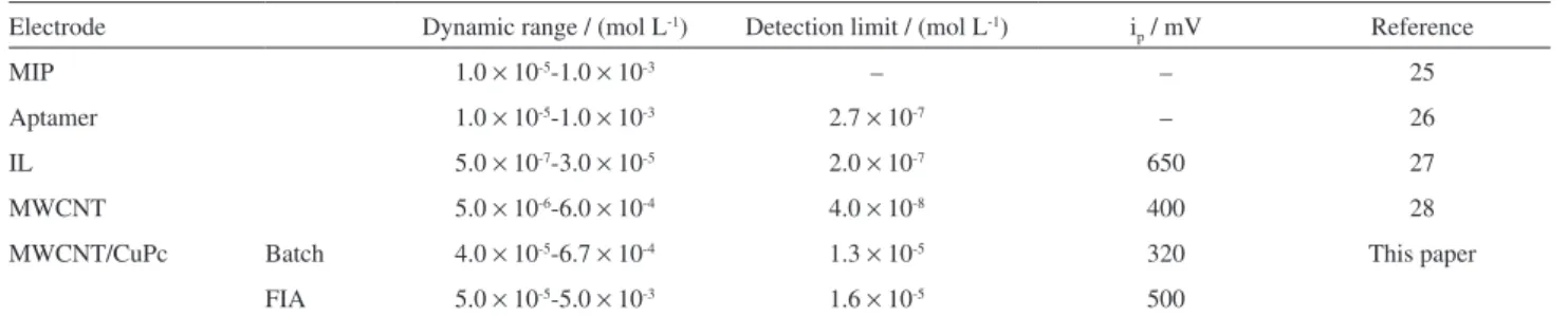
Table 1 shows a comparison of the proposed sensor with different kinds of electrochemical sensors that have been employed for the determination of diclofenac.25-28
The proposed sensor showed linear response range similar to other electrochemical sensors. Its advantages lie in its lower analysis potential, which allows an increase in its selectivity and the possibility to conduct analyses in situ and in real time by the flow injection analysis (FIA) system.
Figure 1. Chemical structure of iron protoporphyrin IX.
Table 1. Electrochemical sensors used for the detection of diclofenac
Electrode Dynamic range / (mol L-1) Detection limit / (mol L-1) i
p / mV Reference
MIP 1.0 × 10⁻⁵-1.0 × 10⁻³ – – 25
Aptamer 1.0 × 10⁻⁵-1.0 × 10⁻³ 2.7 × 10⁻⁷ – 26
IL 5.0 × 10⁻⁷-3.0 × 10⁻⁵ 2.0 × 10⁻⁷ 650 27
MWCNT 5.0 × 10⁻⁶-6.0 × 10⁻⁴ 4.0 × 10⁻⁸ 400 28
MWCNT/CuPc Batch 4.0 × 10⁻⁵-6.7 × 10⁻⁴ 1.3 × 10⁻⁵ 320 This paper
FIA 5.0 × 10⁻⁵-5.0 × 10⁻³ 1.6 × 10⁻⁵ 500
The concept of FIA was proposed by Ruzicka and Hansen in 1975 as a new concept for chemical analysis. This mode of analysis has been exploited with success and provides numerous advantages, such as versatile use of instrumentation, low cost of the components of the system, high frequency sampling, higher sensitivity and selectivity, reduced consumption of reagents and consequently reduction in waste generation. In these analyses the risk of exposure of the analyst is low when compared with other analysis methodologies. FIA refers to all analytical techniques that are based on the introduction, processing, and detection of liquid samples in flow.29
The aim of this work is to develop a biomimetic sensor using copper(II) 1,4,8,11,15,18,22,25-octabutoxy-29H,31H-phthalocyanine complex and multi-walled carbon nanotubes (MWCNT) for the detection of diclofenac (Figure 2) in batch and FIA systems. This article will further provide description regarding the application of biomimetic sensor in batch and FIA systems, its optimization and application as an alternative methodology for the analysis of diclofenac in pharmaceutical drugs and human serum.
Experimental
Reagents
The reagents used in this work are of analytical or HPLC grade, and standard solutions of sodium diclofenac (1.0 × 10⁻³ mol L-1) were prepared by dissolving 7.4 mg
and diluting to 25 mL with deionized water. Sodium diclofenac, copper(II) 1,4,8,11,15,18,22,25-octabutoxy, N,N-dimethylformamide (DMF) were acquired from Sigma-Aldrich, NaOH and phosphate buffer solution (PBS) from Synth and functionalized multi-walled carbon nanotubes from Dropsens®.
Apparatus
The voltammetry measurements were performed initially in batch system with a micro-Autolab type III (Autolab/Eco Chemie) potentiostat, fitted with an electrochemical cell containing 3 electrodes, commercial Ag|AgCl|KClsat (Analion®) as the reference electrode, a
platinum spiral wire as the counter electrode and glassy carbon electrode with area of 0.19 cm2 as working electrode (WE).
The electrochemical measurements in FIA system were performed using a PalmSense potentiostat (Palm Instruments BV, The Netherlands), interfaced with a microcomputer for potential control and data acquisition. For coupling to a flow system, the biomimetic sensor was inserted into a flow-through wall-jet amperometric cell, and used as the working electrode (Supplementary Information Figure S1). A homemade Ag|AgCl|KClsat electrode was used as the reference, and a platinum wire as the auxiliary electrode. The electrodes were connected to the PalmSense potentiostat. The flow rate of the 0.1 mol L-1 phosphate buffer carrier solution was controlled using an Ismatec® peristaltic pump, and the standards and samples containing diclofenac were injected into the carrier using a sliding central bar sampling valve.
Preparation of the modified glassy carbon electrode
Initially, the surface of the glassy carbon electrode was polished with alumina (1, 0.5 and 0.1 µm) using a polishing machine (Fortel). The electrode was rinsed with deionized water and placed in the ultrasonic apparatus for one minute to remove residual alumina that may have been incorporated onto the electrode surface. After that, the electrode was immersed in a 0.1 mol L-1 solution of phosphate buffer (pH 7.0) and was further bubbled with N2 for 10 minutes in an electrochemical cell prior to the voltammetric measurements. A solution of K3Fe(CN)6 and K4Fe(CN)6 was then added, where verification took place afterwards regarding the difference between the potentials of the anodic and cathodic peaks, which was found to be less than 100 mV, indicating that the cleaning was indeed satisfactory.30
The film synthesis was performed with 2.0 mg MWCNT-COOH and 1.0 mg copper(II) 29H,31H-phthalocyanine, 1,4,8,11,15,18,22,25-octabutoxy mixed with 1 mL of DMF and sonificated for 20 minutes.31 Twenty µL of the solution was then placed on the surface of the glassy carbon electrode and drying occurred at room temperature.
Functionalization of multi-walled carbon nanotubes
The functionalization method consisted of mixing 100 mg of multi-walled carbon nanotubes in sulfonitrile solution (3 H2SO4:1 HNO3). This mixture was then stirred for 12 hours and was subsequently washed with ultra-pure water until reaching pH 7.0. Afterwards, the MWCNT was heated at 70 °C for 12 hours to enable the drying of the material.32,33
Study of selectivity
The selectivity of the proposed sensor was observed based on the analysis of seven drugs (secnidazol, piroxicam, ranitidine, ciprofloxacin, lidocaine, tetracycline and diclofenac) under optimized analysis conditions. Drug solutions were prepared with deionized water in concentration of 2.0 × 10⁻⁴ mol L-1 for electrochemical
analysis in square wave voltammetry (SWV).
Application of the biomimetic sensor in human serum samples
To determine the presence of diclofenac in a human serum sample, solutions containing 2.5 mL of untreated serum samples and 2.5 mL of diclofenac in the concentration of 3.0 × 10⁻⁴ mol L-1 were prepared. Conventional filtration
of the samples was performed to remove coarse insoluble substances. Square wave voltammetry (SWV) was used for the determination of diclofenac in the samples in order to verify a possible matrix effect.34 The analyses were carried out in batch system and the results were compared with HPLC.
Application of the biomimetic sensor in pharmaceutical samples
The quantification of diclofenac in pharmaceutical formulation was carried out by SWV. For the preparation of each sample solution, five tablets were ground and homogenized in a mortar, then a suitable amount of triturated sample was weighed out and dissolved in deionized water, followed by a filtration step in order to remove any insoluble substances present in the tablets.19
A stock solution of 100 mL of standard diclofenac in the concentration 1.0 × 10⁻² mol L-1 was prepared for analysis
in batch and FIA systems as well as HPLC.
Analysis of samples in HPLC
The chromatographic analyses were performed using a Shimadzu Model 20A liquid chromatograph, coupled to an SPD-20A UV-Vis detector, an SIL-20A autosampler and a DGU-20A5 degasser. The chromatography system was controlled by a microcomputer and the C18 column (250 mm × 4.6 m) was used in the analysis. The chromatographic conditions used were: mobile phase composed of a mixture of acetonitrile:water deionized:formic acid in the ratio 65:34.5:0.5, flow rate of 1.0 mL min-1, sample injection volume of 10 µL and wavelength of 225 nm.35
Results and Discussion
Electrochemical detection of diclofenac in biomimetic sensor
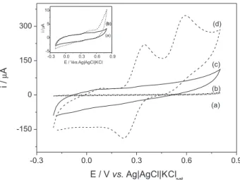
The electrochemical detection of diclofenac using the biomimetic sensor modified with MWCNT-COOH was carried out by the cyclic voltammetry technique as shown in Figure 3. The cyclic voltammetry exhibits behavior related to the irreversible anodic oxidation of the diclofenac in the potential of 600 mV. The sweep was initiated in the positive direction (–0.2 to 0.9 V vs. Ag|AgCl|KClsat). When the reverse sweep occurred, an anodic peak at 320 mV and a cathodic peak at 220 mV were found to appear, featuring a reversible system. The explanation concerning these peaks can be said to be as a result of the strong adsorption of the reaction products of diclofenac on the electrode surface, which was strongly favored with the use of MWCNT.36 These peaks are best visualized when compared with the electrochemical signal of the electrolyte.
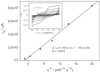
In Figure S2, it can be seen in the cyclic voltammogram graph that the change in the anodic current is a linear function of the square root of the scan rate, in the range between 10 and 400 mV s-1, indicating that the oxidation of diclofenac on the surface of the sensor at the 320 mV potential is controlled by a diffusion process.37
The electroactive area of the modified glassy carbon electrode (GCE) was estimated in 0.1 mol L-1 KCl in the presence of 1.0 × 10⁻³ mol L-1 [Fe(CN)6]4− (data not shown)
according to the Randles-Sevick equation:37
ip = 2.695n3/2AD1/2Cν1/2 (1)
where C is the [Fe(CN)6]4− concentration in bulk solution (1.0 × 10⁻⁶ mol cm-3); D is the diffusion coefficient of
[Fe(CN)6]4− in solution (7.6 × 10⁻⁶ cm2 s-1) and A is the electroactive area (cm2).
The values obtained for the electroactive areas of the modified GCE and the bare GCE were of 0.3 and 0.9 cm2, respectively. It can be seen that the modification of the electrode surface with MWCNT-COOH promoted a three-fold increase in area compared to the bare electrode.
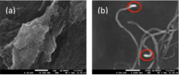
The analyses of the surface morphology of the biomimetic sensor modified with MWCNT-COOH were registered using scanning electron microscopy (SEM) imaging in a JEOL JSM 7500F scanning electron microscope. Figure 4a shows SEM image of copper(II) 29H,31H -phthalocyanine-1,4,8,11,15,18,22,25-octabutoxy and MWCNT-COOH and Figure 4b depicts the functionalization of the nanotubes with carboxylic group which is present at the extremities of this material.
Copper (Cu) atoms that constitute the complex molecule were observed in the analysis of energy-dispersive X-rays (EDX). The C and O are from the MWCNT and Si is from the substrate (Figure S3).
Verification of biomimetic character
The biomimetic character that is similar to theenzymatic characterwas checked using SWV response profile. The hyperbolic profile obtained (inset in Figure 5), is characteristic of enzymatic biosensors and biomimetic sensors, which is in agreement with Michaelis-Menten kinetics.18,38 A Lineweaver-Burk graph (Figure 5) was constructed in order to calculate the apparent Michaelis-Menten constant (KMM
app) for diclofenac for the proposed sensor. The value of 1.2 × 10⁻³ mol L-1 indicates the degree
of affinity between the catalyst (copper complex) and the analyte. This (KMM
app) value is higher when compared with the Km of the P450 enzyme (CYP 2D6), which was 4.3 × 10⁻⁶ mol L-1. These values indicate the affinity of
the enzyme for diclofenac, which is greater than the
biomimetic complex, although satisfactory results with this biomimetic compound were visualized in electrochemical measurements.39,40
Study of diclofenac in SWV
A square wave voltammetry (SWV) study was performed to verify the response profile of the unmodified and modified electrode. The optimization of the parameters of SWV can be found in Table 2.
In Figure 6, the anodic peak current of diclofenac on the electrode modified with MWCNT-COOH at 360 mV, and the response with the electrode modified with MWCNT-COOH and copper(II) 1,4,8,11,15,18,22,25-octabutoxy-29H,31H -phthalocyanine at 320 mV can be observed. The modification of the electrode promoted an electrocatalysis, slightly decreasing the redox potential while increasing the electrochemical response (electrocatalysis), contributing to the improvement of selectivity and sensitivity of the biomimetic sensor.
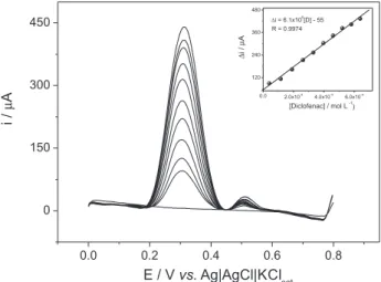
The SWV technique was employed in order to obtain the best limits of detection and quantification. Figure 7 shows the SWV obtained with the biomimetic sensor for different diclofenac concentrations. Using SWV, the sensor
Figure 4. SEM image of the MWCNT-COOH and copper(II) complex on the surface of the glassy carbon electrode (a) and MWCNT with carboxylic group highlighted in red (b).
Figure 5. Lineweaver-Burk plot obtained with the biomimetic sensor in the oxidation of diclofenac.
Table 2. Optimized parameters of square wave voltammetry (SVW)
Frequency / Hz
ipa / µA
Amplitude / mV
ipa / µA
Increment / mV
ipa / µA
10a 212 25 42 2 184
20 137 50 75 4a 220
40 77 100a 132 6 200
60 16 150 130 10 180
showed a linear response for diclofenac concentrations between 4.0 × 10⁻⁵ and 6.7 × 10⁻⁴ mol L-1, and a sensitivity
of 6.1 × 10⁵ (± 1.5 × 10⁴) µA L mol-1. The limits of detection
and quantification were 1.3 × 10⁻⁵ and 4.0 × 10⁻⁵ mol L-1,
respectively, calculated as recommended by ANVISA (the Brazilian National Health Surveillance Agency).41
Study of selectivity
In the study of selectivity, seven drugs were analyzed and only diclofenac presented an anodic peak current (ipa) in square wave voltammetry (SWV). The drugs analyzed included secnidazol, piroxicam, ranitidine, ciprofloxacin, lidocaine, tetracycline and diclofenac (Figure 8). These results clearly show that the proposed sensor, in addition to
being highly sensitive, is also highly selective, as expected for a device designed to emulate an enzymatic biosensor.
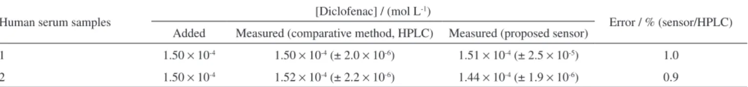
Aplication of the biomimetic sensor in human serum samples
The proposed amperometric sensor was evaluated using analyses of enriched samples of human serum. The measurements were analyzed by square wave voltammetry (SWV). The results obtained were found to be close to the reference value, indicating that the system analyzed showed no matrix effect (Table 3). The method used in the determination of diclofenac in the matrices analyzed was the standard addition. In these analyses standard solution aliquots are added to the electrochemical cell, and the current obtained is extrapolated in the analytical curve, in the order to obtain the respective concentration of diclofenac in the sample (Figure S6).
Analyses of biomimetic sensor in FIA
Optimization of the FIA system
The pH and potential applied to the wall-jet electrochemical cell was varied, and the highest sensitivity was achieved at pH 7.5 and potential of 500 mV vs. Ag|AgCl|KClsat. This potential was therefore used in subsequent FIA measurements (Figure S4).
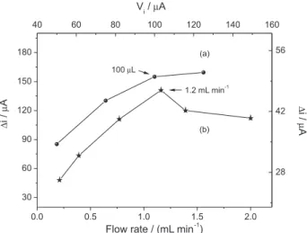
Figure S5 shows the optimization of flow rates and injected sample volume. The flow rates between 0.2 and 2.3 mL min-1 were tested. An optimum value of 1.2 mL min-1 was selected since this did not affect the sampling frequency, while the current value was close to that obtained at the lowest flow rate.
Figure 6. Study of the electrochemical response in square wave voltammetry (SWV) of electrode not modified in the absence (a) and presence (b) of diclofenac, sensor modified with MWCNT-COOH (c) and biomimetic sensor with MWCNT-COOH (d) in the presence of diclofenac. Analysis conditions: f = 10 Hz, a = 150 mV, ∆E = 4 mV,
0.1 mol L-1 phosphate buffer (pH 7.5), [diclofenac] = 3.5 × 10⁻⁴ mol L-1.
Figure 7. Response profile in SWV for diclofenac in 0.1 mol L-1 phosphate buffer (pH 7.5) and analytical curve in the optimized conditions (inset). Analysis conditions: f = 10 Hz, a = 100 mV, ∆E = 4 mV.
Figure 8. Study of selectivity carried out in square wave voltammetry (SWV): (a) electrolyte; (b) secnidazol, piroxicam, ranitidine, ciprofloxacin, lidocaine and tetracycline; (c) diclofenac. All analytes were added to the concentration of 2.0 × 10⁻⁴ mol L-1. Analysis conditions: f = 10 Hz,
The injected sample volume (Vi) was investigated using loop volumes of 50, 75, 100 and 125 µL. The signal increased with increasing Vi, reaching saturation at 100 µL. The volume of 100 µL was selected, since this provided a good current signal without affecting the sampling frequency.
Analytical characteristics of the FIA system
Once the FIA parameters had been optimized (Table 4), a calibration curve was plotted using the data shown in Figure 9. It can be seen that the sensor did not exhibit any memory effects.
The analytical curves showed linear fits for both increasing and decreasing concentrations of diclofenac, as described by equations 2 and 3, respectively:
∆i/µA = 1.3(±0.7) + 1.6 × 10⁴(±3.0 × 10²)[Diclofenac]/
(mol L–1) (2)
R = 0.9991 (n = 7);
∆i/µA = (0.4 ± 1.0 × 10⁻²) + 1.6 × 10⁴(±4.5 × 10²)
[Diclofenac]/(mol L–1) (3)
R = 0.9981 (n = 7);
Limits of detection and quantification, calculated according to ANVISA recommendations41 from the standard deviation of seven independent measurements were 1.7 × 10⁻⁵ and 5.0 × 10⁻⁵ mol L-1, respectively.
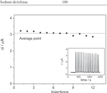
The repeatability of the sensor was investigated by using injections of 5.0 × 10⁻⁵ mol L-1 diclofenac solution, for
which a relative standard deviation (RSD)of 3.5% (n = 12) was obtained (Figure 10).
Application of biomimetic sensor in pharmaceutical formulations
Application in the batch and FIA systems was tested using two commercial pharmaceutical formulations. The standard additions procedure was used for diclofenac quantification, and the results obtained were compared with the method based on HPLC (Table 5).
After the assessment of the results, the two methods were not found to be significantly different, thus leading to
the conclusion that the proposed methodology is an efficient and rapid alternative for the determination of diclofenac in pharmaceutical preparations.
Conclusions
The biomimetic sensor showed satisfactory sensibility and selectivity, indicating that the modifiers can be considered as a biomimetic catalyst for the monitoring of
Table 3. Results obtained in the analyses of human serum samples in batch system
Human serum samples [Diclofenac] / (mol L
-1)
Error / % (sensor/HPLC) Added Measured (comparative method, HPLC) Measured (proposed sensor)
1 1.50 × 10⁻⁴ 1.50 × 10⁻⁴ (± 2.0 × 10⁻⁶) 1.51 × 10⁻⁴ (± 2.5 × 10⁻⁵) 1.0
2 1.50 × 10⁻⁴ 1.52 × 10⁻⁴ (± 2.2 × 10⁻⁶) 1.44 × 10⁻⁴ (± 1.9 × 10⁻⁶) 0.9
Table 4. Optimized parameters and analytical characteristics of the biomimetic sensor in the FIA system
Parameter Value
Applied E / V vs. Ag|AgCl|KClsat 0.5
Flow rate / (mL min-1) 1.2
Vi / µL 100
Linear range / (mol L-1) 5.0 × 10⁻⁵-5.0 × 10⁻³
Sensitivity / (µA L µmol-1) 1.6 × 10⁴ ± 3.0 × 10²
Limit of quantification / (µmol L-1) 50.0 Limit of detection / (µmol L-1) 16.6
Measurement repeatability (RSD, n = 14); [diclofenac] = 10-3 mol L-1
3%
Analytical frequency 45 samples h-1
RSD: relative standard deviation.
Figure 9. FIA signals obtained using the biomimetic sensor for different diclofenac concentrations (mol L-1): (a) 5.0 × 10⁻⁵; (b) 5.0 × 10⁻⁴; (c) 1.0 × 10⁻³; (d) 1.5 x 10⁻³; (e) 2.0 × 10⁻³; (f) 3.5 × 10⁻³; (g) 5.0 × 10⁻³.
Table 5. Results obtained with the proposed sensor and the comparative method (HPLC) in the analysis of drugs containing diclofenac in batch and FIA mode
Pharmaceutical formulations
Value obtained / (mg tablets-1)
Nominal value Comparative method (HPLC) Batch system FIA system
Sodium diclofenac 50 49.7 (± 0.2) 50.5 (± 2.5) 49.0 (± 3.5)
Sodium diclofenac 100 98.4 (± 0.1) 95.0 (± 0.7) 98.0 (± 1.0)
Figure 10. Profile of the repeatability for 16 consecutive injections of 5.0 × 10⁻⁵ mol L-1 diclofenac solution.
sodium diclofenac. The sensor has numerous advantages, such as easy preparation, sensibility, selectivity and especially low cost.
The coupling of the biomimetic sensor to the FIA system proved to be a simple and versatile tool for the determination of diclofenac.
The results obtained in the batch and FIA systems for both the pharmaceutical and human serum samples were found to be similar to the compared method (HPLC), thus making its application possible in other matrices.
Supplementary Information
Supplementary data are available free of charge at http://jbcs.sbq.org.br as PDF file.
Acknowledgements
The authors would like to gratefully acknowledge the financial support provided by FAPESP (Proc. 2011/03008-7) and CNPQ (Proc. 471231/2011-8).
References
1. Shalini, K.; Anwer, Z.; Sharma, P. K.; Garg, V. K.; Kumar, N.;
Int. J. PharmTech. Res. 2010, 2, 2265.
2. Zhang, Y.; Geissen, S. U.; Gal, C.; Chemosphere 2008, 73, 1151.
3. Stulten, D.; Zühlke, S.; Lamshöft, M.; Spiteller, M.; Sci. Total Environ. 2008, 405, 310.
4. Xu, M.; Chen, L.; Song, J.; Anal. Biochem. 2004, 329, 21. 5. Hoeger, B.; Kollner, B.; Dietrich, D. R.; Hitzfeld, B.; Aquat.
Toxicol. 2005, 75, 53.
6. Perez-Estrada, L. P.; Malato, S.; Gernajak, W.; Aguera, A.; Thurman, E. M.; Ferrer, I.; Fernandez-Alba, A. R.; Environ. Sci. Technol. 2005, 39, 8300.
7. Solangi, A. R.; Menon, S. Q.; Mallah, A.; Menon, N.; Khuhawar, M.; Bhanger, M.; Pak. J. Pharm. Sci. 2011, 24, 539. 8. Song, X.-Y.; Shi, Y.-P.; Chen, J.; Talanta 2012, 100, 153. 9. Faber, H.; Melles, D.; Brauckmann, C.; Wehe, C. A.;
Wentker, K.; Karst, U.; Anal. Bioanal. Chem. 2012, 403, 345. 10. Souza, R. L.; Tubino, M.; J. Braz. Chem. Soc. 2005, 16, 1068.
11. Ensafi, A. A.; Izadi, M.; Karimi-Maleh, H.; Ionics 2013, 19, 137.
12. Souza, M. D. B.; Quim. Nova 1997, 20, 195.
13. Liu, L.; Gou, L. P.; Bo, X. J.; Bai, J.; Cui, X. J.; Anal. Chim. Acta2010, 673, 88.
14. Sotomayor, M. D. P. T.; Kubota, L. T.; Quim. Nova 2002, 25, 123.
15. Sotomayor, M. D. P. T.; Tanaka, A. A.; Freire, R. S.; Kubota, L. T. In Encyclopedia of Sensors; Grimes, C. A.; Dickey, E. C.; Pishko, M. V., eds.; American Scientific Publishers: California, 2006.
16. Danielson, P. B.; Curr. Drug Metab. 2002, 3, 561.
17. Sono, M.; Roach, M. P.; Coulter, E. D.; Dawson, J. H.; Chem. Rev. 1996, 96, 2841.
18. Wong, A.; Sotomayor, M. D. P. T; Sens. Actuators, B 2013, 181, 322.
19. Wong, A.; Lanza, M. R. V.; Sotomayor, M. D. P. T.; Comb. Chem. High Throughput Screening 2010, 13, 666.
20. Boni, A. C.; Wong, A.; Dutra, R. A.; Sotomayor, M. D. P. T.;
Talanta 2011, 30, 2067.
21. Boni, A. C.; Sotomayor, M. D. P. T.; Lanza, M. R. V.; Tanaka, S. M. C. N.; Tanaka, A. A.; J. Braz. Chem. Soc. 2010, 21, 1377. 22. Sotomayor, M. D. P. T.; Sigoli, A.; Lanza, M. R. V.; Tanaka,
A. A.; Kubota, L. T.; J. Braz. Chem. Soc. 2008, 19, 734. 23. Batista, I. V.; Lanza, M. R. V.; Dias, I. L. T.; Tanaka, S. M. C. N.;
Tanaka, A. A.; Sotomayor, M. D. P. T.; Analyst2008, 133, 1692.
24. Wong, A.; Lanza, M. R. V.; Sotomayor, M. D. P. T.;
25. Blanco-Lopez, M. C.; Lobo-Castanon, M.-J.; Miranda-Ordieres, A. J.; Tunon-Blanco, P.; Anal. Bioanal. Chem. 2003, 377, 257. 26. Kashefi-Kheyrabadi, L.; Mehrgardi, M. A.; Biosens.
Bioelectron. 2012, 33, 184.
27. Razmi, H.; Sarhang-Zadeh, K.; Mohammad-Rezaei, R.; Anal. Lett. 2013, 46, 1885.
28. Mokhtar, A.; Karimi-Maleh, H.; Ensafi, A. A.; Beitollahi, H.;
Sens. Actuators, B 2012, 169, 96.
29. Ruzicka, J.; Hansen, E. H.; Flow Injection Analysis; John Wiley: New York, 1988.
30. Oliveira, M. C. Q.; Lanza, M. R. V.; Tanaka, A. A.; Sotomayor, M. D. P. T.; Anal. Methods 2010, 2, 507.
31. Moraes, F. C.; Mascaro, L. H.; Machado, S. A. S.; Brett, C. M. A.; Talanta 2009, 79, 1406.
32. Jiuling, C.; Qinghai, C.; Qing, M.; J. Colloid Interface Sci.
2012, 370, 32.
33. Rezaei, B.; Damiri, S.; Sens. Actuators, B 2008, 134, 324. 34. Emami, J.; Ghassami, N.; Talari, R.; Daru, J. Fac. Pharm.,
Tehran Univ. Med. Sci. 2007, 15, 3.
35. Yilmaz, B.; Asci, A.; Palabiyik, S. S.; J. Chromatogr. Sci. 2011,
49, 422.
36. Arvand, M.; Gholizadeh, T. M.; Zanjanchim, M. A.; Mater. Sci. Eng., C 2012, 32, 1682.
37. Andrieux, C. P.; Savéant, J. M.; J. Electroanal. Chem.1978,
93, 163.
38. Wang, K.; Li, H.-N.; Wu, J.; Ju, C.; Yan, J.-J.; Liu, Q.; Qiu, B.;
Analyst 2011, 136, 3349.
39. Fantuzzi, A.; Mak, L. H.; Capria, E.; Dodhia, V.; Panicco, P.; Collins, S.; Gilardi, G.; Anal. Chem. 2011, 83, 3831. 40. Reminek R.; Glatz, Z.; J. Sep. Sci. 2010, 33, 3201.
41. http://portal.anvisa.gov.br/wps/wcm/connect/733b41004745 86a1900fd43fbc4c6735/Consolidado+de+normas+COBIO. pdf?MOD=AJPERES accessed in May 2014.
Submitted: December 17, 2013 Published online: May 20, 2014
Supplementary Information
0103 - 5053 $6.00+0.00S
I
*e-mail: [email protected]
Monitoring of Diclofenac with Biomimetic Sensor in Batch and FIA Systems
Ademar Wong,* Luiz D. Marestoni and Maria D. P. T. Sotomayor
Departamento de Química Analítica, Instituto de Química, Universidade Estadual Paulista (UNESP), 14801-970 Araraquara-SP, Brazil
Figure S1. Schematic diagram of the flow injection system for amperometric determination of diclofenac. WE: working electrode (biomimetic sensor); AE: auxiliary electrode (platinum); RE: homemade reference electrode (Ag|AgCl|KClsat).
Figure S2. Linear dependence of the anodic peak current (∆i) vs. square root of the scan rate (v1/2). Inset: study of scan rate with the proposed sensor from cyclic voltammetry in 0.1 mol L-1 phosphate buffer (pH 7.5) containing 1.0 × 10⁻⁴ mol L-1 of diclofenac.
Figure S3. EDX spectrum of the copper complex and MWCNT-COOH.
Figure S4. Influence of the parameters in the proposed FIA system: (a) effect of pH on the response to diclofenac using a flow rate of 1.2 mL min-1; (b) effect of potential on the response to diclofenac, using
a sample volume (Vi) of 100 µL. The experiments were carried out using
a carrier of 0.1 mol L-1 PBS buffer at pH 7.5, and applying a potential of
Figure S5. Influence of the parameters in the proposed FIA system: (a) effect of injected sample volume (Vi) on the response to diclofenac using
a flow rate of 1.2 mL min-1; (b) effect of flow rate on the response to
diclofenac, using a Vi of 100 µL. The experiments were carried out using
a carrier of 0.1 mol L-1 PBS buffer at pH 7.5, and applying a potential of
500 mV vs. Ag|AgCl|KClsat.