S
ho
rt
R
e
p
o
rt
J. Braz. Chem. Soc., Vol. 20, No. 2, 383-386, 2009. Printed in Brazil - ©2009 Sociedade Brasileira de Química 0103 - 5053 $6.00+0.00
*e-mail: [email protected]
A New β-Lapachone Derivative from
Distictella elongata
(Vahl) Urb.
Erdal Bedir,a Ana M. S. Pereira,*,b Shabana I. Khan,c Amar Chittiboyina,e
Rita M. Moraesc and Ikhlas A. Khanc,d
aDepartment of Bioengineering, Faculty of Engineering, Ege University, Bornova, Izmir, 35100 Turkey
bDepartamento de Biotecnologia, Universidade de Ribeirão Preto, 14096-900 Ribeirão Preto-SP, Brazil
cNational Center for Natural Products Research, Research Institute of Pharmaceutical Sciences;
dDepartment of Pharmacognosy, School of Pharmacy and eDepartment of Medicinal Chemistry, School of Pharmacy,
University of Mississippi, University, MS 38677
O presente trabalho descreve a elucidação estrutural de uma nova β-lapachona tipo naftoquinona, isolada a partir das raízes de Distictella elongata. A nova substância, apresentando fórmula molecular C16H16O6, foi identificada como sendo
4,7-diidróxi-10-metóxi-2,2-dimetil-3,4-diidro-2H-benzo[h]cromeno-5,6-diona, de acordo com os dados obtidos por meio de análises espectrométricas. A elucidação estrutural foi realizada utilizando-se as técnicas espectrométricas
[HRESIMS, 1D RMN (1H e 13C) e 2D RMN (g-DQF-COSY, g-HMQC e g-HMBC]. O extrato
clorofórmico das raízes de D. elongata demonstrou significante inibição no crescimento de células do tipo SK-MEL (melanoma) e SK-OV-3 (carcinoma de ovário), com valores de IC50 de
40 µg mL-1 e 56 µg mL-1, respectivamente. Entretanto, a naftoquinona não foi responsável pela
atividade citotóxica exibida pelo extrato.
The present study describes the structure elucidation of the new β-lapachone type naphthoquinone isolated from the roots of Distictella elongata. Its structure, according to the molecular formula C16H16O6, was identified as 4,7-dihydroxy-10-methoxy-2,2-dimethyl-3,4-dihydro-2H-benzo[h]chromene-5,6-dione. The structure was assigned by spectrometric methods [HRESIMS, 1D NMR (1H and 13C), and 2D NMR (g-DQF-COSY, g-HMQC and g-HMBC].
Root chloroform extract of D. elongata showed significant inhibition of the growth of SK-MEL (melanoma) and SK-OV-3 (ovary adenocarcinoma) cells with IC50 values of 40 µg mL-1 and
56 µg mL-1, respectively. However, the naphthoquinone was not responsible for the cytotoxic
activity exhibited by the extract.
Keywords: cerrado, naphthoquinone, Bignoniaceae, Distictella elongata
Introduction
Many Bignoniaceae species have been investigated for
their medicinal value.1 The anti-cancer activity of some
endemic Bignoniaceae, native from Cerrado regions, like
Tabebuia avellanedae, Anemopaegma arvense; Zeyheria montana, Kigelia pinnata and Jacaranda caucana have
been reported in the literature.2-6 The Bignoniaceae family,
represented by more than 100 genera and about 800
species, including Distictella elongata, is mainly tropical,
with most accessions dispersed in tropical America.7 This
species occurs in Cerrado areas, within the states of Goiás,
São Paulo and Minas Gerais.8 The Cerrado is a highly
endangered ecosystem, due to the intensive introduction of soybean farming and cattle ranching in this area over the last two or three decades. It represents the second largest biome in South America after the rain forests and originally covered one fourth of Brazil. No phytochemical or pharmacological investigation has yet been carried out on D. elongata.
Results and Discussion
Fractionation of chloroform root extract from D.
elongata resulted in the isolation of a new naphthoquinone
A New β-Lapachone Derivative fromDistictella elongata (Vahl) Urb. J. Braz. Chem. Soc.
384
formula of 1 was determined as C16H16O6 by HRESIMS,
which exhibited ions at m/z 305.1031 [M+H]+ and 327.0846
[M+Na]+ (in the positive-ion mode).
The 1H NMR (CDCl
3) spectrum of 1 exhibited a
deshielded hydrogen at d 12.7 (s, O-H), two aromatic
hydrogens overlapping at d 7.29, an O-methyl at d 3.97
(s, 3H), as well as a triplet at d 4.96 (t, J 6.0 Hz). In
addition, the 1H NMR spectrum showed two tertiary methyl
hydrogens at d 1.45 and d 1.55, and a gem-methylene
signals at d 2.05 and d 2.12 (dd, J 6.0 and14.2 Hz, 2H).
The 13C NMR spectrum displayed 16 signals including
six aromatic, two olefinic and two carbonyl signals. Total 6 double bonds, attributable to aforementioned carbon signals, when considered with 9 unsaturation degrees, the
remaining 3 degrees required that compound 1 consists
of tri-ring systems. The presence of β-lapachone type
pyranonaphthoquinone skeleton (compound 1), which is
common in Bignoniaceae family, was easily deduced from
the proton and carbon chemical shifts.9-11
Two overlapping aromatic hydrogens (7.29, 2H) did not provide any information about substitution pattern of the
aromatic ring. Thus, the 1H NMR spectrum of compound
1 was obtained using acetone-d6 as a solvent, in which
aromatic hydrogens were observed as doublets at d7.52
(d, 9.0 Hz, 1H), and d 7.30 (d, 9.0 Hz, 1H), indicative of
the presence of ortho-coupling hydrogens.
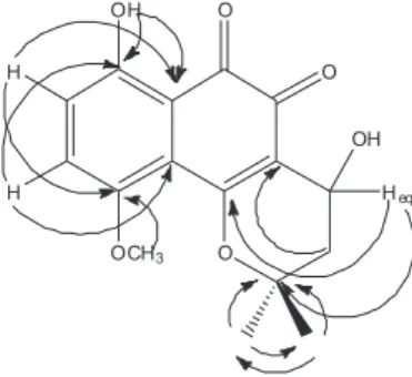
The combined use of g-DQF-COSY, g-HMQC and g-HMBC techniques permitted the complete assignment
of the pyranonaphthoquinone skeleton and the substitution
pattern for the compound 1. Thus, the deshielded 1H NMR
signal at d 12.7 [C-7(OH)] showed correlations with a
methine carbon at d 127.9 and two quaternary carbons
at d 156.6 and d 114.1, attributed to C-8, C-7 and C-6a,
respectively. Also, a methine carbon at d 127.9 (C-8)
showed correlation with the aromatic hydrogen at d7.30
(d, 9.0 Hz, in acetone-d6), which is assigned to H-8. The
aromatic hydrogen observed at d 7.52 (in acetone-d6, H-9)
indicated a long-range correlation in the G-HMBC spectrum
with a quaternary carbon at d 117.5, attributed to C-10a.
The carbon signal at d 155.1 displayed a correlation with
O-methyl hydrogen (d 3.97, s) in the g-HMBC spectrum,
hence allowing it to be assigned unambiguously to C-10, and locating the methoxyl group to C-10. Moreover, the position of the hydroxyl group located on pyrane ring was deduced from g-HMBC correlations from quaternary carbons C-10b (155.9) and C-2 (80.4) to a methine proton
at d 4.96 (t, J 6.0 Hz) attributed to H-4.
Based on these results, the structure of 1, a new natural product, was established as 4,7-dihydroxy-10-methoxy-
2,2-dimethyl-3,4-dihydro-2H-benzo[h]chromene-5,6-dione. Having established the structure of 1, the relative
stereochemistry of chiral center (C-4) remained to be
solved. The absolute stereochemistry of 1 was established
by NMR data and molecular modeling analysis after calculation of the minimal conformational energy for each of the two possible enantiomers using Accelrys Discovery
Studio v2.0.0.7264. Only the stereoisomer, (S)-4-hydroxy
Figure 1. Structure of new naphthoquinone, compound 1.
Figure 2. Key HMBC of compound 1.
Bedir et al. 385 Vol. 20, No. 2, 2009
isomer of 1 showed agreement with the coupling constants
observed in the 1H NMR spectrum and the dihedral angles
measured after the calculation of the minimal conformation energy for each of the two possible stereoisomers. The key coupling constant analyzed was that measured between H3a-H4 = H3b-H4 = 6.0 Hz. Therefore, the dihedral angles H3a-C3-C4-H4 and H3b-C3-C4-H4 must be identical.
Whereas, the dihedral angles for the R-isomer should
be non identical and should have at least two different coupling constants. When a comparison was made of the current analysis data with reported spectral data of similar
systems,12,13 we concluded that the absolute stereochemistry
at C-4 position must be in S configuration.
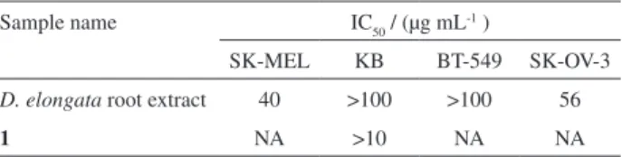
The chloroform root extract of D. elongata was tested
for in vitro growth inhibition activity against a panel of four cancer cell lines. Results included in Table 1 showed that the extract significantly inhibited the growth of SK-MEL and
SK-OV-3 cells with IC50 values of 40 µg mL-1 and 56 µg mL-1,
respectively. However, 4,7-dihydroxy-10-methoxy-2,2-dimethyl-3,4-dihydro-2H-benzo[h]-chromene-5,6-dione
(1) was not responsible for the anticancer activity exhibited
by the whole extracts (Table 1).
Experimental
General
NMR spectra were recorded on a Bruker Avance DRX
500 FT spectrometer operating at 500 and 125 MHz for
1H and 13C NMR, respectively. The chemical shift values
are reported as parts per million (ppm) units relative to tetramethylsilane (TMS); and the coupling constants are in
Hz (in parentheses). For the 13C NMR spectra, multiplicities
were determined by a distortionless enhancement by polarization transfer (DEPT) experiment. HRESIMS (High Resolution Electrospray Ionization Mass Spectrometry) were obtained using a Bruker BioApex FT-MS in ESI mode. For TLC, precoated Si 250F layers (Baker) were used. Column chromatography was performed on silica gel 230-400 mesh (Merck).
Plant material
Roots of Distictella elongata (Bignoniaceae) were
collected from preserved areas of a Brazilian Cerrado, at the Biological Reserve of Mogi-Guaçu Ecological and Experimental Station, São Paulo, Brazil. A voucher specimen was deposited in the herbarium of the University of Ribeirão Preto (HPM-482). The roots were dried at 50 ºC, powdered and kept until ready for extraction.
Extraction and isolation
Powdered roots (330 g) were extracted in CHCl3 for
7 days. The CHCl3 extract was concentrated to a small
volume at reduced pressure to yield 1.3 g of oily residue. This residue was fractionated on a silica gel column eluting with hexane, and hexane/ethyl acetate gradient elution to ethyl acetate 100%. Using 50% hexane/ethyl acetate solvent a red crystal was obtained (naphthoquinone) and further purified by preparative TLC using ethyl acetate/hexane (4:1) as solvent system. The amount of 30 mg of the naphthoquinone was removed from the layer. The final purification performed on Sephadex column (LH-20, 30 mg) eluted with MeOH, to
afford 9 mg naphthoquinone 1.
Biological activity
The in vitro anticancer activity was tested against a panel of four human cancer cell lines that included SK-MEL (malignant, melanoma), KB (epidermal carcinoma, oral), BT-549 (ductal carcinoma, breast), and SK-OV-3 (ovary
carcinoma).14 All the cell lines were from ATCC (Manassas,
VA). The cells were cultured in 75 cm2 culture flasks in
RPMI-1640 medium (GibcoTM, Invitrogen Corp.) supplemented with
bovine calf serum (10%) and amikacin (60 mg L-1), at 37 oC,
95% humidity, 5% CO2 using standard cell culture techniques.
The assay was performed in 96-well microplates. Cells were seeded to the wells of the plate at a density of 25,000 cells/
well and grown for 24 hours at 37 oC. Samples were added
to the cells and again incubated for 48 h. The number of viable cells was determined according to Neutral Red assay
procedure.15 IC
50 (the concentration of the test sample that
caused a growth inhibition of 50% after 48 h exposure of the cells) was calculated from the dose curves generated by plotting percent growth versus the test concentration on a logarithmic scale using Microsoft Excel®.
Compound 1
Reddish oil; 1H NMR data (500 MHz, CDCl
3): d 12.7
(1H, s, OH-7), 7.29 (s, 2H, overlapping), 4.96 (1H, t, J
6.0 Hz, H-4), 3.97 (3H, s, O-CH3), 2.12 (1H, dd, J 6.0 and
Table 1. In vitro anticancer activity of Distictella elongata root extract and compound 1
Sample name IC50 / (µg mL-1 )
SK-MEL KB BT-549 SK-OV-3
D. elongata root extract 40 >100 >100 56
1 NA >10 NA NA
A New β-Lapachone Derivative fromDistictella elongata (Vahl) Urb. J. Braz. Chem. Soc.
386
14.2 Hz, H-3a), 2.05 (1H, dd, J 6.0 and 14.2 Hz, H-3b),
1.55 (3H, s, Me-12), 1.45 (3H, s, Me-11); 1H NMR data
(500 MHz, acetone-d6): d 12.8 (1H, s, OH-7), 7.52 (1H, d,
J 9.0 Hz, H-9), 7.30 (1H, d, J 9.0 Hz, H-8), 4.93 (1H, d, J
5.0 Hz, H-4), 2.31 (1H, dd, J 5.0 and 14.0 Hz, H-3a), 2.26
(1H, dd, J 5.0 and 14.0 Hz, H-3b), 1.51 (3H, s, Me-12), 1.46
(3H, s, Me-11). 13C NMR data (125 MHz, CDCl
3): d191.4
6), 177.9 5), 156.6 7), 155.9 10b), 155.1 (C-10), 127.9 (C-8), 123.1 (C-9), 118.6 (C-4a), 117.5 (C-10a),
114.1 (C-6a), 80.4 (C-2), 60.0 (C-4), 56,2 (O-CH3), 39.9
(C-3), 27.3 (x2, Me-11 and Me-12); HRESIFTMS: m/z
305.1031 [M+H]+ (calculated for C
16H16O6: 305.1026).
Acknowledgments
This work was supported in part by the United States Department of Agriculture, ARS Specific Cooperative Research Agreement and by the FAPESP BIOTA project No. 99/10610-1.
References
1. Gentry, A. H.; Ann. Missouri Bot. Gard. 1992, 79, 53. 2. de Santana, C. F.; de Lima, O.; d’ Albuquerque, I. L.; Lacerda,
A. L.; Martins, D. G.; Rev. Inst. Antibiot. 1968, 8, 89. 3. Ogura, M.; Cordell, G. A.; Farnsworth, R.; Lloydia 1977, 40,
157.
4. Jácome, R. L. R. P.; Oliveira, A. B. ; Raslan, D. S.; Muller, A.; Wagner, H. Quim. Nova 1999, 22, 175.
5. Jackson, S. J.; Houghton, P. J.; Retsas, S.; Photiou, A.; Planta Med. 2000, 66, 758.
6. Uchino, T.; Kawahara, N.; Sekita, S.; Satake, M.; Saito, Y.; Tokunaga, H.; Ando, M.; Toxicol. In Vitro 2004, 18, 255. 7. Cronquist, A.; An Integrated System of Classification of
Flowering Plant; Columbia University Press: New York, 1981.
8. Scudeller, V. V.; Carvalho-Okano, R. M. de; Iheringia-Série Botânica 1998, 51, 79.
9. McDonald, I. A.; Simpson, J. T.; Sirekowski, A. F.; Aust. J. Chem. 1977, 30, 1727.
10. Oliveira, A. B.; Raslan, D. S.; Miraglia, M. C. M.; Mesquita, A. A. L.; Quim. Nova 1990, 13, 302.
11. Schaffner-Sabba, K.; Schmidt-Ruppin, K. H.; Wehrli, W.; Schuerch, A. R.; Wasley, J. W. F.; J.Med. Chem.1984, 27, 990.
12. Burgard, A.; Lang, H.-J.; Gerlach, U.; Tetrahedron 1999, 55, 7555.
13. Yamada, T.; Nagata, T.; Sugi, K. D.; Yorozu, K.; Ikeno, T.; Ohtsuka, Y.; Miyazaki, D.; Mukaiyama, T.; Chem. Eur. J. 2003,
9, 4485.
14. Mustafa, J.; Khan, S. I.; Ma, G.; Walker, L. A.; Khan, I. A.;
Lipids 2004, 39, 659.
15. Borenfreund, E.; Babich, H.; Martin-Alguacil, N.; Toxicol. In Vitro 1988, 1,1.
Received: October 19, 2007
Web Release Date: December 18, 2008