Cardiopulmonary exercise testing in the
early-phase of myocardial infarction
Teste de exercício cardiopulmonar na fase precoce do infarto do miocárdio
Vandeni C. Kunz1, Karina B. S. Serra2, Érica N. Borges2, Paulo E. S. Serra3, Ester Silva1,2
Abstract
Objective: To evaluate and to compare the cardiorespiratory and metabolic variables at the ventilatory anaerobic threshold level (AT) and at submaximal cardiopulmonary exercise testing (CPET) in both, healthy volunteers and in patients in the early phase after acute myocardial infarction (AMI). Method: Twenty-six volunteers underwent a submaximal or symptom-limited cardiopulmonary exercise testing (CPET) on a cycle ergometer and were divided into AMI group (AMIG=12, 56.33±8.65 years) and healthy group (CG=14, 53.33±3.28 years). The primary outcome measures were the cardiorespiratory and metabolic variables obtained at the peak workload and at the AT of the CPET. Statistical test: independent Student’s t-test, α=5%. Results: The AMIG presented lower values at the AT and the peak workload of the CPET compered to the CG: power in watts (91.06±30.10 and 64.88±19.92; 154.93±34.65 and 120.40±29.60); VO2 mL.kg
-1.min-1 (17.26±2.71 and 12.19±2.51; 25.39±5.73 and 19.41±5.63); VCO 2 L/min
-1 (1.43±0.31 and 0.93±0.23; 2.07±0.43 and 1.42±0.36), VO2 L/min
-1 (1.33±0.32 and 1.00±0.23; 1.97±0.39 and 1.49±0.36); VE L/min-1 (42.13±8.32 and 27.51±5.86; 63.07±20.83 and 40.82±11.96); HR (bpm) (122.96±14.02 and 103.46±13.38; 149.67±13.77 and 127.60±10.04), double product (DP) (bpm.mmHg.min-1) (21835.86±3245.93 and 17333.25±2716.51; 27302.33±3053.08 and 21864.00±2051.48), respectively. The variable oxygen uptake efficiency slope (OUES L/min) was lower in the AMIG (1.79±0.51) than the CG (2.26±0.37). The AMIG presented neither ECG alterations nor symptoms that limited the CPET. Conclusion:The results suggest that patients with AMI Killip class I presented lower functional capacity and DP compared to the CG without presenting ischemic alterations. Thus, the study suggests that submaximal CPET can be applied at an early stage to evaluate cardiorespiratory status since it is both safe and highly sensitive to detect changes.
Keywords: oxygenconsumption; myocardial infarction; physical exercise; physical therapy.
Resumo
Objetivo:Avaliar e comparar as variáveis cardiorrespiratórias e metabólicas no nível do limiar de anaerobiose ventilatório (LAV) e no pico do teste de exercício cardiopulmonar (TECP) submáximo em voluntários saudáveis e em pacientes na fase precoce após o infarto agudo do miocárdio (IAM). Método: Vinte e seis voluntários realizaram TECP submáximo ou sintoma limitante em cicloergômetro e foram divididos em grupo IAM (G-IAM=12, 56,33±8,65 anos) e grupo saudável (GC=14, 53,33±3,28 anos). As medidas dos desfechos principais foram as variáveis cardiorrespiratórias e metabólicas obtidas no pico e no LAV do TECP. Teste estatístico: t-Student não pareado, α=5%.
Resultados: O G-IAM apresentou menores valores no LAV e no pico do TECP que o GC (p<0,05): potência em Watts (91,06±30,10 e
64,88±19,92; 154,93±34,65 e 120,40±29,60); VO2mL.kg
-1.min-1 (17,26±2,71 e 12,19±2,51; 25,39±5,73 e 19,41±5,63); VCO 2L/min
-1 (1,43±0,31 e 0,93±0,23; 2,07±0,43 e 1,42±0,36), VO2L/min
-1 (1,33±0,32 e 1,00±0,23; 1,97±0,39 e 1,49±0,36); VEL/min-1 (42,13±8,32 e 27,51±5,86; 63,07±20,83 e 40,82±11,96); FC (bpm) (122,96±14,02 e 103,46±13,38; 149,67±13,77 e 127,60±10,04); duplo produto (DP) (bpm.mmHg.min -1) (21835,86±3245,93 e 17333,25±2716,51; 27302,33±3053,08 e 21864,00±2051,48), respectivamente. A variável Oxygen Uptake Efficiency
Slope (OUES L/min) do G-IAM foi 1,79±0,51 e do GC 2,26±0,37, p<0.05. O G-IAM não apresentou alterações eletrocardiográficas ou sintomas que limitassem o TECP. Conclusão: Os resultados mostram que os pacientes com IAM Killip I apresentaram menor capacidade funcional e DP em relação ao GC, sem apresentar alterações isquêmicas. Assim, o estudo sugere que o TECP submáximo pode ser aplicado precocemente para a avaliação cardiorrespiratória por apresentar alta sensibilidade para detectar alterações de forma segura.
Palavras-chave: consumo de oxigênio; infarto do miocárdio; exercício físico; fisioterapia.
Received: 12/16/2011 – Revised: 03/13/2012 – Accepted: 04/23/2012
Introduction
The recommendation for physical therapy treatment based on physical training within a cardiac rehabilitation program for patients after acute myocardial infarction (AMI) depends on its evolution based on clinical criteria and results of invasive and non-invasive exams1-3. Among the non-invasive examinations, the cardiopulmonary ex-ercise testing (CPET) performed in an early-phase after the coronary event has become routine in several medical centers3-5. From this exam, it is possible to obtain more precise information regarding the integration among the pulmonary, cardiovascular and musculoskeletal systems and the changes of the functional aerobic capacity due to pathological conditions, such as in the AMI5-7. In addition, it is possible to evaluate the prognosis of the patient in rela-tion to risk stratificarela-tion for new cardiac events2,3.
he literature reports that the maximal oxygen consump-tion (VO2max) is one of the most investigated variables
8,9.
However, the VO2max is hardly achieved by cardiac patients, whose the exercise performance is limited by peripheral muscle fatigue, dyspnea, and the presence of signiicant car-diac alterations10. Moreover, the exercise performance depends on the motivation and perception of the subjects. In this case, the determination of the ventilatory anaerobic threshold (AT), which is an important physiologic parameter, has been recom-mended since it provides information regarding the physiologi-cal changes from the aerobic to the anaerobic metabolism11. herefore, it has been considered more objective for the pre-scription of aerobic physical training, demonstrating good reproducibility9,12,13.
Furthermore, in the AT level there is a balance between the supply and consumption of O2 in the working muscles, pre-venting acidosis; the sympathetic nervous system is not over stimulated, and thus minor changes occur in the release of the epinephrine and norepinephrine hormones, which allows that, in this exercise level, these patients become able to maintain a working frequency for a prolonged period of time14.
Additionally, from the CPET it is possible to evaluate the variables VE/VCO2 slope and the Oxygen Uptake Efficiency Slope (OUES), which indicate the ventilatory efficiency for the production of carbon dioxide and oxygen consump-tion, respectively. Thus, from these variables it is possible to obtain clinical information about the functional status, disease severity15-17, and also information about the prog-nosis of coronary diseases, without exposing the patient to maximal CPET9,18,19. Moreover, it can be used to evaluate and prescribe the intensity of physical exercise in a cardiac rehabilitation program19,20.
he submaximal CPET has been used to evaluate the variables of cardiac patients, since during this test the oxygen supply to tissues while performing the exercise is not limited. Submaximal tests are more appropriate for patients with car-diovascular disorders because they provide greater eicacy and safety for the prescription of physical training in physical therapy treatment programs14,21,22.
The hypothesis of the present study was that patients may be submitted to submaximal CPET, after hospital discharge due to AMI, to evaluate clinical, hemodynamic, metabolic and electrocardiographic responses to submaxi-mal CPET. Therefore, the aim of this study was to evalu-ate and to compare the cardiorespiratory and metabolic variables at the AT level and at the peak of the submaximal CPET in healthy subjects and in patients in the early phase after the AMI.
Method
Study design and ethic approval
his was a cross-sectional study, with the approval of the Ethics Committee of Research of the Universidade Metodista de Piracicaba (UNIMEP), Piracicaba, SP, Brazil, no 63/06. he participants agreed and signed an informed consent form according to the Resolution no 196/96 of the Brazilian Health National Council.
Sample size calculation
he sample size was calculated for the variable VO2 in mL.kg-1.min-1, with a 95% conidence level and statistical power of 80%, capable to detect a diference between-groups (two-tailed test) suggested a number of 12 subjects for each group,
(Graph Pad Stat Mate, version 1.01i, 1998).
Participants
he participants of the AMI group (AMIG) were selected in the Coronary Units of the Hospital dos Fornecedores de Cana de Piracicaba, SP and Santa Casa de Misericórdia de Limeira, SP, Brazil. he patients from the AMIG underwent to the dop-pler echocardiogram and cardiac catheterization with chemi-cal or mechanichemi-cal reperfusion in the irst hours after the AMI; were taking beta-blockers (atenolol, 46±9.4mg/day); with left ventricular ejection fraction (LVEF) within the limits of nor-mality (0.61±0.06); and with clinical classiication of Killip I. To compose the healthy group (CG), 34 subjects were evaluated (CG), being included 14 subjects whom had not participated in physical training programs; presented an aerobic classiication as weak23, did not have any indication of cardiovascular, respira-tory, musculoskeletal and/or metabolic abnormalities, were not taken medications and were not smokers or alcohol drinkers.
Subjects from both groups underwent clinical and cardio-vascular evaluation and also performed blood biochemical tests (total and fraction cholesterol (HLD, LDL) blood glucose, triglycerides, creatinine and uric acid).
he diagnosis of AMI had been conirmed by the presence of two or more criteria: 1) chest or retrosternal pain (constricting or burning pain), with or without radiation for the upper limbs, neck and upper back, lasting >30 minutes with no relief of symptoms to the vasodilator; 2) ST-segment elevation >1 mV in at least two or more contiguous electrocardiogram (ECG), 3) elevation of myocar-dial necrosis markers CK-MB and CPK, twice the normal values24.
Experimental procedure
All the participants underwent clinical evaluation per-formed by a cardiologist before the performance of the CPET.
he experimental procedures were held in acclimatized laboratory, with temperature and relative humidity around 23ºC and 60% respectively. he tests were performed in the afternoon to minimize the interferences of the circadian cycle on the cardiovascular responses. he participants were previ-ously familiarized with the laboratory environment, with the
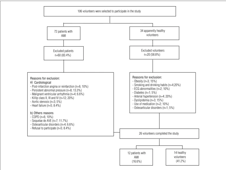
72 patients with AMI
34 apparently healthy volunteers
Excluded patients n=60 (83.4%)
Excluded volunteers n=20 (58.8%)
Reasons for exclusion: - Obesity (n=3; 15%)
- Smoking and drinking habits (n=4;20%) - ECG abnormalities (n=2; 10%) - Diabetes (n=1; 5%)
- Arterial hypertension (n=4; 20%) - Dyslipidemia (n=3; 15%) - Use of medication (n=2; 10%) - Osteoarticular disorders (n=1; 5%)
26 volunteers completed the study
12 patients with AMI (16.6%)
14 healthy volunteers (41.2%) 106 volunteers were selected to participate in the study
Reasons for exclusion: a) Cardiological
- Post-infarction angina or reinfarction (n=6; 10%) - Persistent abnormal pressure (n=8; 13.3%) - Malignant ventricular arrhythmia (n=4; 6.6%) - Killip class II, III and IV (n=12; 20%) - Aortic stenosis (n=3; 5%) - Heart failure (n=5; 8.4%)
b) Others reasons - COPD (n=6; 10%) - Sequelae de AVE (n=7; 11.7%) - Osteoarticular disorders (n=4; 6.6%) - Refusal to participate (n=5; 8.4%)
experimental procedures and with the equipments that were used in the experiment. All subjects were instructed not to in-gest cafeine, alcoholic beverages and do not perform physical exercise on the day before and in the day of the experiments. During the experiments, the AMIG should maintain the medi-cation in use. Before beginning the tests, the volunteers were asked about the occurrence of a normal night’s sleep and, in addition, examined in order to guarantee that they were with their basal conditions within the normality limits25.
Experimental protocol
he experiment consisted of a CPET, ramp-type continuous physical exercise, performed in a cycle ergometer with elec-tromagnetic breaking (QuintonCorival 400, Seattle, WA, USA), with seat height adjusted to allow knee lexion of 5-10 degrees. he participants were instructed not to perform an isometric contraction of the upper limbs, while holding the handlebars of the bicycle, and to maintain pedaling cadence at 60 rpm.
he protocol consisted of a pre-test rest sitting on a cycle ergometer for a period of 60 seconds; starting the exercise with free load during a period of 240 seconds, followed by an increase of power on every 60 seconds, individually calculated accord-ing to the formula described by Wasserman et al.14, being of 10 watts (W) for the patients of the AMIG and 15W for the volun-teers of the CG. For both groups, the CPET was submaximal, being interrupted when the submaximal heart rate (HR) was reach or in the presence of signs and/or symptom-limiting26.
he HR was captured through a heart rate monitor of one channel (MINISCOPE II Instramed-Porto Alegre, RS, Brazil) and processed through an analog to digital converter Lab. PC+ (National Instruments Co. Austin, TX, USA), which represents an in-terface between the heart rate monitor and a microcomputer. he signal was recorded in real time, after A/D conversion, in a sampling rate of 500 Hz27; and the blood pressure (BP) was measured using the auscultatory method based on Korotkof sound on every 2 min-utes, using a mercury sphygmomanometer (WanMed, São Paulo, SP, Brasil) and a stethoscope (Littman, St. Paul, MN, USA).
Ventilatory and metabolic variables, such as pulmonary ventilation (VE) in BTPS L/min-1, VO
2mL.kg
-1.min-1, and
VCO2 in L/min
-1, respiratory exchange ratio (R) and HR were
recorded simultaneously during the entire CPET, breath by breath, through a measurement system of the expired gases (CPX/D Med Graphics – Breeze, St. Paul, Minesota, USA), which was calibrated before each test. hese variables were subse-quently processed and calculated as moving means every eight respiratory cycles for better kinetic observation of responses during physical exercise.
Analysis of ventilatory and metabolic variables
he determination of AT was performed through the graphic visual analysis method of the responses of the metabolic and ventilatory variables, performed by three observers with previ-ous experience in the administration of the procedures used for such purpose. he criterion for the quantiication of the AT was the moment that a disproportional increase of VCO2 in relation to a linear increase of VO2 was observed by analyz-ing the graphic in the ergospirometer monitor11,27. For this analysis, it was selected the slope interval between the early response of the ventilatory and metabolic variables at increas-ing power until the respiratory compensation point (RCP) or until the end of the exercise, if the participant did not pres-ent the RCP. his analysis was based in the method V-slope described by Wasserman and McIlroy28, which is considered a gold-standard. From this, there were veriied the power values in watts, HR in bpm, VO2 in mL.kg
-1.min-1 and L/min, VCO 2 and L/min R, VE in L/min, respiratory exchange rate (R) and corre-spondening to the AT and the peak of exercise. he AT value was considered as the mean of the data obtained from the analysis of the three observers. he inter-rater reliability measured by the intraclass correlation coeicient (ICC) was of 0.9629.
he slope VE/VCO2 was calculated by linear regression models, from the beginning of increase of power during the test until the peak of exercise, using the values of the increased minute ventilation in relation to the carbon dioxide produc-tion, obtained during the CPET30.
he OUES, which represents the relation between VO2 and VE during the incremental exercise test, was calculated by logarithm expression of ventilation, in which OUES is de-ined as the regression slope “a” in VO2=a.logVE+b. A high or sharpened OUES represents greater eiciency of VO2, whereas a low OUES represents a greater VE in relation to VO2
20. For
the calculation of the predicted values of OUES, we used the equation published by Hollenberg and Tanger31, in which men is represented by OUES (L/min)=[1320-(26.7 x age)+(1394 x body surface area)]/1000.
Statistical analysis
characteristics were expressed in number of volunteers and percentage. he values of cardiorespiratory variables obtained in the CPET were expressed as mean and standard deviation, mean diference and its 95% conidence intervals (CI).
Results
Sample characteristics
Anthropometric characteristics, age and cardiovascu-lar variables during rest did not differ between the groups studied (Table 1). The risk factors present at AMIG were tabagism, hyperglycemia, hypercholesterolemia, hypertri-glyceridemia, systemic arterial hypertension. On the other hand, the CG did not present any of the risk factors men-tioned above. The clinical characteristics related to the AMIG data concerning the localization and clinical classi-fication of the AMI, left ventricle ejection fraction (LVFE), type of myocardial reperfusion and the use of medications are presented in Table 1.
Analysis of the ventilatory and metabolic variables
he ventilatory and metabolic variables obtained during peak exercise and the AT, concerning to power, VO2, VCO2, VE, HR e DP of the AMIG were signiicantly lower compared to CG (p<0.05). he values of the variables slope VE/VCO2 and OUES predicted did not present signiicant between-group dif-ference. he OUES obtained in the AMIG was lower than the CG (p<0.05). However, the variables R, SBP and DBP at peak exercise and at AT did not present between-group diference (p>0.05) (Table 2).
Signs and symptoms during the cardiopulmonary
exercise testing
The electrocardiographic data during rest and during CPET are described in Table 3. Patients pre-sented an abnormal resting ECG related to AMI, and in the effort ECG any patient presented alterations in the ST-segment and the changes observed dur-ing restdur-ing was maintained. The data related to the symptoms of AMIG present at the peak of exercise and at the AT during the CPET is described in Table 3. In the AT, all patients reported absence of symptoms during effort
and at peak of exercise, two patients had dyspnea associ-ated to hypertension reactive to the effort, one patient and presented dyspnea and nine patients had fatigue of the lower limbs, evaluated based on the non-maintenance of the rpm.
Discussion
he early evaluation of the aerobic functional capacity in patients sufering from AMI using beta blockers is of funda-mental importance for the risk stratiication, recommendation about the limitations to perform physical activity, as well as to prescribe the appropriate physical training intensity14,32. In this
Table 1. Age, anthropometric characteristics, cardiovascular variables and
clinical characteristics of the acute myocardial infarction group (AMIG) and the control group (CG) expressed as mean and standard deviation, number of volunteers and percentages of the sample.
AMIG (n=12) CG (n=14)
mean±SD mean±SD
Age and anthropometric characteristics
Age (years) 56.33±8.65 53.33±3.28
Height (cm) 166.08±4.94 170±6.68
Body mass (kg) 76.25±12.45 79.46±7.96
BMI (kg/m2) 27.42±3.82 27.4±3.28
Cardiovascular variables
HR (bpm) 66.08±10.08 69.5±9.43
SBP (mmHg) 126.67±13.03 116.25±6.44
DBP (mmHg) 78.33±11.15 76.25±6.44
n (%) n (%)
Clinical characteristics
Smokers 6 (50) 0 (0)
Hypertension≥140/90 mmHg 2 (16.6) 0 (0)
Fasting hyperglycemia≥126 mg/dL 4 (33.3) 0 (0)
Hypercholesterolemia >240 mg/dL 3 (25) 0 (0)
Hypertriglyceridemia >200 mg/dL 4 (33.3) 0 (0)
AMI location
Anterior 4 (33.3)
-Postero-inferior 8 (66.6)
-Killip I clinical classification 12 (100)
LVEF>40% 12 (100)
-Reperfusion
Chemical 6 (50)
-Mechanical 6 (50)
-Medications
Betablockers 12 (100)
-ACEI 2 (16.6)
-kg/m2=kilograms per square meter; HR=heart rate; bpm=beats per minute; SBP=systolic blood
pres-sure; DBP=diastolic blood prespres-sure; mmHg=millimeters of mercury; n=number of volunteers; mg/ dL=milligram per deciliter, LVEF=left ventricle ejection fraction; ACEI=angiotensine-converting enzyme
sense, the AMIG underwent to sub-maximal CPET 22±4 days (on average) after the AMI, as well as in the studies of Tabet et al.33 and Duru et al.34, which evaluated the patients at 18 and 36 days (on average) after the AMI, respectively. During the CPET, the AMIG have not presented any signs or symptoms that lim-ited its performance.
In our study, we observed that the AMIG showed lower aerobic functional capacity both in the AT and at peak ex-ercise compared to the CG, demonstrated by the power, HR, VO2, VCO2 and VE, obtained during the CPET. he AMIG presented VO2 values in the AT of 12.19±2.51 mL.kg
-1.min-1,
being this result lower that the indings by Tabet et al.33 (15.9±5.1 mL.kg-1.min-1) which have evaluated sedentary infarction patients using atenolol (69±4 mg). At peak ex-ercise, the values obtained from VO2 were 17.27±2.71 and 25.39±5.73 mL.kg-1.min-1 for the AMIG and CG, respectively, being these values similar to those found in studies of Gial-lauria et al.30 and Tabet et al.33.
he lowest functional capacity values presented by AMIG, in both at the AT and at peak exercise, can be attributed to two
distinct mechanisms: 1) the presence of pathology and 2) to the use of betablocker therapy.
Intolerance to exercise in patients with AMI is a com-mon problem. These patients usually present a reduction in the central and peripheral blood flow during exercise, since the increased peripheral vascular conduction is strongly as-sociated to cardiac output response. Thus, after the AMI, the changes in ventricular function that compromise car-diac output may be responsible for the decline in aerobic functional capacity of these patients35-37. Therefore, the low-est values of VO2 found in the AMIG, both in the AT and in the peak of the sub-maximal CPET, may be attributed to reduced cardiac output and increased peripheral vascular conductance. This condition may lead the patient, during exercise, to a situation of lactate accumulation and lower limb fatigue38-40, which contributes for lower exercise toler-ance and possibly justify the findings of this study.
Another important aspect in this study is related to beta-blocker therapy used by AMIG, being these medications are routine and first choice for treatment of this pathology41.
Table 2. Values in means and standard deviation of the ventilatory and metabolic variables obtained at the peak of the cardiopulmonary exercise test (CPET) and the
anaerobic threshold (AT) of the acute myocardial infarction (AMIG) and control groups (CG).
Variables AMIG (n=12) CG (n=14) Diff. b/t
means
95%CI
(diff. b/t means) p-value
PEAK
Power (W) 91.06±30.10 154.93±34.65 -65.46 -90.14 to -0.79 0.00
VO2 (mL.kg-1.min-1) 17.26±2.71 25.39±5.73 -7.93 -11.29 to -4.58 0.00
VCO2 (L/min) 1.43±0.31 2.07±0.43 -0.64 -0.93 to -0.35 0.00
VO2 (L/min) 1.33±0.32 1.97±0.39 -0.65 -0.93 to -0.38 0.00
VE (L/min) 42.13±8.32 63.07±20.83 -20.39 -32.12 to -8.65 0.00
R 1.03±0.04 1.05±0.06 -0.01 -0.04 to 0.02 0.26
HR (bpm) 122.96±14.02 149.67±13.77 -26.80 -37.20 to -16.40 0.00
SBP (mmHg) 177.08±10.54 182.33±9.98 -5.19 -12.75 to 2.37 0.08
DBP (mmHg) 95.00±9.05 90.67±7.99 4.13 -2.28 to -10.55 0.09
DP (bpm.mmHg.min-1) 21835.86±3245.93 27302.33±3053.08 -5466.50 -7971.21 to -2961.78 0.00
AT
Potency (W) 64.88±19.92 120.40±29.60 -55.47 -74.72 to -36.23 0.00
VO2 (mL.kg-1.min-1) 12.19±2.51 19.41±5.63 -7.07 -10.32 to -3.81 0.00
VCO2 (L/min) 0.93±0.23 1.42±0.36 -0.49 -0.73 to -0.26 0.00
VO2 (L/min) 1.00±0.23 1.49±0.36 -0.50 -0.74 to -0.27 0.00
VE (L/min) 27.51±5.86 40.82±11.96 -13.03 -20.10 to -5.96 0.00
R 0.92±0.03 0.94±0.03 -0.01 -0.04 to 0.01 0.12
HR (bpm) 103.46±13.38 127.60±10.04 -23.71 -32.56 to -14.87 0.00
SBP (mmHg) 167.08±10.10 171.33±8.34 -4.56 -11.44 to 2.30 0.09
DBP (mmHg) 88.33±7.18 85.33±5.50 2.83 -1.85 to 7.52 0.11
DP (bpm.mmHg.min-1) 17333.25±2716.51 21864.00±2051.48 -4527.75 -6416.40 to 2639.09 0.00
VE/VCO2 slope 26.89±3.54 28.21±4.79 -1.31 -4.73 to -2.09 0.21
OUES (L/min) predicted 2.38±0.35 2.53±0.23 -0.15 -0.37 to 0.07 0.09
OUES (L/min) obtained 1.79±0.51 2.26±0.37 -0.36 -0.71 to -0.01 0.02
AHA Very weak Weak
W=watts; VO2=oxygen consumption; mL.kg.min-1=milliliters per kilogram per minute; VCO2=carbon dioxide production; L/min=liters per minute; R=respiratory exchange ratio; VE= pulmonary ventilation; HR=heart
These drugs improve survival and reduce hospitalization in this group of patients, and its action on exercise tolerance is still contradictory30,33,43,42.
The beta-blockers are able to partially antagonize the sympathetic activity, which reduces HR, the myocar-dial oxygen consumption and increases the time of left ventricular filling, with improve myocardial perfusion44. The beta-blocker may also partially inhibit inflammatory activity, with effects on apoptosis and hypertrophy of cardiomyocytes, leading to increase left ventricular ejec-tion fracejec-tion and, consequently, increase cardiac output. Therefore, the beta-blocker therapy may influence the he-modynamic adjustment needed to maintain the increas-ing needs of muscle metabolic demand44,45, although some studies suggest that these drugs improve exercise toler-ance, but do not improve the performance of the exercise and the consumption of oxygen9,45. It is likely that, in our study, patients after AMI have been benefited by the use of beta-blocker therapy, however presenting lower values of oxygen consumption in relation to the CG.
Although the SBP and the DBP at the peak and at AT has not differ during the CPET, the DP, which is estimated by multiplying the HR by SBP, was significantly higher for the CG. This indicates an increased myocardial oxygen consumption, which may be explained by the fact that the CG have reached higher power (watts), indicating greater functional aerobic capacity. In addition, the AMIG was in beta-blocker therapy and showed lower HR values at peak exercise and at AT. Accordingly, it is important to concur-rently measure the HR and BP to safely assess the cardio-vascular stress during exercise, since high values of DP at the peak of the exercise test must be related to preserved ventricular function and absence of ischemia46.
In addition to the variables already discussed, we em-phasize that the assessment of the slope VE/VCO2 and OUES is of fundamental importance, since we can obtain information from pulmonary perfusion capability and car-diac output in an indirect way. Our results show median val-ues of VE/VCO2 of 26.89±3.54 for AMIG and 28.21±4.79 for CG, which are considered within the normal range. Previous
Table 3. Symptoms, signs and electrocardiographic alterations presented by the AIMG during the cardiopulmonary exercise test (CPET).
AMIG patients
Symptoms presented at the AT
Signs and symptoms at CPET peak workload
Electrocardiographic alterations presented in repose
Electrocardiographic
alterations presented during the CPET
1 Asymptomatic Dyspnea and light
effort-induced hypertension
Sinus rhythm, inactive area of the anteroseptal wall, diffuse alteration of ventricular repolarization
Absence of ST-segment alterations and absence of arrhythmias
2 Asymptomatic LL fatigue Sinus rhythm, inactive area of the inferolateral wall Absence of ST-segment alterations and
absence of arrhythmias
3 Asymptomatic LL fatigue Sinus rhythm, inactive area of the anterior wall Absence of ST-segment alterations and
absence of arrhythmias
4 Asymptomatic LL fatigue Sinus rhythm, inactive inferolateral area Absence of ST-segment alterations and
absence of arrhythmias
5 Asymptomatic LL fatigue Sinus rhythm, inactive area of the lower wall Absence of ST-segment alterations and
absence of arrhythmias
6 Asymptomatic LL fatigue Sinus rhythm, inactive area of the anteroseptal
wall, diffuse alteration of ventricular repolarization
Absence of ST-segment alterations and absence of arrhythmias
7 Asymptomatic LL fatigue Sinus rhythm, left anterior hemiblock and
altera-tion in ventricular regularizaaltera-tion
Absence of ST-segment alterations and absence of arrhythmias
8 Asymptomatic Dyspnea Sinus rhythm, inactive inferolateral area, isolated
polymorphic ventricular extrasystoles
Absence of ST-segment alterations and ventricular arrhythmias
9 Asymptomatic LL fatigue Sinus rhythm, inactive area of the lower wall Absence of ST-segment alterations and
absence of arrhythmias
10 Asymptomatic Dyspnea and light-to-
moderate effort-induced hypertension
Sinus rhythm, inactive inferolateral area Absence of ST-segment alterations and
absence of arrhythmias
11 Asymptomatic LL fatigue Sinus rhythm, inactive inferolateral area Absence of ST-segment alterations and
ventricular arrhythmias
12 Asymptomatic LL fatigue Sinus rhythm, inactive area of the lower wall
studies have determined values of normality for VE/VCO2 slope lower than 30 and changed values between 30 and 70, which are usually found in patients with cardiac altera-tions9,42. In this way, the results suggest that both groups evaluated did not present reduced pulmonary perfusion capability and cardiac output, since this variable present a significant correlation with preserved function of mecanor-receptores and ergormecanor-receptores, thereby contributing to the normal respiratory test response to exercise. However, in the study of Giallauria et al.30 in patients after 36 days of AMI, the relationship of VE/VCO2 was 33.80±4.00. These results differ from those observed in our study. On the other hand, for healthy individuals aged 50-60 years-old, Sun et al.47 found VE/VCO
2 values of 27.20±3.00, which is similar to our findings.
Addition to the evaluated parameters described above, it is also possible to estimate the cardiopulmonary func-tional reserve of patients with AMI through the OUES variable20 without therefore submitting them to maximal exercise testing, The OUES is physiologically based on the development of metabolic acidosis, which is controlled by the redistribution of blood flow to the activate muscles. The OUES is also based on physiologic dead space, which is affected by pulmonary perfusion9. In this study, we found OUES values for AMIG below the predicted, being the ob-tained value of 1.79±0.51 L/min and the predicted values of 2.38±0.35 L/min. However, the results of the AMIG are sim-ilar to the findings of the studies from Van Laethem et al.9, Davies et al.20 (1.60±0.70 L/min) and Van de Veire et al.48 (1.30±0.43 L/min).
herefore, the AMIG presents a reduction of cardiopulmo-nary functional reserve index in addition to the reduction of functional capacity, which perhaps may be justify by the fact that the presence of pathologies and in elderly people occur changes in the ventilatory responses to exercise due to ab-normalities in the musculoskeletal system (increased ergor-relex activity, reduced eiciency of muscle ibers of type 1) and decreased lung capacity and volumes, resulting in lower OUES values.
he results of the present study reinforce the importance of determining the functional capacity and the aerobic cardio-pulmonary reserve of coronary patients, as these variables will contribute to guide the physical activity prescription for this population37.
Moreover, during the CPET, is also possible to determine clinical changes that usually include the presence of chest pain and dyspnea due to physical efort, and also muscle fatigue. During the performance of the CPET the patients from AMIG were asymptomatic at AT reporting only dyspnea and fatigue
of the lower limbs at peak exercise (Table 3). With regards to the electrocardiographic data during the CPET, no patient pre-sented changes in the ST-segment, indicating the absence of ischemic event during the efort. here was also no changes were observed for the electrocardiogram alteration that al-ready existent during rest. hese indings demonstrate that even those patients that have presented reduced functional aerobic capacity, they have not presented clinical changes regarding the symptomatology that could limited the CPET. herefore, these patients can be safely included in physical training programs to improve functional capacity and exercise tolerance, aiming to reduce modiiable risk factors for cardio-vascular disease, to stabilize and slow down or even reverse the progression of atherosclerotic processes, reducing morbidity and mortality.
he impossibility to generalize our results can be consid-ered a limitation of the present study, since only AMI Killip class I patients, asymptomatic, clinically stable and using beta-blockers were evaluated. More detailed studies with samples that involve patients with Killip II clinical classiication, as well as patients with AMI not receiving beta-blocker therapy, would be relevant to assess whether they can perform physical activ-ity safely without the supervision of a specialist in the area of cardiac rehabilitation.
Conclusion
he results show that patients with AMI Killip I had lower functional capacity in relation to CG, without presenting isch-emic changes. he results of the VE/VCO2 slope and OUES indicate that the AMIG has normal ventilatory response to exercise, however with reduced cardiorespiratory functional response. hus, the study suggests that the sub-maximal CPET can be early administered to cardio-respiratory assess-ment since it ofers high sensitivity to detect changes in a safely manner.
Implications for physical therapy
For some time, the physical therapy area has sought scien-tiic basis to guide clinical practice and subsidize the choice of interventions. Detailed analyses of the CPET may be used as a diagnostic tool for assessing the cardiopulmonary responses to physical exercise.
be directed to minimize the overload to the cardiovascular system, providing better exercise tolerance and reduced symp-toms such as dyspnea and fatigue of the lower limbs.
herefore, the introduction of the CPET executed by health professionals aim to encourage the use of this non-invasive technique in the cardiac rehabilitation iled and, therefore to evaluate and re-evaluate the functional capacity of the AT level before and after physiotherapy interventions.
Acknowledgements
To the Conselho Nacional de Desenvolvimento Cientíico e
Tec-nológico (CNPQ), Brasília DF, Brazil (579408/2008-6, 478601/2010-7
e 308348/2009-5) for the inancial support and to the doctors re-sponsible for the Coronary Units of the Hospital dos Fornecedores de Cana de Piracicaba, SP (Luiz Antônio Gubolino) and of the Santa Casa de Misericórdia de Limeira, SP (Luciano D. Dantas).
References
1. Alpert JS, Thygesen K, Antman E, Bassand JP. Myocardial infarction redefined – A consensus document of The Joint European Society of Cardiology/American College of Cardiology Committee for the Redefinition of Myocardial Infarction. J Am Coll Cardiol. 2000;36(3):959-69.
2. Goto Y, Sumida H, Ueshima K, Adachi H, Nohara R, Itoh H. Safety and implementation of exercise testing and training after coronary stenting in patients with acute myocardial infarction. Circ J. 2002;66(10):930-6.
3. Thompson PD. Exercise prescription and proscription for patients with coronary artery disease. Circulation. 2005;112(15):2354-63.
4. Fuchs ARCN, Meneghelo RS, Stefanini E, De Paola AV, Smanio PEP, Mastrocolla LE, et al. Exercise may cause myocardial ischemia at the anaerobic threshold in cardiac rehabilitation programs. Braz J Med Biol Res. 2009;42(3):272-8.
5. Fletcher GF, Balady GJ, Amsterdam EA, Chaitman B, Eckel R, Fleg J, et al. Exercise standards for testing and training: a statement for healthcare professionals from the American Heart Association. Circulation. 2001;104(14):1694-740.
6. Wasserman K, Hansen JE, Sue DY, Stringer WW, Whipp BJ. Principles of exercise testing and interpretation. 3ª ed. Philadelphia: Wiliams & Wilins; 1999.
7. Regenga MM, Peondini GB, Mafra JMS. Reabilitação precoce do paciente infartado. In: Regenga MM. Fisioterapia em cardiologia: da UTI a Reabilitação. São Paulo: Rocca; 2000.
8. Lavie CJ, Milani RV, Mehra MR. Peak exercise oxygen pulse and prognosis in chronic heart failure. Am J Cardiol. 2004;93(5):588-93.
9. Van Laethem C, Bartunek J, Goethals M, Nellens P, Andries E, Vanderheyden M. Oxygen uptake efficiency slope, a new submaximal parameter in evaluating exercise capacity in chronic heart failure patients. Am Heart J. 2005;149(1):175-80.
10. Braga AMFW, Rondon MUPB, Negrão CE, Wajngarten M. Predictive value of ventilatory and metabolic variables for risk of death in patients with cardiac failure. Arq Bras Cardiol. 2006;86(6):451-8.
11. Higa MN, Silva E, Neves VF, Catai AM, Gallo Júnior L, Silva de Sá MF. Comparison of anaerobic threshold determined by visual and mathematical methods in healthy women. Braz J Med Biol Res. 2007;40(4):501-8.
12. Myers J, Gullestad L. The role of exercise testing and gas-exchange measurement in the prognostic assessment of patients with heart failure. Curr Opin Cardiol. 1998;13(3):145-55. 13. Barmeyer A, Meinertz T. Anaerobic threshold and maximal oxygen uptake in patients with
coronary artery disease and stable angina before and after percutaneous transluminal coronary angioplasty. Cardiology. 2002;98(3):127-31.
14. Wasserman K, Hansen JE, Sue D, Whipp BJ, Casaburi R. Principles of exercise testing and interpretation. 4ª ed. Philadelphia: Williams & Wilkins; 1999.
15. Arena R, Humphrey R, Peberdy MA. Prognostic ability of VE/VCO2 slope calculations using different exercise test time intervals in subjects with heart failure. Eur J Cardiovasc Prev Rehabil. 2003;10(6):463-8.
16. Defoor J, Schepers D, Reybrouck T, Fagard R, Vanhees L. Oxygen uptake efficiency slope in coronary artery disease: clinical use and response to training. Int J Sports Med. 2006;27(9):730-7. 17. Pinkstaff S, Peberdy MA, Kontos MC, Fabiato A, Finucane S, Arena R. Usefulness of decrease
in oxygen uptake efficiency slope to identify myocardial perfusion defects in men undergoing myocardial ischemic evaluation. Am J Cardiol. 2010;106(11):1534-9.
18. Arena R, Brubaker P, Moore B, Kitzman D. The oxygen uptake efficiency slope is reduced in older patients with heart failure and a normal ejection fraction. Int J Cardiol. 2010;144(1):101-2. 19. Arena R, Myers J, Guazzi M. The clinical importance of cardiopulmonary exercise testing and
aerobic training in patients with heart failure. Rev Bras Fisioter. 2008;12(2):75-87.
20. Davies LC, Wensel R, Georgiadou P, Cicoira M, Coats AJS, Piepoli M, et al. Enhanced prognostic value from cardiopulmonary exercise testing in chronic heart failure by non-linear analysis: oxygen uptake efficiency slope. Eur Heart J. 2006;27(6):684-90.
21. Paschoal MA. Avaliação fisioterapêutica cardiovascular realizada durante o esforço físico. In: Paschoal MA. Fisioterapia cardiovascular: avaliação e conduta na reabilitação cardíaca. 1ª ed. Barueri: Manole; 2010. P. 122-44.
22. Sacilotto MC, Del Grossi RT, Sirol FN, Pessotti ER, Catai AM, Sakabe DI, et al. Relação da freqüência cardíaca e da potência pico do teste ergométrico e no nível do limiar de anaerobiose de homens de meia-idade saudáveis e de hipertensos. Fisioter Mov. 2007;20(4):43-53. 23. American Heart Association. Committee on exercise. Exercise testing and training of apparently
health individuals. A handbook for physicians. Dallas: American Heart Association; 1972. 24. Piegas LS, Feitosa G, Mattos LA, Nicolau JC, Rossi Neto JM, Timerman A, et al. Sociedade
Brasileira de Cardiologia. Diretriz da Sociedade Brasileira de Cardiologia sobre tratamento do infarto agudo do miocárdio com supradesnível do segmento ST. Arq Bras Cardiol. 2009; 93(6 Supl 2):e179-264.
25. Mion Jr D, Kohlmann Jr O, Machado CA, Amodeo C, Gomes MAM, Praxedes JN, et al. V Diretrizes brasileiras de hipertensão arterial. Arq Bras Cardiol. 2007;89(3):e24-79.
26. Andrade J, Brito FS, Vilas Boas F, Castro I, Oliveira JA, Guimarrães JI, et al. II Diretrizes da Sociedade Brasileira de Cardiologia sobre teste ergométrico. Arq Bras Cardiol. 2002;78(Supl 2):1-17. 27. Silva E, Catai AM, Trevelin LC, Guimarães JO, Silva Jr LP, Silva LMP, et al. Design of a
computerized system to evaluate the cardiac function during dynamic exercise [abstract]. Phys Med Biol. 1994;33:409.
28. Wasserman K, McIlroy MB. Detecting the threshold of anaerobic metabolism in cardiac patients during exercise. Am J Cardiol. 1964;14:844-52.
29. Weir JP. Quantifying test-retest reliability using the intraclass correlation coefficient and the SEM. J Strength Cond Res. 2005;19(1):231-40.
30. Giallauria F, Galizia G, Lucci R, D’Agostino M, Vitelli A, Maresca L, et al. Favourable effects of exercise-based Cardiac Rehabilitation after acute myocardial infarction on left atrial remodeling. Int J Cardiol. 2010;136(3):300-6.
31. Hollenberg M, Tanger IB. Oxygen uptake efficiency slope: an index of exercise performance and cardiopulmonary reserve requiring only submaximal exercise. J Am Coll Cardiol. 2000;36(1):194-201.
32. Pithon KR, Martins LEB, Gallo Jr L, Catai AM, Silva E. Comparação das respostas cardiorrespiratórias entre exercício de carga constante e incremental abaixo, acima e no limiar de anaerobiose ventilatório. Rev Bras Fisioter. 2006;10(2):163-9.
33. Tabet JY, Meurin P, Ben Driss AB, Thabut G, Weber H, Renaud N, et al. Determination of exercise training heart rate in patients on B-blockers after myocardial infarction. Eur J Cardiovasc Prev Rehabil. 2006;13(4):538-43.
35. Koike A, Shimizu N, Tajima A. Relation between oscillatory ventilation at rest before cardiopulmonary exercise testing and prognosis in patients with left ventricular dysfunction. Chest. 2003;123(2):372-9.
36. Ogawa T, Spina RJ, Martin WH 3rd, Schechtman KB, Holloszy JO, Ehsani AA. Effects of
aging, sex, and physical training on cardiovascular responses to exercise. Circulation. 1992;86(2):494-503.
37. Motohiro M, Yuasa F, Hattori T, Sumimoto T, Takeuchi M, Kaida M, et al. Cardiovascular adaptations to exercise training after uncomplicated acute myocardial infarction. Am J Phys Med Rahabil. 2005;84(9):684-91.
38. Sakate Y, Yoshiyama M, Hirata K, Fujita H, Takeuchi K, Tachibana K, et al. Relationship between doppler-derived left ventricular diastolic function and exercise capacity in patients with myocardial infarction. Jpn Circ J. 2001;65(7):627-31.
39. Wilson JR, Martin JL, Schwartz D, Ferraro N. Exercise intolerance in patients with chronic heart failure: role of impaired nutritive flow to skeletal muscle. Circulation. 1984;69(6):1079-87. 40. Tajima A, Itoh H, Osada N, Omiya K, Maeda T, Ohkoshi N, et al. Oxygen uptake kinetics during
and after exercise are useful markers of coronary artery disease in patients with exercise electrocardiography suggesting myocardial ischemia. Circ J. 2009;73(10):1864-70. 41. Shakar SF, Lowes BD, Lindenfeld J, Zolty R, Simon M, Robertson AD, et al. Peak oxigen
consumption and outcome in heart failure patients chronically treated with beta-blockers. J Card Fail. 2004;10(1):15-20.
42. Guimarães GV, Silva MSV, D’Ávila VM, Ferreira SMA, Silva CP, Bocchi EA. Peak VO2 and VE/ VCO2 slope in betablockers era in pathients with heart failure: a Brazilian experience. Arq Bras Cardiol. 2008;91(1):39-48.
43. Zugck C, Haunstetter A, Krüger C, Kell R, Schellberg D, Kübler W, et al. Impact of beta-blocker treatment on the prognostic value of currently used risk predictors in congestive heart failure. J Am Coll Cardiol. 2002;39(10):1615-22.
44. Wolk R, Johnson BD, Somers VK, Allison TG, Squires RW, Gau GT, et al. Effects of beta-blocker therapy on ventilatory responses to exercise in patients with heart failure. J Card Fail. 2005;11(5):333-9.
45. Corrà U, Mezzani A, Bosimini E, Scapellato F, Temporelli PL, Eleuteri E, et al. Limited predictive value of cardiopulmonary exercise indices in patients with moderate chronic heart failure treated with carvedilol. Am Heart J. 2004;147(3):553-60.
46. Fornitano LD, Godoy MF. Duplo produto elevado como preditor de ausência de coronariopatia obstrutiva de grau importante em pacientes com teste ergométrico positivo. Arq Bras Cardiol. 2006;86(2):139-44.
47. Sun XG, Hansen JE, Garatachea N, Storer TW, Wasserman K. Ventilatory efficiency during exercise in healthy subjects. Am J Respir Crit Care Med. 2002;166(11):1443-8.
48. Van de Veire NR, Van Laethem C, Philippé J, De Winter O, De Backer G, Vanderheyden M, et al. VE/VCO2 slope and oxygen uptake efficiency slope in patients with coronary artery disease and