v. 11 n. 3, 2007ISSN 1809-9246 Chondral lesion and peak torque 209 Rev. bras. fisioter., São Carlos, v. 11, n. 3, p. 209-212, May/June 2007
©Revista Brasileira de Fisioterapia
RELATIONSHIP BETWEEN CHONDRAL LESION AND PEAK TORQUE
FOLLOWING ANTERIOR CRUCIATE LIGAMENT RECONSTRUCTION:
CASE STUDY
T
RAETERF
1, P
INTOKNZ
2& M
ATTIELLO-R
OSASM
11 Physical Therapy Department, São Carlos Federal University - UFSCar , São Carlos, SP-Brazil
2 Morphology and Pathology Department, UFSCar
Correspondence to: Stela Márcia Mattiello Gonçalves Rosa, Departamento de Fisioterapia, Universidade Federal de São Carlos, Rodovia Washington Luís, Km 235, CEP 13565-905, São Carlos, SP – Brazil, e-mail: [email protected]
Received: 09/03/2006 - Revised: 25/01/2007 - Accepted: 24/04/2007
ABSTRACT
Context: The association between anterior cruciate ligament (ACL) injury and the development of secondary osteoarthritis has been the subject of several studies. Ligament injury predisposes joint cartilage lesions, thus influencing motor control and peak torque. Objective: To evaluate the influence of the degree of chondral lesion on peak torque of the anterior and posterior musculature of the thigh following ACL injury and reconstruction. Method: Six male subjects with total unilateral ACL injury were evaluated 24 hours before surgery and at two times after surgery (4.66 ± 1.03 and 15.83 ± 2.63 months, respectively). Isokinetic evaluations of knee flexion and extension were made in concentric mode, at velocities of 60°/s and 180°/s, and the variables of hamstring peak torque (HPT) and quadriceps peak torque (QPT) and hamstring/quadriceps ratio (H/Q) were analyzed. The modified Mankin histological scale was used to evaluate the macroscopic degree of chondral injury. The ANOVA statistical test (p≤ 0.05) was used to compare the results between the three times studied, with the Newman-Keuls post-hoc test. Spearman’s correlation test was then applied to evaluate the influence of the degree of chondral injury on the peak torque of the thigh musculature. Results: There were statistically significant differences in the H/Q ratio at the velocity of 60°/s (p= 0.01). The degree of chondral injury degree ranged from 1 to 5, although there was no correlation with the isokinetic findings. Conclusion: There was an imbalance in agonist/antagonist relationship, with regard to torque, between muscle groups of the knee following ACL reconstruction. However, the degree of secondary chondral lesion did not influence the progressive gains in this muscle groups over the study period.
Key words: chondral injury; ACL; peak torque.
INTRODUCTION
The association between anterior cruciate ligament (ACL) injury and the development of secondary osteo-arthritis (OA) has been investigated in several studies1,2,3. The ACL-deficient knee lacks a mechanical stabilizer and this can lead to joint instability and repeated micro-traumas. These factors can predispose the joint to degeneration of synovial structures2. Thus mechanical receptors of the ligament itself and of the joint capsule lose their integrity. As a result there can be an attenuation of the afferent proprioceptive information to the central nervous system4. These neuromotor alterations promote deficits in the recruiting pattern of quadriceps motoneurons4. Joint cartilage is one of the synovial structures at the higher risk to develop degenerative lesions after ACL injury2. Chondral lesion is the main feature in the early stages of OA1. As a result of cartilage lesion there is a decrease in activation
of the thigh muscles, probably caused by deficits in afferent information coming from the degenerated joint5,6,7. This arthrogenic muscular inhibition may partially explain the decreased recruitment of muscle fibers6. Furthermore, joint instability, effusion, pain and biochemical and metabolic changes in the synthesis and degradation of collagen in the osteoarthritic knee also contribute for alterations of motor control6,7.
Several studies have related knee OA with deficits in isokinetic torque generation of the thigh muscles. Muscular inhibition would be one of the causes of such deficit7,8. According to Slemenda et al.8 individuals with chondral lesions of the knee, even if asymptomatic, would have a lower capacity to produce torque than healthy individuals.
210 Traete RF, Pinto KNZ & Mattiello-Rosa SM Rev. bras. fisioter.
reestablishes the anterior/posterior joint mechanical stability, the synovial structures may have already been impaired by the evolution of the degenerative process at the time of surgery. Therefore, the inhibitory process6 may be perpetuated. The objective of this study was to evaluate the influence of the severity of chondral lesion on peak torque of anterior and posterior muscles of the thigh after injury and reconstruction of the ACL by means of isokinetic asessments of hamstrings peak torque (HPT), quadriceps peak torque (QPT) and the relation between them (H/Q).
MATERIALS AND METHODS
Six male individuals with a mean age of 32.66 ± 5.98 years were assessed. All volunteers had been diagnosed with complete ACL unilateral rupture. They were submitted to reconstruction with a graft from the middle third of the patellar tendon performed by the same surgeon. Non-sedentary individuals or individuals that were using anti-inflammatory drugs or had any joint pain or hemorrhage the date of the isokinetic assessment were excluded from the study. This research project was approved by the Ethics Committee of The Federal University of São Carlos (Protocol number: 138/03), according to resolution 196/96 of the Health National Council, and all the subjects signed the Free and Informed Consent Form.
Each individual performed three isokinetic assessments on the BIODEX computerized dynamometer (Biodex Multi Joint System 2). Individuals were assessed for the first time 24 hours before surgery (pre-surgical). The second assessment took place at a mean time of 4.66 ± 1.03 months after surgery (post-surgical 1). The third assessment was performed at 15.83 ± 2.63 months after surgery (post-surgical 2).
The variables QPT, HPT and the H/Q ratio were determined in five maximal continual and reciprocal concentric isokinetic contractions at 60 and 180º/s from 0º to 90º of knee flexion. For the variables QPT and HPT, the uninjured lower limb was assessed for comparison and results of peak torque were expressed in percent difference between limbs. Test order between limbs was randomized for each subject. Severity of chondral lesion was assessed immediately before ligament reconstruction through arthroscopy. Determination of severity was based on the modified histological classification by Mankin9. Classification categories
include normal cartilage (0); fibrillation and surface irregularities (1); surface irregularities and development of pannus tissue (2); formation of surface fissures (3); profound but circumscribed fissures down to sub-chondral bone (4); large defects of the joint surface with exposition of the sub-chondral bone (5); complete loss of joint cartilage in the areas of weight loading (6). The higher score for any of the four regions of the femoral or tibial condyles was used as the score of severity of tissue destruction.
After surgery all patients were submitted to the same physical therapy protocol for a maximal time of six months. The statistical test ANOVA was used to analyze differences between the three assessments in the involved limb in QPT and HPT (expressed as percentages of uninvolved limb values) and the H/Q ratio. The Newman Keuls test was used as a post-hoc test.
The influence of the severity of chondral lesion on PT of thigh muscles was tested with the Spearman’s Rank Correlation Coefficient. The test determined the correlation between the severity of macroscopic chondral lesion and the QPT, HPT (expressed as percentages of uninvolved limb values) and H/Q ratio of the involved limb. Correlations were determined for all three assessments.
For all statistical analyses a significance level of 5% (p
≤
0.05) was used.RESULTS
No statistically significant differences between the pre-surgical, post-surgical 1 and post-surgical 2 periods were found for QPT percent difference at 60º/s (p= 0.08) and 180º/ s (p= 0.23) or for HPT at 60º/s (p= 0.59) and 180º/s (p= 0.34) (Figure 1A).
Statistically significant differences between the pre-surgical, post-surgical 1 and post-surgical 2 periods were found for the H/Q ratio of the involved limb at 60º/s (p= 0.01). However, no statistically significant differences between assessments were found for this variable at 180º/s (p= 0.27), (Figure 1B).
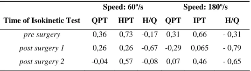
Severity of chondral lesion of individuals included in the sample varied from 1 to 5. However, a low correlation was observed between the severity of macroscopic chondral lesion and QPT, HPT and H/Q ratio on the three assessments at 60º and 180º/s (Table 1).
Table 1. Spearman Correlation (rs value) between severity of chondral lesion and QPT and HPT difference between involved and uninvolved limbs, and H/Q ratio of the involved limb for all three assessments at 60°/s and 180°/s.
Speed: 60º/s Speed: 180º/s
Time of Isokinetic Test QPT HPT H/Q QPT IPT H/Q
pre surgery 0,36 0,73 -0,17 0,31 0,66 - 0,31
post surgery 1 0,26 0,26 -0,67 -0,29 0,065 - 0,79
v. 11 n. 3, 2007 Chondral lesion and peak torque 211
0 5 10 15 20 25 30
QPT 60º/s HPT 60º/s QPT 180º/s HPT 180º/s
%
d
if
fe
re
nc
e
be
tw
e
e
n l
im
bs
PRE SURGERY
POST SURGERY 1
POST SURGERY 2
A
0 10 20 30 40 50 60 70 80 90
H/Q RATIO INVOLVED LIMB 60º/s
H/Q RATIO INVOLVED LIMB 180º/s
% H
/Q
r
a
ti
o
PRE SURGERY
POST SURGERY 1
POST SURGERY 2
B
*
Figure 1. A) Mean percent difference between involved and uninvolved limbs for QPT and HPT for all three assessments at 60°/s and
180°/s; B) Mean H/Q ratio of involved limb for all three assessments at 60°/s and 180°/s.
* Statistically significant difference
DISCUSSION
In the present study no correlations between severity of macroscopic chondral lesion and peak torque of the knee muscles were found in the studied cases. However there was a trend of association between HPT and severity of chondral lesion in the pre-surgical period at 60º/s (rs= 0.73) and 180º/s (rs= 0.66). No data to support such finding were found in the literature. However, Solomonow et al.10 describe a muscle-ligament reflex that would avoid excessive tibial translation by inhibition of quadriceps and activation of hamstrings. Hamstrings would function thus as a dynamic agonists of the ACL in ACL-deficient individuals.
One possible explanation for the lack of influence of severity of macroscopic chondral lesion on peak torque of knee muscles at other assessment times in this study may be the fact that volunteers did not have clinical signals of OA: they had normal radiological exams, no pain or effusion, and performed daily tasks with no complaints. Furthermore, mean age of the individuals included in the sample was 32 years, and perhaps they were not yet suffering the effects of physiological sarcopenia, which is common in OA8.
According to Felson et al.3, cartilage lesions smaller than 2 cm2 remain asymptomatic for a long time. For Shelbourne et al.2, little is known about the natural course of chondral lesions and the time when clinical symptoms first appear. Nevertheless, Slemenda et al.8 studied the relation between decreases in quadriceps strength and OA and found that individuals with clinical symptoms of the disease as well as asymptomatic individuals produced lower quadriceps torque
values. These results suggest that quadriceps weakness may develop even in clinically normal individuals, that is, at the early stages of the disease.
The literature relates ACL rupture and lesions of the joint cartilage and other synovial structures to inhibition of knee muscles. Hurley et al.6 investigated the effects of electrical stimulation applied during maximal voluntary contraction and found evidence of muscle inhibition in all subjects with OA. After an isometric and isokinetic strengthening program there was no statistically significant difference in the degree of inhibition between the involved and noninvolved lower limbs. Additionally, after the program inhibition was not correlated with deficits in the peak isokinetic torque. Lewek et al.5 studied patients with unilateral OA and observed that 50% of the patients in the experimental group failed to totally activate the quadriceps, although the group reached a mean of 93% of total activation. These findings suggest that the cause of weakness remains uncertain. Nevertheless, muscle inhibition, pain, fear that contraction may cause pain and aging effects and the related disuse have also been found to contribute to strength deficits1,7.
212 Traete RF, Pinto KNZ & Mattiello-Rosa SM Rev. bras. fisioter.
and not for the flexors or in the ration between flexors and extensors. Loentzon et al.11, however, studied ACL-deficient individuals and did not find any relationship between joint cartilage degeneration and muscle isokinetic performance. In a prospective study in which individuals were followed for 5 to 9 years after ACL reconstruction, Jarvelan et al.12 reported that manifestations of OA in the individuals were rare, and that results of functional scales reached normal levels. Although there were no statistically significant differences between the three assessment times in torque generation capacity expressed by QPT and HPT at both velocities, it was possible to observe that the deficits in relation to the healthy limb tended to be larger at the post-surgical period 1, both for the quadriceps and the hamstrings. This observation is clinically important since the subjects were receiving of physical therapy intervention at this period. After the rehabilitation period, QPT and HPT tended to return to acceptable values of bilateral difference4. However, there was a statistically significant difference in the H/Q ration of the involved limb at 60º/s between the pre-surgical, post-surgical 1 and post-surgical 2 periods. This difference could be explained by the high quadriceps deficit after ACL injury, in association with the minimum hamstring deficit, leading to an imbalanced H/Q ratio of the involved limb.
Results presented in this study of cases, demonstrate an imbalance in the knee agonist/antagonist relationship after ACL reconstruction. However, the severity of the secondary chondral lesion did not influence the progressive increase in the capacity of knee muscles to generate torque during the studied period. Due to the small sample size more studies should be conducted to investigate whether there is a relationship between chondral lesion and peak torque after injury and reconstruction of ACL.
REFERENCES
1. Tiderius CJ, Olsson LE, Nyquist F, Dahlberg L. Cartilage glycosaminoglycan loss in the acute phase after an anterior cruciate Ligament Injury. Arthritis Rheum. 2005;52(1):120-7.
2. Shelbourne KD, Jari S, Gray T. Outcome of untreated traumatic articular cartilage defects of the knee: A natural history study. J Bone Joint Surg. 2003;85-A Suppl 2:S8-16.
3. Felson DT. Risk factors for osteoarthritis: understanding joint vulnerability. Clin Orthop Relat Res. 2004;427 Suppl:S16-21. 4. Konishi Y, Fukubayashi T, Takeshita D. Possible mechanism of quadriceps femoris weakness in pacients with ruptured anterior cruciate ligament. Medicine & Science in Sports & Exercice. 2002;34(9):1414-8.
5. Lewek MD, Rudolph KS, Snyder-Mackler L. Quadriceps femoris muscle weakness and activation failure in patients with symptomatic knee osteoarthritis. J Orthop Res. 2004;22: 110-5.
6. Hurley MV, Newham DJ. The influence of arthrogenous muscle inhibition on quadriceps rehabilitation of patients with early, unilateral osteoarthritic knees. Br J Reumatol. 1993;32:127-31.
7. Becker R, Berth A, Nehring M, Awiszus F. Neuromuscular quadriceps dysfunction prior to osteoarthritis of the knee. J Orthop Res. 2004;22:768-73.
8. Slemenda C, Brandt KD, Heilman DK, Mazzuca S, Braunstein EM, Katz BP, et al. Quadriceps weakness and osteoarthritis of the knee. Ann Intern Med. 1997;127(2):97-104.
9. Messner K, Gillquist J, Bjornsson S, Lohmander LS. Proteo-glycan fragments in rabbit joint fluid correlated to arthrosis stage. Acta Orthop Scand. 1993;64(3):312-6.
10. Solomonow M, Krogsgaard M. Sensorimotor control of knee stability. A review. Scand J Med Sci Sports. 2001;11(2):64-80. 11. Lorentzon R, Elmqvist L, Sjostrom M, Fagerlund M, Fuglmeyer A. Thigh musculature in relation to chronic anterior cruciate ligament tear: Muscle size, morphology, and mechanical output before reconstruction. Am J Sports Med. 1989;17(3):423-9.