Printed in Brazil - ©2006 Sociedade Brasileira de Química 0103 - 5053 $6.00+0.00
A
r
ti
c
le
*e-mail: [email protected]
Reverse Saturable Absorption in 5,10,15,20-Tetra(4-pyridyl)-21H,23H-porphyrin with
Ruthenium Outlying Complexes
Newton M. Barbosa Neto,*,a Samuel L. Oliveira,b Ilde Guedes,b,c Luis R. Dinelli,d Lino Misoguti,b
Cleber R. Mendonça,b Alzir A. Batista e and Sergio C. Zíliob
a
Instituto de Física, Universidade Federal de Uberlândia, CP 593, 38400-902 Uberlândia-MG, Brazil
b
Instituto de Física de São Carlos, Universidade de São Paulo, CP 369, 13560-970 São Carlos-SP, Brazil
c
Departamento de Física, Universidade Federal do Ceará, CP 6030, 60455-970 Fortaleza-CE, Brazil
d
Fundação Educacional de Barretos, Av. Prof. Frade Monte, 389, 14783-226 Barretos-SP, Brazil
e
Departamento de Química, Universidade Federal de São Carlos, BR-13565-905 São Carlos-SP, Brazil
Modificações no sinal de absorção de estado excitado da 5,10,15,20-tetra(4-piridil)-21H,23H-porfirina, provocadas tanto pela substituição do íon central quanto pela adição de grupos periféricos, são investigadas empregando-se pulsos laser de 5 ns com 532 nm, juntamente com medidas de absorção linear e tempo de decaimento da fluorescência. A absorção saturada reversa (RSA) aumenta consideravelmente quando os íons H+ são substituídos pelo Zn2+ na posição
central do anel porfirínico. Contudo, esta diminui quando os grupos RuCl2(CO)(PPh3)2 e RuCl2(CO)(dppb) são ligados através dos anéis piridínicos às posições periféricas da porfirina, tanto para a base livre quanto para a zinco porfirina. Um modelo teórico baseado no diagrama de Jablonski foi usado para verificar qual a principal causa da diminuição do sinal de RSA provocada pelos grupos de rutênio.
Investigations on the excited state absorption signal of 5,10,15,20-tetra(4-pyridyl)-21H,23H-porphyrin, modified by either changing the central ion or adding peripheral groups, were performed using 5 ns laser pulses at 532 nm, in association with linear absorbance and fluorescence decay time measurements. The reverse saturable absorption (RSA) increases remarkably when Zn2+ replaces H+ at the central position of the porphyrin ring. However, it
decreases when RuCl2(CO)(PPh3)2 and RuCl2(CO)(dppb) groups are attached at outlying positions
through the pyridine rings, for both the free base and zinc porphyrins. A theoretical model based on Jablonski diagram was used to verify the main reason for the decrease on the RSA signal caused by ruthenium groups.
Keywords: porphyrins, ruthenium complexes, optical properties, reverse saturable absorption
Introduction
In the last few decades, special attention has been given to the investigation of nonlinear optical properties of
π-conjugated macrocyclic molecules, due to their potential application in photonic devices.1 Since these compounds
present reverse saturable absorption (RSA), they are potential candidates to be used as optical limiting materials.2-4 RSA is a resonant nonlinear absorption effect
that arises when the absorption cross-section from an excited state up to a higher excited state is larger than the
absorption cross-section from the ground state to it.2 RSA
efficiency depends mainly on the ratio between excited and ground state absorption cross-sections (σexc/σg) and
on the time the population remains in the excited state. Among macrocyclic molecules, porphyrin complexes have deserved special attention because they present strong nonlinear signal, which can be further enhanced by modifying their structures. This can be accomplished by changing their central ion,5 adding groups to outlying
positions6,7 or adding axial ligands to ions at the central
part of the ring.8 Porphyrins are aromatic macrocyclics,
linked to the nitrogen atoms of the ring.9 In particular,
5,10,15,20-tetra(pyridyl)-21H,23H-porphyrin is formed by four pyridyl groups attached to mesocarbons of the macrocycle, as shown in Figure 1.
The RSA effect has been investigated in several porphyrins: basket handle porphyrins,10
tetraphenyl-porphyrins,11 and metalloporphyrins.5,12 Among the
metalloporphyrins, those modified by ruthenium ions have been extensively studied due to their application as biological models. Most of these studies were performed with RuII placed at the center of the porphyrin ring, either
with or without axial ligands.13,14 This produces strong
metal-to-ligand charge transfer bands in the visible region,13 which deeply influence the photophysical
properties of porphyrins. There are some papers reporting on the basic characterization of porphyrins with ruthenium groups linked to outlying positions.15,16 However, none of
them has reported on the influence of RuII complexes at
peripheral positions on the RSA performance of porphyrins.
Recently, we studied a class of porphyrins named 5,10,15,20-tetra(4-pyridyl)-porphyrin (TPyP) with different central ions.17,18 We observed that the free base
(H2TPyP) and the zinc species (ZnTPyP) present a RSA process due to both singlet and triplet excited states, with fluorescence and intersystem crossing times in the nanosecond time scale. This fact is of great importance for optical limiting purpose. Here we use 5 ns laser pulses at 532 nm in an optical limiting configuration,4 in
association with linear absorbance and fluorescence decay time measurements. We investigate the influence of RuII
complexes, placed at the peripheral position of the ring, on the RSA efficiency of H2TPyP and ZnTPyP. Two RuII
species were used: RuCl2(CO)(PPh3)2 and RuCl2(CO) (dppb), hereafter groups A and B, respectively. PPh3 stands
for triphenylphosphine and dppb for 1,4-bis(diphenyl-phosphine)butane. We observed that the RSA signal doubles for Zn-modified porphyrins, while it decreases when any of the RuII groups is added. In order to determine
the main factor responsible for the reduction on RSA signal caused by the presence of RuII groups, we fitted the
experimental data with a simplified five-energy-level model using as adjustable parameters the absorption cross-sections of the first excited singlet and triplet states and the intersystem crossing time.
Experimental
Samples
H2TPyP was synthesized by the following procedure: freshly distilled pyrrole (9.9 g, 0.15 mol) and 4-pyridinecarboxaldehyde (16.1 g, 0.15 mol) were added to 800 mL of refluxing reagent grade propionic acid. After refluxing for 1.5 h, the solution was cooled down to room temperature and filtered, and the filter cake was washed thoroughly with methanol. After a hot water wash, the resulting purple crystals were dried in vacuum to remove absorbed acid. The H2TPyP was purified on an alumina column using chloroform as a solvent and 5% methanol-chloroform as an eluent to yield 2.68 g (14%).
ZnTPyP was synthesized according to the procedure described in reference 19 with the following modifications: 0.216 g (3.5 × 10⁻⁴ mol) of the H
2TPyP and 0.173 g (8.7 ×
10-4 mol) of zinc acetate were dissolved in a mixture of
20 mL of acetic acid and 20 mL of dimethylformamide under reflux. Yield: 0.238 g (92%).
The RuII porphyrin complexes were synthesized under
argon by reaction of H2TPyP with [RuCl2(CO) (PPh3)2(DMF)]20 or [Ru
2Cl4(CO)2(dppb)3]
21 to give group A
and group B complexes, respectively. The solutions were stirred for 4 h and their volume was then reduced by approximately 90%. The solutions were then filtered and the filter cake was washed thoroughly with ethyl ether. Later, the powder obtained was dried in vacuum to give the desired compound. Thus, using the described procedure we synthesized [H2TPyP{RuCl2(CO)(PPh3)2}4] using 20 mg (3.23 × 10⁻⁵ mol) of H
2TPyP with 95 mg (1.29 × 10 -4 mol)
of [RuCl2(CO)(PPh3)2(dmf)] in 20 mL of dichloromethane. Yield: 64% (70 mg); Exp. (Calc.), %C = 62.68 (63.71); %H= 4.33 (4.24); %N = 3.72 (3.23); ν(CO), 1957 cm-1; 31P{1H}, δ 30.4, singlet, in CH
2Cl2 as solvent.
[H2TPyP{RuCl2(CO)(dppb)}4] was synthesized using 20 mg (3.23 × 10⁻⁵ mol) of H
2TPyP and 119.5 mg (6.7 ×
10-5 mol) of [Ru
2Cl4(CO)2(dppb)3]. Yield: 65% (65 mg);
Exp. (Calc), %C = 59.74 (59.93); %H = 4.55 (4.51); Figure 1. Molecular structure of the
%N = 3.71 (3.58); ν(CO), 1956 cm-1; 31P{1H}, δ 44.20
and δ 25.93, doublet, in CH2Cl2 as solvent.
ZnTPyP-A was obtained from the reaction of 10 mg of ZnTPyP (1.47 × 10⁻⁵ mol) with 43.1 mg (5.88 × 10⁻⁵
mol) of [RuCl2(CO)(PPh3)2(dmf)] in 20 mL of CH2Cl2. The mixture was stirred for 4 h, after what the volume of the reaction mixture was reduced to about 2 mL and ether was added to precipitate a violet solid, which was well washed with ether. Yield: 36.10% (19 mg); Exp. (Calc.), %C = 61.40 (61.36); %H= 3.98 (3.95); %N=3.14 (3.03);
ν(CO), 1960 cm-1; 31P{1H}, δ 30.1, singlet, in CH 2Cl2 as
solvent.
Zn-TPyP-B was obtained by the same procedure, using the binuclear [Ru2Cl4(CO)2(dppb)3]in1:2 proportion. Yield: 79% (42 mg); Exp.(Calc.), %C = 58.60 (58.74); %H = 4.36 (4.40); %N = 3.51 (3.72); ν(CO), 1956 cm-1; 31P{1H}, δ
44.61 and δ 25.36, doublet, in CH2Cl2 as solvent.
Fluorescence decay times
Fluorescence decay times were obtained pumping the samples with a 70-ps laser pulse at 532 nm, delivered by a Q-switch Nd:YAG laser, and collecting the signal with a fast detector with a rise time of 0.5 ns, coupled to an optical fiber. The signal was acquired and processed by a 1GHz digital oscilloscope.
Linear and nonlinear absorption measurements
UV-Vis absorption spectra were obtained with a CARY-17 spectrophotometer, with samples dissolved in chloroform and placed in a 0.2 cm cell. Nonlinear absorption measurements were performed, with the solutions placed in a 1 cm cell, using the second harmonic of the Q-switched Nd:YAG laser system producing 5 ns pulses at a 5 Hz repetition rate. The 532 nm laser beam was focused using a 27 cm focal-length lens in an f/135 configuration. The 1 cm cell was used in nonlinear optical measurements to assure that the focal volume was completely located inside the sample. After passing through the sample, the beam was collected by a 6 cm focal-length lens into a photodiode detector, which avoids self-defocusing, plasma and/or cavity bubble scattering contributions. The incoming laser energy was adjusted using a half-wave plate placed between two crossed polarizers; the maximum incoming energy was around 3.5 mJ. The linear transmittance, in the nonlinear absorption measurements, was adjusted at around 60% for all samples. This was performed to assure the same molecular population transfer from the ground to the excited state.
Results and Discussion
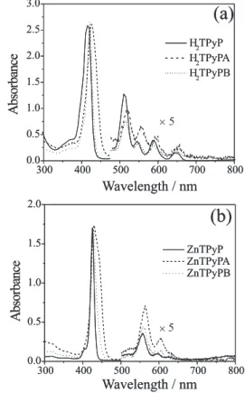
The absorption spectra of the H2TPyP, H2TPyP-A, H2TPyP-B, ZnTPyP, ZnTPyP-A and ZnTPyP-B complexes are shown in Figure 2a and 2b, where the characteristic B band (Soret band) located around 400 nm and Q bands located between 470 and 700 nm are observed. The Soret band is related to a strongly allowed π-π* electronic transition, while the Q band to a quasi-allowed one. In fact, the Q-band is enhanced by a vibronic coupling to the B band.9 For H
2TPyP, H2TPyP-A and H2TPyP-B, the
Q-band presents four peaks: Qx(0,0) and Qy(0,0) with their respective vibronic overtones Qx(1,0) and Qy(1,0). On the other hand, a considerable modification on the spectrum is observed for zinc porphyrins, with the Q band presenting just two peaks: Q(0,0) and its respective vibrational overtone Q(1,0). This results from the increase of the overall molecular symmetry that changes from D2h to D4h when Zn+2 substitutes for 2H+.9 Also, just a small red shift
in Soret and Q bands, caused by ruthenium outlying groups, is observed.
when Zn2+ ion replaces for 2H+, while it decreases when
RuII outlying groups are present, exhibiting a worthless
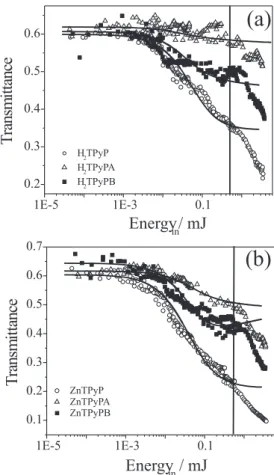
signal for porphyrins with group A, for both H2TPyP and ZnTPyP. This can be observed from the figure of merit (FM) in Table 1, which is defined as the ratio between the minimum and the linear transmittance values.
The RSA mechanism is a resonant absorption effect involving ground and excited states, which can be understood, at nanosecond time scale, considering a five-energy-level diagram known as the Jablonski diagram,9 shown in Figure
4. It indicates the possible nonradiative and radiative processes that include intersystem crossing, internal conversion, radiative decay and excited state absorption.
Initially, light promotes the molecules from the ground state S0 to the first excited singlet state (S1). From this state, the molecule can return back to the state S0, be excited to the state S2, or yet undergo an intersystem crossing relaxation to the state T1. From T1, the molecule can return to S0 or, by absorbing another excitation photon, can be promoted to T2. If sufficient population is transferred to S1 or T1, and the excited state absorption
cross-sections of these states are larger than the ground ones, RSA arises. In particular, for nanosecond pulses, RSA in porphyrin molecules is related to both singlet and triplet excited state absorption. Therefore, the efficiency of the RSA process depends on high σexc/σg values and on
the time the population remains in the lower excited state. To determine the main factor responsible for the reduction on RSA signal caused by the presence of RuII groups, we
adjusted the experimental results with a simplified five-energy-level model based on the Jablonski diagram.
In this model, the decay from state T1 to S0 is neglected once the triplet state (T1) lifetime is in the order of some microseconds for porphyrins, being much slower than the pulse duration. Moreover, as the relaxation times of the higher excited state (S2 and T2) are very fast, the population buildup is neglected for these states. Consequently, the rate equations used to describe the population dynamics are
(1a)
(1b)
(1c)
where is the upward transition rate, τisc is
the intersystem crossing time, and τS
10 is the relaxation
time from the first excited singlet state to the ground state. Besides, τS
10 is related to τisc through the relation τf –1 = τisc
–1 + τS 10
–1, where τ
f is the measured fluorescence time.
Beer´s law equation, which governs the change in the pulse intensity as it propagates through the sample, is given by
(2) Figure 4. Jablonski diagram. Si represents the singlet states while Ti represents the triplet states. σS
exc, σ T
exc are singlet and triplet excited state
absorption cross-sections, respectively. σg is the ground state absorption
cross-section and τisc is the intersystem crossing time.
where N is the molecular concentration and I is the temporal Gaussian intensity profile. σg, σ
S exc and σ
T exc are
the absorption cross-sections for ground, first singlet and first triplet excited state, respectively. The transmittance is obtained from the ratio between incoming and outgoing pulse intensities.
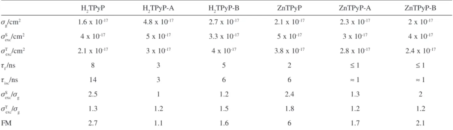
After performing numerical calculations with equations 1 and 2, we were able to obtain the excited state absorption cross-sections and the intersystem crossing time as adjustable parameters. The ground state absorption cross section and fluorescence decay time were obtained from absorbance spectra and fluorescence decay time measurements. The spectroscopic parameters values are listed in Table 1.
In order to test the reliability of the procedure we performed fittings for H2TPyP and ZnTPyP with no ruthenium groups using the spectroscopic parameters obtained from references 17 and 18. The fittings (solid lines) are also shown in Figure 3. A discrepancy between experimental and theoretical fittings for all samples is observed for incoming energies above 0.5 mJ. This is explained by the strong cavitation bubble and thermal lens refraction contributions present in high energy regime.22
The increase in RSA performance caused by introducing Zn2+ in the porphyrin ring was expected and
has already been reported for other porphyrins.4,5,11 This
enhancement is attributed to the well known heavy atom effect.23-25 Adding heavy atoms to the porphyrin structure
enhances the spin-orbit coupling, increasing the intersystem crossing rate. This effect leads to an increase of the excited triplet state population, which in general presents a higher excited state absorption cross-section.23
From Table 1 we observe that Zn2+ increases the ratio
between the first triplet excited and ground state absorption cross-sections. However, the increase in the ratio of absorption cross-sections is smaller than that observed for the figure of merit. The explanation for this difference
comes from the fact that in calculating the figure of merit we considered the minimum transmittance that has contributions from other effects.
On the other hand, when RuII complexes are attached
to the pyridyl groups of both H2TPyP and ZnTPyP, we observe a deleterious effect on the RSA signal, being worst for group A. The results obtained from the theoretical fitting show that the RuII complexes reduce the σ
exc/σg ratio
at 532 nm, mainly due to the population of the first singlet excited state. Although the outlying groups reduce the fluorescence time, they still are in the order of a few nanoseconds, and this points out that the principal contribution comes from the reduction on the absorption cross-section ratios. Moreover, the fact that the RSA signal decreases for both H2TPyP and ZnTPyP indicates that the deleterious influence of ruthenium groups is independent of the central ion, at least for closed shell central ions in regular porphyrins.9
Conclusions
We investigated the influence of peripheral RuII
complexes on the RSA performance of H2TPyP and ZnTPyP, both regular type porphyrins. We observed that the RSA signal decreases when RuII groups are attached
to peripheral position of the porphyrins. The increase in the RSA observed for ZnTPyP without ruthenium groups agrees with other works reported in the literature, being explained by means of the heavy-atom effect. We observed from a simple theoretical model that the reduction of RSA signal due to the presence of peripheral groups is caused by the reduction of the ratio between excited and ground states absorption cross-sections, mainly for singlet excited state.
Theoretical fittings also show that for high energy regime the optical limiting behavior of porphyrins should have contributions from additional effects besides RSA. Table 1. Spectroscopic parameters and figure of merit for H2TPyP and ZnTPyP complexes
H2TPyP H2TPyP-A H2TPyP-B ZnTPyP ZnTPyP-A ZnTPyP-B
σg/cm
2 1.6 x 10⁻¹⁷ 4.8 x 10⁻¹⁷ 2.7 x 10⁻¹⁷ 2.1 x 10⁻¹⁷ 2.3 x 10⁻¹⁷ 2 x 10-17
σS
exc/cm
2 4 x 10-17 5 x 10-17 3.3 x 10⁻¹⁷ 5 x 10-17 3 x 10-17 4 x 10-17
σT
exc/cm
2 2.1 x 10⁻¹⁷ 3 x 10-17 4 x 10-17 3.8 x 10⁻¹⁷ 2.8 x 10⁻¹⁷ 2.4 x 10⁻¹⁷
τf/ns 8 3 5 2 ≤ 1 ≤ 1
τisc/ns 14 3 6 6 ≈ 1 ≈ 1
σS
exc/σg 2.5 1 1.2 2.4 1.3 2
σT
exc/σg 1.3 1.2 1.5 1.8 1.2 1.2
Probably, two of the most important additional processes to the optical limiting mechanisms are the scattering caused by cavitation bubbles and the thermal nonlinear refraction. The results also indicate that both ZnTPyP and H2TPyP are promising candidates to be applied as optical limiting materials, once they have a very small energy threshold. However, molecular engineering strategies are required in order to increase the ratio between the absorption cross-sections.
Acknowledgments
The authors thank Fundação de Amparo à Pesquisa do Estado de São Paulo (FAPESP) and Conselho Nacional de Pesquisa (CNPq) for financial support. Dr. I. Guedes is grateful to the Departamento de Física of Universidade Federal do Ceará and FAPESP (grant # 2004/02067-6) for support during his stay at the Instituto de Física de São Carlos – USP.
References
1. Potenza, G.; Sona, A.; Nuovo Cimento 1965, 38, 1438. 2. Perry, J. W. In Nonlinear Optics of Organic Molecules and
Polymers, Nalwa, H. S.; Miyta, S., eds.; CRC Press: Boca Raton, 1997, pp. 813.
3. Shirk, J. S.; Pong, R. G. S.; Bartolli, F. J.; Snow, A. W.; Appl. Phys. Lett. 1993, 63, 1880.
4. Calvete, M.; Yang, G. Y.; Hanack, M.; Synth. Met. 2004, 141, 231.
5. Mishra, S. R.; Rawat, H. S.; Laghate, M.; Opt. Commun. 1998, 147, 328.
6. Su, W.; Cooper, T. M.; Brant, M. C.; Chem. Mater. 1998, 10, 1212.
7. Mcewan, K. J.; Bourhill, G.; Robertson, J. M.; J. Nonlinear Phys. Mater. 2000, 9, 451.
8. Kiran, P. P.; Reddy, D. R.; Maiya, B. G.; Dharmadhikari, A. K.; Kumar, G. R.; Desai, N. R.; Appl. Optics 2002, 41, 7631.
9. Kalyanasundaram, K.; Photochemistry of Polypyridine and Porphyrin Complexes, Academic Press: San Diego, 1992. 10. Sevian, A.; Ravikanth, M.; Kumar, G. R.; Chem. Phys. Lett.
1996, 263, 241.
11. Blau, W.; Byrne, H.; Dennis, W. M.; Kelly, J. M.; Opt. Commun. 1985,56, 25.
12. Martin, R. B.; Li, H.; Gu, L.; Kumar, S.; Sanders, C. M.; Sun, Y.-P.; Opt. Mater. 2005, 27, 1340.
13. Rodriguez, J.; McDowell, L.; Holten, D.; Chem. Phys. Lett. 1988, 147, 235.
14. Antipas, A.; Buchler, J. W.; Gouterman, M.; Smith, P.D.; J. Am. Chem. Soc. 1978, 100, 3015.
15. Steiger, B.; Shi, C.; Anson, F.C.; Inorg. Chem. 1993, 32, 2107. 16. Araki, K.; Angnes, L.; Azevedo, C.M.N.; Toma, H. E.; J.
Electroanal. Chem. 1995, 397, 205.
17. Barbosa Neto, N. M.; De Boni, L.; Rodrigues Jr., J. J.; Misoguti, L.; Mendonça, C. R.; Dinelli, L. R.; Batista, A. A.; Zílio, S. C.; J. Porphyr. Phthalocyanines 2003, 7, 452.
18. Barbosa Neto, N. M.; De Boni, L.; Mendonça, C. R.; Misoguti, L.; Queiroz, S. L.; Dinelli, L. R.; Batista, A. A.; Zílio, S. C.; J. Phys. Chem. B 2005, 109, 17340.
19. Fleischer, E. B.; Inorg. Chem. 1962, 1, 493.
20. Wohnrath, K.; Batista, A. A.; Ferreira, A. G.; Zukerman-Schpector, J.; De Oliveira, L. A. A.; Castellano, E. E.; Polyhedron 1998, 17, 2013
21. Bressan, M.; Rigo, P.; Inorg. Chem. 1975, 14, 2286. 22. Sendhil, K.; Vijayan, C.; Kothiyal M. P.; Opt. Laser Technol.
2006, 38, 512.
23. Perry, J. W.; Mansour, K.; Marder, S. R.; Perry, K. J.; Alvarez Jr., D.; Choong, I.; Opt. Lett. 1994, 19, 625.
24. Shirk, J. S.; Pong, R. G. S.; Bartolli, F. J.; Snow, A. W.; Appl. Phys. Lett. 1993, 63, 1880.
25. Brannon, J. H.; Magde, D.; J. Am. Chem. Soc. 1980, 102, 62.
Received: October 4, 2005
Published on the web: September 26, 2006