The Compensatory Immune-Regulatory Reflex System (CIRS)
in Depression and Bipolar Disorder
Michael Maes1,2,3 &Andre F. Carvalho4,5
Received: 12 January 2018 / Accepted: 16 March 2018
#Springer Science+Business Media, LLC, part of Springer Nature 2018
Abstract
Here, we review a novel concept namely the compensatory immune-regulatory reflex system (CIRS) as applied to the patho-physiology of major depressive disorder (MDD) and bipolar disorder (BD). There is evidence that a substantial subset of individuals with MDD and BD exhibit an activation of the immune-inflammatory response system (IRS), as indicated by an increased production of macrophagic M1 and T helper (Th)-1 pro-inflammatory cytokines, interleukin (IL)-6 trans-signaling, positive acute phase proteins (APPs), and complement factors. These immune aberrations appear to be evident during the course of major affective episodes of either depressive or (hypo) manic polarity. Here, we review (a) the current state of the art of CIRS functions in both mood disorders and (b) the possible role of CIRS-related biomarkers for the understanding of affective disorders within the framework of precision psychiatry that could also provide novel drug targets for both MDD and BD. CIRS-related abnormalities in mood disorders include elevated Th-2 and T regulatory (Treg) activities with increased IL-4 and IL-10 produc-tion, classical IL-6 signaling, increased levels of sIL-1R antagonist (sIL-1RA), soluble IL-2 (sIL-2R) and tumor necrosis factor–α- receptors, and positive APPs, including haptoglobin, hemopexin,α1-acid glycoprotein,α1-antitrypsin, and cerulo-plasmin. It is concluded that CIRS is involved in MDD and BD by regulating the primary immune-inflammatory response, thereby contributing to spontaneous and antidepressant-promoted recovery from the acute phase of illness. Signs of activated IRS and CIRS pathways are observed in the remitted phases of both disorders indicating that there is no return to the original homeostasis after an acute episode, while later episodes of mood disorders are characterized by sensitized IRS and CIRS responses. New z-unit weighted composite biomarker scores are proposed, which reflect different aspects of IRS versus CIRS activation and may be used to estimate different IRS/CIRS activity ratios in mood and other neuroimmune disorders.
Keywords Major depression . Bipolar disorder . Inflammation . Neuroimmune . Cytokines . Oxidative and nitrosative stress
Introduction
In 1987, the first author of this conceptual analysis started to explore why major depressive disorder and bipolar disorder are accompanied by signs of immunosuppression, including but not limited to lowered lymphocyte transformation (stimulation) tests (LTT) and blunted natural killer cell activity (NKCA) [1,2]. In a first attempt to delineate the factors that may modulate lowered LTT outcomes in depression, we mea-sured lymphocyte responses to different mitogens, including pokeweed mitogen (PWM), phytohaemagglutinin A (PHA), and concanavalin A (CON A), and examined possible effects of increased activity of the hypothalamic–pituitary–adrenal (HPA)- axis as well as noradrenergic imbalances in depres-sion. Lymphocyte proliferation in response to PWM, PHA, and CON A was frequently used methods to measure ex vivo activity of cell-mediated immunity (CMI), namely * Michael Maes
[email protected];https://scholar.google.com.br/ citations?user=1wzMZ7UAAAAJ&hl=pt-BR&oi=ao
1
Department of Psychiatry, Faculty of Medicine, Chulalongkorn University, Bangkok, Thailand
2 Department of Psychiatry, Medical University of Plovdiv, Plovdiv, Bulgaria
3
IMPACT Strategic Research Centre, School of Medicine, Deakin University, PO Box 281, Geelong, Vic 3220, Australia
4
Department of Psychiatry, Faculty of Medicine, University of Toronto, Toronto, ON, Canada
5 Centre for Addiction and Mental Health (CAMH), Toronto, ON M6J 1H4, Canada
the functional activity of T cells [3]. Two major pathways that regulate immune cell functions constitute neuroendocrine immunomodulation via activation of the HPA-axis and catechol-amine spillover during sympatho-adrenal stress (SAS) [4]. Therefore, we assessed the possible intercorrelations between LTT results and post-dexamethasone cortisol (DST) levels, an index that proxies HPA-axis activity, and 3-methoxy-4-hydroxyphenylglycol (MHPG) excretion in 24-h urine. The assay of MHPG in 24-h urine (a major metabolite of noradrenaline) is a measure of SAS activity in patients with severe depression [4]. Glucocorticoids and catecholamines are known to exert negative feedback upon the immune system by as yet incompletely elucidated mechanisms [5,6]. It was evi-denced that a large proportion of the variance of lymphocyte responses to these three mitogens could be attributed to both glucocorticoid and MHPG levels and that lowered lymphocyte transformation in depression could be at least partly explained by increased HPA-axis activity [4]. Based on these and other find-ings, we suggested that an increase in HPA-axis activity observed in a subset of patients with depression could exert a negative feedback on CMI functions thereby explaining lowered lympho-cyte transformation test results in depression.
In 1988, we started new research projects, namely the as-says of cytokines, which were known to modulate CMI and activate HPA-axis activity [5, 7]. The cytokines of interest were interleukin-2 (IL-2) and IL-1β since it was already known that T cell proliferation is in part regulated by IL-1β
and IL-2, while the same cytokines also activate HPA-axis activity [5]. IL-1βis produced by activated immune cells and plays a key role in the initiation of inflammatory process-es, with an acknowledged role in the pathophysiology of immune-inflammatory and neurodegenerative disorders [8, 9]. Since at that time IL-1βwas very difficult to measure in serum/plasma, we assayed IL-1β in culture supernatants of stimulated lymphocytes or whole blood [5]. Furthermore, plasma IL-2 levels were/are difficult to assay in plasma, and therefore, we also measured plasma sIL-2 receptor (sIL-2R) levels in patients with MDD, a surrogate marker of IL-2-related T cell activation [5,7]. The production and re-lease of sIL-2Rs into plasma are strongly correlated with the activation state of lymphocytes and the expression of IL-2Rs on immune cells [10,11]. Our a priori hypotheses were that (a) both stimulated whole blood IL-1βproduction and plasma sIL-2R and IL-2 levels could be decreased in depression in tandem with lowered LTT tests and (b) glucocorticoids could down-regulate the production rates of IL-1βand sIL-2R.
In contrast to our primary hypotheses, our results showed an increased production of IL-1βand sIL-2R and an increased rate of measurable plasma IL-2 levels in MDD, pointing to peripheral immune activation rather than immunosuppression [5, 7]. In addition, we observed that lymphocyte responses were significantly lower in patients with MDD than in indi-viduals with less severe forms of depression (adjustment
disorder and dysthymia) and that dexamethasone administra-tion reduced the producadministra-tion of IL-1βand sIL-2R in healthy controls, but not in participants with depression [5,7]. The latter findings provided evidence that the production of IL-1β
and sIL-2R in depression could be resistant to the immune-suppressive effects of dexamethasone. This was ex-plained by the knowledge that resistance of immune cells to suppression by dexamethasone is a marker of T cell activation and because activated lymphocytes are generally less sensitive to suppression by glucocorticoids [12,13]. Moreover, we found that this glucocorticoid-resistant state in depression could be ex-plained by aberrations in IL-1βand IL-2-related mechanisms, which are known to attenuate glucocorticoid-induced inhibition of immuno-proliferative responses [13,14].
These findings opened relevant as yet unanswered ques-tions namely (a) are depression and bipolar disorder really accompanied by immune activation or an inflammatory re-sponse? and (b) how could one reconcile findings that sug-gested immunosuppression (i.e., lowered LTT and blunted NKCA) to those that pointed to an increase in immune acti-vation (i.e., increased IL-1βand sIL-2R production) in de-pression? Since 1989, we therefore performed targeted re-search projects aimed to further assess the balance of immune activation in relation to immunosuppression.
In different projects (1990–2001), we showed that depres-sion is accompanied by immune activation, including a chron-ic mild immune-inflammatory response as indchron-icated by in-creased levels of the pro-inflammatory cytokines IL-1β, IL-6, tumor necrosis factor (TNF)-α, increased levels of acute phase proteins, including haptoglobin (Hp), ceruloplasmin (Cp), fibrinogen (Fb), hemopexin (Hpx), α1-antitrypsin (α1AT) andα1-acid glycoprotein (α1S), and increased levels of complement factors [15–18]. There are now many meta-analyses showing that depression and BD are accompa-nied by an immune-inflammatory response with increased levels of pro-inflammatory cytokines, acute phase proteins, a n d o t h e r c o m p o u n d s r e l e a s e d b y a n a c t i v a t e d immune-inflammatory response system (IRS) [19–28].
novel conceptual framework in an attempt to incorporate more recent findings in this expanding field of research.
Compensatory Immune-Regulatory Reflex
System
Figure1 shows that sepsis, burn, and trauma-induced tissue injuries are accompanied by acute inflammation initially aimed to eliminate triggering factors (e.g., infectious agents), a condition referred to as the systemic inflammatory response syndrome (SIRS) [30]. The SIRS is accompanied by an in-creased susceptibility to infections coupled to lowered lym-phocytic functions, named autoimmunosuppression [30]. The latter response which tends to deactivate the SIRS and which aims to restore immune homeostasis is conceptualized as the compensatory anti-inflammatory response syndrome (CARS) [30,31]. In other words, SIRS is an acute pro-inflammatory syndrome, while the CARS is an anti-inflammatory response, which aims to dampen an overzealous inflammatory response [30]. One of the most important actors of the CARS is IL-10, an immunosuppressive cytokine, while other CARS-related biomarkers include lowered mitogen-induced lymphocyte re-sponses, T cell anergy and macrophage paralysis, increased negative feedback exerted by cortisol levels, a shift from Th-1 to Th-2 cells, increased transforming growth factor (TGF)-βand prostaglandin E2 (PGE2) production, and in-creased Treg cells [30,32–35].
In individuals suffering from sepsis, imbalances between SIRS and CARS-related pathways, through an overzealous immune activation and a failure or dysregulation of
compensatory immune mechanisms, may underpin uncon-trolled inflammation that could ultimately cause death [30]. One complication of SIRS/CARS is that some patients may suffer from a persistent inflammation, immunosuppression, and catabolism syndrome (PICS) condition that could be par-ticularly associated with detrimental outcomes [36, 37]. Interestingly, a meaningful group of patients with PICS also exhibit co-occurring neuroendocrine alterations, pain, depres-sion, psychological distress, fatigue, delirium, and a cachexia phenotype [38]. Potential markers of PICS may include per-sistently increased levels of C-reactive protein (CRP), lowered creatinine height index, lowered albumin and prealbumin, and retinol binding protein (RBP) [36].
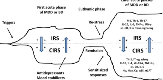
In 1995, we proposed that depression is not only character-ized by a mild immune-inflammatory response with increased production of M1 macrophagic and Th-1 cytokines, acute phase proteins, and complement factors, but also by mild im-munosuppression (attenuated LTT and NKCA), as well as changes in adaptive immune biomarkers which could exert negative immunoregulatory effects and signs of protein catab-olism [15]. Biomarkers of this negative immunoregulatory response in depression are (among others) increased levels of sIL-2R, haptoglobin (Hp), prostaglandin E2 (PGE2), and cortisol. Subsequently, we and other research groups detected more immune-regulatory biomarkers, which are released dur-ing the immune-inflammatory response in depression, includ-ing sIL-1R antagonist (sIL-1RA) and sTNF-R levels (see be-low). All these compounds have immunosuppressive func-tions thereby attenuating the primary immune-inflammatory response [15,29]. Indicants of protein catabolism in depres-sion are anorexia and weight loss, lowered plasma levels of
Fig. 1 The compensatory anti-inflammatory response syndrome (CARS). Sepsis, burn, and trauma-induced tissue injuries are accompa-nied by a systemic inflammatory response syndrome (SIRS) character-ized by M1 macrophagic activation with increased production of tumor necrosis factor (TNF)-α, interleukin (IL)-1βand IL-6, increased release of HMGB1, a T helper (Th)-1 response with increased levels of interferon
albumin, transferrin and retinol-binding protein (RBP) as well as significant associations between lowered albumin and anorexia-weight loss [39–41].
All in all, there are some parallels between the SIRS/CARS/PICS in sepsis, burn, and severe trauma and the immune-inflammatory response syndrome (IRS), signs of im-munosuppression, and protein catabolism which are frequent-ly observed in patients with mood disorders, although in a significantly lowered magnitude compared to aberrations ob-served in patients with SIRS/CARS/PICS. Based on the CARS concept in acute sepsis and the recent findings of a similar, albeit much less severe, process in depression, we formulated the CARS concept in depression and named this concept Bthe compensatory inflammatory reflex system (CIRS)^ [29] or Bcompensatory immune-regulatory reflex system.^ We will now review the current state of the art of the immune-arm of the CIRS.
Interleukin-1 and the Interleukin-1 Receptor
Antagonist
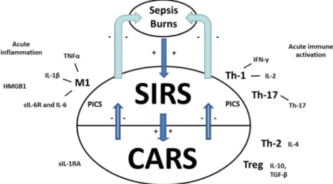
Figure2shows that macrophages exist in functionally distinct states, including M1 and M2 macrophages [42,43]. M1 mac-rophages produce pro-inflammatory cytokines, including IL-1β, IL-6, and TNF-α, and promote a Th-1 response to produce interferon (IFN)-γ. In contrast, M2 macrophages have negative immunoregulatory properties and are involved in Th-2 like responses while inhibiting IL-1 release and in-creasing the production of the IL-1RA [43]. IL-1βis produced by activated immune cells and plays a key role in the onset of inflammatory processes and the acute phase response thereby increasing the production of positive acute phase proteins in-cluding Hp and CRP. Moreover, IL-1 activates the production
of IFN-γand TNF-αand appears to play a pivotal role in the onset of many diseases with a meaningful inflammatory com-ponent including rheumatoid arthritis, diabetes, and cardio-vascular disorders. IL-1RA is produced by cells that also produce IL-1 and is stimulated by IL-1 and other cyto-kines including IL-6 and IFN-γ[41]. The IL-1RA is secreted into the serum by activated immune cells, and the soluble form of IL-1RA (sIL-1RA) may competitively inhibit IL-1β bind-ing to its receptor thereby attenuatbind-ing IL-1βsignaling [41]. Serum sIL-1RA is an endogenous inhibitor of IL-1 signaling and plays a key role in the promotion of tissue repair in dis-orders accompanied by increased IL-1βas well as in the res-olution of the inflammatory response [44]. As such, IL-1RA attenuates the pro-inflammatory effects of IL-1βand therefore plays a role in IL-1β-associated conditions including inflam-matory and autoimmune disorders [43].
In 1991, we published the first paper showing increased stimulated production of IL-1β in major depression [5]. Increased stimulated production was consequently de-scribed in dysthymic disorder [45]. Also, other studies reported increased IL-1β production in depression, early-onset depression, or depression in elderly individuals [46–49]. In major depression, increased levels of serum IL-1 are significantly associated with the number of pre-vious depressive episodes, suggesting that a higher produc-tion of IL-1 may contribute to the recurrence of major depressive episodes and to neuroprogression, which ap-pears to be evident in at least a subset of patients with MDD [43]. Administration of IL-1 to rodents may induce a number of depressive-like behaviors including anorexia and weight loss, psychomotor retardation, fatigue, anxiety, impaired cognition, memory disturbances, altered sleep patterns, impaired social behaviors, lowered sexual behav-iors and interest, and anhedonia [43].
Fig. 2 Interplay between macrophage polarization and T helper (Th) and T regulatory cells. M1/M2: M1/M2 macrophages; IL: interleukin; IL-1RA: IL-1 re-ceptor antagonist; TNF: tumor necrosis factor; TGF:
In 1995, we published the first paper that sIL-RA is elevat-ed in patients with MDD [50]. Interestingly, the elevations in serum sIL-1RA and IL-6 levels were strongly related suggest-ing that the release of IL-1RA into the serum could be at least partly due to immune activation [51]. Recently, it was pro-posed that increased sIL-1RA levels could be a trait-related biomarker of major depression (i.e., levels of this immune mediator were elevated even during affective remission), while there were no significant differences between the ele-vated sIL-1RA levels among patients with depression and bipolar disorder [52,53]. In BD, increased sIL-1RA levels were evident in either bipolar depression or acute mania, while sIL-1RA levels in the euthymic phase were somewhat but not significantly higher than in controls [53]. A first meta-analysis study reported that depression is associated with both in-creased serum IL-1 and IL-1RA concentration levels [19]. Recent meta-analyses show that sIL-1RA levels are consis-tently increased in patients with acute phases of MDD [26] or BD [27].
All in all, these and other results underscore that both MDD and BD appear to be accompanied by increased IL-1β pro-duction and sIL-1RA release [54–57] and thus that both dis-orders are accompanied by M1 activation and simultaneous reflex inhibition of IL-1 signaling [29,43]. However, there is a paucity of data to indicate whether the IL-1β/sIL-1RA ratio could be increased, decreased, or unchanged among individ-uals with mood disorders compared to healthy controls. Such information would be of paramount importance to know whether mood disorders are accompanied by increased IL-1 signaling or increased regulation of IL-1 signaling. There is only one study showing that in males with depression, the IL-1RA/IL-1βratio may be significantly increased, thus sug-gesting increased negative feedback [58]. Clearly, the field awaits replicated evidence of this interesting finding.
TNFα
and Soluble TNF Receptors
TNF-αis a pro-inflammatory cytokine produced and released during immune-inflammatory responses especially by mono-cytes and macrophages, but also by T cells and fibroblasts [59]. TNF-αplays a key role in the host response to infectious pathogens and cancer by inducing necrosis or apoptosis [59, 60]. Increased levels of TNF-αmay drive monocytic differ-entiation, activation of IL-1 and IL-6 production, T cell liferation, an acute phase response, increased antibody pro-duction, modifications of neuronal synaptic transmission, in-creased angiogenesis and hypervascularization, fibroblast ap-optosis, cardiac myocyte death, and activation of adipocytes [59–62]. Elevated levels of TNFαmay exert host-damaging effects as observed in autoimmune disorders, sepsis, as well as cancer-related cachexia and have been regarded as potential biomarkers of progression of autoimmune diseases including
rheumatoid arthritis, inflammatory bowel disease, and multi-ple sclerosis [59,60].
TNF-αexerts its effects by binding to 55 kDa (TNF-R1) or 75 kDa (TNF-R2) cell membrane receptors [59]. Both TNF receptors are shed into the peripheral blood after proteolytic cleavage and readily assayed as soluble receptors, namely sTNF-R1 and sTNF-R2 [63]. The serum/plasma concentra-tions of both soluble receptor levels seem to be significantly i n c r e a s e d i n d i s p a r a t e c h r o n i c a n d a c u t e immune-inflammatory conditions, including infections and some cancers [63]. Increased levels of sTNF-Rs may be a reliable surrogate marker of the magnitude of the immune-inflammatory response, and hence has been pro-posed as a biomarker related to the severity of a number of immune disorders accompanied by tissue injury [64]. Important immune-inflammatory signals that cause shedding of those receptors thereby increasing sTNF-Rs are TNFα it-self, IL-1, IL-2, IL-6, IL-10, and LPS [63].
sTNF-R1 and sTNF-R2 may bind free circulating TNF-α
and act as decoy receptors reducing TNF-αbioactivity and signaling [65,66]. sTNF-R1 and sTNF-R2 compete with the TNF receptors expressed on cells for TNF binding thereby attenuating TNF signaling. In addition, by shedding the extra-cellular TNF-R parts, immune cells may become less sensitive to TNF-α binding [63]. Increased levels of sTNF-Rs may increase TNF-αclearance from the peripheral blood through kidney excretion of the TNF-α/sTNF-R complex. Finally, in-creased sTNF-R2 levels may block the entry of TNF-αinto the brain [67]. As such, sTNF-R1 and sTNF-R2 may protect against TNF-α-induced toxicity during sepsis. For example, a recombinant soluble TNF-R may protect mice from the detri-mental effects of endotoxemia [68].
have sTNF-R2 levels which are not significantly different from controls [53]. These findings suggest that during the acute phase of illness, the negative immunoregulatory feed-back exerted by sTNF-R2 may counteract the IRS and thus could play a role in the achievement of remission of manic and depressive episodes. The findings suggest that elevated sTNF-R1 and sTNF-R2 levels could be state markers for ma-jor depression and bipolar disorder and severity and staging of illness.
All in all, increased levels of sTNF-R1 and sTNF-R2 in MDD and BD may exert an increased negative feedback upon TNF-αsignaling. Nevertheless, whether the TNF-α/sTNF-R1 or TNF-α/sTNF-R2 ratios are changed in MDD and BD re-mains unknown. Therefore, it is unclear whether pro-inflammatory IRS signals or CIRS-related signals prevail in TNF signaling pathways in patients with affective disorders.
Interleukin-2 and Interleukin-2 Receptor
Following activation by macrophages or antigens, interleukin-2 (IL-2) is produced by Th cells, especially Th-1 cells, and by NK cells. IL-2 is a major immunomodulatory cytokine that acts in an autocrine fashion and promotes the proliferation and differentiation of T cells into memory cells and effector T cells [74–77]. As such, IL-2 is a main growth factor for T cells that play a key role in T cell memory and cytotoxicity [74–77]. IL-2 stimulates not only Th cells, but also monocytes, NK, and B cells, and also activates T cells to produce other cytokines, including IFN-γ. Furthermore, IL-2 has direct immune-regulatory functions through the acti-vation of Treg cells and increased production of IL-4, while in the thymus, IL-2 may prevent the onset of autoimmunity by activating Treg cells [74–77]. As such, IL-2 is a major regu-lator of CMI and a key mediator in the achievement of im-mune tolerance, and hence may play relevant roles not only in mounting but also in dampening immune responses [74–76, 78].
The effects of IL-2 are mediated by its binding to high-affinity IL-2 receptors (IL-2Rα, CD25), which are expressed on the cell membrane of activated T cells [77,78]. Proteolytic cleavage of the membrane IL-2Rαmay release IL-2R in the circulation, and, as a consequence, its soluble counterpart (sIL-2R, sTAC, sCD25) may be measured in plas-ma. The plasma levels of sIL-2R are proportional to the mem-brane expression of IL-2R and production of IL-2, and there-fore, their plasma levels may be used as a surrogate marker for T cell activation. Moreover, increased sIL-2R levels are a bio-marker of T cell activation that is useful in different (auto) immune disorders, including sarcoidosis, rheumatoid arthritis and systemic lupus erythematosus, and cancers, including na-sopharyngeal carcinoma and malignant melanoma [74–77]. Plasma sIL-2R levels are also associated with disease
activity and progression and response to treatment in these disorders [74–77].
sIL-2Rs may bind circulating IL-2 levels and form a sIL-2R-IL-2 complex thereby lowering IL-2 concentrations a n d e x e r t i n g n e g a t i v e ( i . e . , c o m p e n s a t o r y ) immune-regulatory effects [74,75]. Early studies showed that sIL-2R levels may suppress IL-2-induced proliferation and cy-totoxicity and NKC functions [79]. Moreover, the sIL-2R–IL-2 is biologically active and may enhance some regulatory func-tions of IL-2, including promoting differentiation into immu-nosuppressive Treg cells, upregulating the expression of FoxP3 on CD4 + T cells and inhibiting CD8+ T cell functions [80–82]. On the other hand, sIL-2R could also enhance IL-2-mediated T cell proliferation and serve as a carrier protein thereby decreasing proteolytic IL-2 degradation and prolonging the half-life and thus biological activity of IL-2 [82]. As such, sIL-2R levels may have immunosuppressant, immunoregulatory, and immunostimulatory effects depending on the underlying disease process [82]. Nevertheless, most studies report that sIL-2Rs have inhibitory effects, including lowered cytotoxicity and T cell proliferation and increased ap-optosis, thereby attenuating an ongoing inflammatory process. In 1990, the first report was published that major depres-sion is accompanied by increased expresdepres-sion of CD25+ on peripheral T lymphocytes and increased plasma IL-2 and sIL-2R levels [7]. In a follow-up study, other indicants of T cell activation were found in depression, such as an increased number and percentage of CD2 + HLADR+ T, CD7 + CD25+ cells, CD4 + CD45RA memory cells, HLA-DR, and CD25+ T cells [83,84]. In fact, using machine learning techniques, it was shown that major depression is a qualitatively different class than healthy controls with regard to these biomarkers of immune activation [83,84]. sIL-2R levels are also significant-ly elevated in patients with BD during mania [17]. There are now different meta-analyses showing that peripheral blood sIL-2R levels are increased in patients with depression and bipolar disorder [21,26–28,85]. Interestingly, in an early study, increased sIL-2R levels in depression were significantly correlated with antinuclear and anticardiolipin antibodies, suggesting that increased levels of sIL-2Rs may play a role in the onset of autoimmune responses and that this could pos-sibly contribute to the frequent overlap between MDD and autoimmune disorders [86]. Moreover, in depressed patients, significant correlations were established between increased plasma neopterin and IL-6 levels and increased sIL-2R con-centrations [16,87], suggesting that elevated sIL-2R levels are associated with immune-inflammatory responses and CMI activation.
IL-2 is difficult to measure in plasma and culture supernatants [88]. For example, adequate estimation of IL-2 production by ex vivo stimulated peripheral blood mononuclear cells (PBMCs) requires the addition of anti-Tac antibodies to prevent enhanced IL-2 consumption by activated T cells [88]. As with increased sIL-1RA, sTNF-R1, and sTNF-R2, the increased sIL-2R levels in depression and bipolar mania may exert immune-regulatory effects on IL-2 signaling. However, there is no information on possible changes in the IL-2 or IL-2Rα
membrane expression/sIL-2R ratios in both mood disorders.
Classical Interleukin-6 Signaling and IL-6
Trans-signaling
M1 cytokines, such as TNF-αand IL-1β, stimulate nuclear factor kappa B (NF-κB) and enhance the production of IL-6, which together with IL-1βand TNF-αelicit the acute phase response in the liver characterized by increased levels of pos-itive acute phase proteins and lowered levels of negative acute phase proteins [89]. Moreover, IL-6 promotes the recruitment of mononuclear cells, inhibits T cell apoptosis, and enhances the activation of Treg cells by TGF-β, while enhancing the differentiation of Th-17 cells, which produce IL-17 [89]. Skewing Th responses towards Th-17 away from Treg is as-sociated with inflammation and autoimmune responses. Nevertheless, IL-6 has also regulatory and homeostatic effects by increasing the production of sIL-1RA and sTNF-Rs and by STAT activation with proliferation of intestinal epithelial cells and inhibition of their apoptosis. Moreover, IL-6 displays re-generative effects by stimulating remyelination and has neu-rotrophic effects thereby promoting neuronal survival and neurogenesis [89,90].
Leukocytes, hepatocytes, and megakaryocytes express IL-6 receptors (IL-6Rs) with two functional membrane recep-tors, namely the IL-6-binding IL-6R (CD126) and glycopro-tein 130 (gp130, CD130), which is highly expressed in most cells. IL-6 binds to the membrane IL-6R receptor thereby forming a complex with gp130, which activates classical IL-6 signaling [89]. When activated, immune cells may shed the IL-6R into the peripheral blood as a result of cleavage, and the soluble IL-6R (sIL-6R) may bind IL-6 forming an IL-6-sIL-6R complex. In cells without the IL-6R but express-ing gp130, the IL-6-sIL-6R complex can drive BIL-6 trans-signaling^ by forming a complex with gp130 [89]. While classical IL-6 signaling is confined to cells expressing the IL-6R, the IL-6-IL-6R complex induces IL-6 trans-signaling in most cell types. Classical IL-6 signaling mediates the regulatory and regenerative effects of IL-6, while IL-6 trans-signaling mediates the pro-inflammatory effects of IL-6. The soluble form of the gp130 molecule (sgp130) is present in high concentrations in serum and may bind the I L - 6 - s I L - 6 R c o m p l e x t h e r e b y a n t a g o n i z i n g I L - 6
trans-signaling, but not the classical pathway [89]. Significantly increased serum levels of both IL-6 and sIL-6R, suggesting increased IL-6 trans-signaling, are ob-served in (auto-)immune disorders, including systemic lupus erythematosus, inflammatory bowel disease, and rheumatoid arthritis [89].
The first report on increased stimulated IL-6 production by stimulated peripheral blood mononuclear cells of depressed was published in 1993 [91]. Follow-up studies showed in-creased IL-6 concentrations in serum of depressed and bipolar patients either in a depressed or manic phase [16, 92]. Increased serum levels of IL-6 are now well established in depression and bipolar mania in different meta-analyses [19–26,85]. As reviewed in Maes et al. [89], increased IL-6 may confer resistance to treatment with antidepressants and is also associated with increased suicidal behaviors. Major de-pression and bipolar disorder are also accompanied by in-creased sIL-6R levels [89,92]. In patients with comorbid de-pression and post-traumatic stress disorder, serum sIL-6R levels and the IL-6/sIL6R interaction were significantly in-creased [93]. In depression, increased sIL-6R levels are a char-acteristic of acute depressive episodes of either melancholic or atypical subtypes [53,73]. Two recent meta-analyses also show increased sIL-6R levels in bipolar disorder [27, 28]. Furthermore, while serum IL-6 and sIL-6R levels are fre-quently and positively correlated in depressed patients, no such correlations were found in controls [16,51]. On the other hand, no significant changes in sgp130 could be detected in depression [94].
Altogether, the elevated serum levels of IL-6 and sIL-6R coupled with unchanged sgp130 levels suggest increased IL-6 pro-inflammatory trans-signaling especially during the acute phase of depression and mania and in melancholic and atyp-ical depression [89]. As such, elevated IL-6 trans-signaling could maintain the chronic inflammatory state by activating T cells, promoting a Th-17 shift and anti-apoptotic effects in T cells, and activating B cells. Nevertheless, it should be underscored that in some, but not all studies, sIL-6R decreased from the acute to the euthymic phase. This could indicate that the process of remission may be accompanied by lowered IL-6 trans-signaling and increased classical IL-6 signaling.
Th-2 and Treg Responses, Interleukin-4
and Interleukin-10 and TGF-β
into Th-2 cells, producing IL-4 and IL-5, which activate B cells, basophils, eosinophils, and mast cells. Th-2 cell activa-tion plays a role in the release of histamine, leukotrienes and serotonin, IgE production, and thus in allergic responses and protection against extracellular parasites.
IL-4 has anti-inflammatory effects and promotes alterna-tively activated macrophages (M2), which attenuate inflam-mation by releasing TGF-β, sIL-1RA, and IL-10 [95]. In ad-dition, IL-4 suppresses the production of IL-1β, IL-6, and TNF-α. Th-2 cells inhibit the differentiation of the Th-1 phe-notype and macrophage-produced IL-12 [96]. TGF-βand IL-10 independently prime T0 cells to differentiate into in-duced Treg (iTreg) cells (CD4+CD25+ Foxp3+). iTreg cells and naturally occurring Treg (nTreg) (CD4+ Foxp3+) T cells produce IL-10 and TGF-βand regulate the immune system, prevent autoimmune responses, and inhibit Th-1, Th-17, and Th-2 cells [97,98]. In addition, Treg cells may induce toler-ance by activating other cell types to produce IL-10 and TGF-β, cause apoptosis and cytolysis in effector cells, and modulate dendritic cells [99]. Moreover, activated Treg cells take up IL-2 thereby depriving effector T cells of IL-2, which may increase their vulnerability to apoptosis. An adequate Th-1/Th-2/Treg balance is important to ensure elimination of the injury (e.g., infectious agents) and resolution of inflamma-tion coupled with tissue repair and regenerainflamma-tion, while atten-uating overzealous Th-1 and Th-2 responses, which could cause more inflammatory damage or allergic responses, re-spectively [100,101].
TGF-βdownregulates the production of pro-inflammatory cytokines by macrophages and monocytes and inhibits acti-vated macrophages and the proliferation of many other cells including B cells [102,103]. TGF-βinduces T0 cells to dif-ferentiate into iTreg cells and Th-17 cells. Although there are some reports that TGF-βmay be increased in depression, a recent meta-analysis was unable to find changes in TGF-β
levels in depression. IL-10 suppresses or regulates monocytes and macrophages, Th-1 cells, dendritic cells, effector and cy-totoxic cells, B and NK cells, inhibits antigen presentation, and activates iTreg cells thereby preventing uncontrolled T cell activation [104, 105]. IL-10 highly significantly sup-presses IFN-γand IL-2 production and LPS-induced produc-tion of IL-1β, IL-12, IFN-γand TNF-αand can stimulate the release of the IL-1RA from macrophages [104,105]. As such, I L - 1 0 m a y c o n t r o l , r e g u l a t e , a n d c o u n t e r a c t t h e immune-inflammatory response and therefore is a major play-er in the CARS mounted in response to sepsis.
A recent meta-analysis performed on 17 studies that mea-sured IL-10 in depression showed that IL-10 is significantly increased in depression as compared with controls [26]. Also, IFN-γ-induced depression is accompanied by inter-related in-creases in the production levels of IL-6, sIL-2R, and IL-10, indicating that increased IL-10 accompanies T cell activation and inflammation [106,107]. Two recent meta-analyses show
that IL-4 levels, indicating a Th-2 response, are frequently observed in patients with bipolar disorder [27,28]. One of these two meta-analyses shows that IL-10 is increased in bi-polar disorder, indicating Treg activation [27]. Moreover, the increments in IL-10 in bipolar disorder [108–110] are signif-icantly associated with functional impairments [111]. It should be added that different types of antidepressants stimulate the production of IL-10 thereby decreasing the IFN/IL-10 produc-tion ratio [112, 113]. Interestingly, depressed patients may show lowered levels of iTreg cells (CD4 + CD25 + FocP3+) w h i l e n Tr e g c e l l s a r e i n v e r s e l y a s s o c i a t e d w i t h pro-inflammatory state of monocytes and treatment with anti-depressants may enhance Treg activities [114, 115]. Thus, increased levels of IL-10 and IL-4 in depression and/or bipolar disorder could play a role in regulating IRS, and therefore, increased levels of IL-10, IL-4, and Treg could be beneficial factors involved in spontaneous or antidepressant-related re-mission of the acute phase of these disorders. In a recent meta-analysis, however, we were unable to detect an associa-tion between IRS/CIRS ratios and a treatment response or remission [25].
The Acute Phase Response
As shown in Fig.2, the IRS induces an acute phase response in the liver with increased levels of positive acute phase pro-teins, including Hp,α1S,α1AT, Cp, Hx, and CRP. IL-6 is the major regulator of the acute phase response, although also IL-1βand TNF-αmodulate acute phase protein synthesis in the liver. An early review [116] showed that some of these positive acute phase proteins (especially Hp) may be used as biomarkers for depression and bipolar disorder or phenotypes of these disorders (e.g., melancholic depression). Importantly, most of these positive acute phase proteins have immune reg-ulatory and immunosuppressant activities.
enhances mortality rates in sepsis models. Moreover, lysed red blood cells release hemoglobin which may synergize with HMGB1 to stimulate the production of pro-inflammatory cy-tokines [121]. Hemoglobin has toxic and oxidative properties ascribed to the iron-containing haem molecules [122]. Hemoglobin is complexed by Hp and the Hp-hemoglobin complex binds to CD163 stimulating the uptake of the com-plex thereby enhancing IL-10 production and macrophage HO-1 activity [119,122,123]. Moreover, Hp has an intrinsic anti-inflammatory activity, induces a Th-2 dominant pheno-type, suppresses effector cells, and inhibits cyclooxygenase 2, an inflammatory mediator [119,124–127]. Hp has also strong anti-oxidant properties by quenching free radicals and protecting Apo A-1 from oxidative damage, while maintain-ing the reverse cholesterol transport. As such, Hp has signif-icant antioxidant and anti-inflammatory activities which pro-mote tissue repair, regeneration, and homeostasis [119, 124–127]. By inference, the increased levels of Hp in depres-sion and bipolar disorder are the consequence of IRS activa-tion with increased M1 cytokine producactiva-tion [15] and exert a negative feedback on the IRS. Interestingly, there is a signif-icant association between depression and Hp genotypes (Hp1–1, Hp1–2, and Hp2–2), with a positive association be-tween Hp-1 gene and depression [128]. Recently, it was shown that the Hp phenotypes have different immune and antioxidant effects with different outcomes of infectious and non-infectious diseases [124,129].
Hemoxepin (Hpx) is another positive acute phase protein, which as Hp, is produced and released in response to IL-1β, IL-6, and TNF-α. Hpx levels are significantly increased in the manic phase of bipolar disorder [130]. As Hp, Hpx binds haem and has anti-inflammatory effects by inhibiting the syn-ergy of HMGB1 and hemoglobin with toll-like receptors in infectious and sterile inflammation and negatively regulating Th-17-associated inflammation [121,122,131].
Other positive acute phase proteins that are elevated in an acute phase of depression or bipolar disorder areα1AT,α1S, and Cp. All three proteins show immune-regulatory effects. Firstly,α1S prevents infection with Gram-negative bacteria (e.g.,Klebsiella pneumonia) and is a major component in resistance to infection [132].α1S inhibits mitogen-induced proliferation of peripheral blood lymphocytes by targeting T cells and inhibits neutrophil activities, including superoxide generation.α1S also promotes wound healing by activating fibroblasts and inducing the formation of fibrous long spacing fibers [132].α1S regulates the production of IL-1 and the IL-1 receptor antagonist, TNF-αand TNF-R and IL-6 [132].α1AT shows a significant anti-protease activity and inhibits many proteases released by neutrophils, including elastase [133, 134].α1AT also inhibits the production of IL-8 by monocytes and reduces bacterial proliferation and LPS-induced lethality [133,134]. Moreover,α1AT regulates neutrophil chemotaxis, T and B cell proliferation, cytokine production by monocytes
and macrophages, and histamine release by mast cells. Finally, Cp attenuates ferrous ion-mediated oxidative stress and myeloperoxidase activity and thus hypochlorous acid produc-tion thereby exerting antioxidant effects [135,136].
Role of IRS and CIRS in the Acute
and Euthymic Phases of Depression
and Bipolar Disorder
Figure3summarizes the major IRS pathways which are acti-vated in mood disorders as a consequence of a wide variety of trigger factors, including psychosocial stressors, increased gut permeability and chronic periodontitis, comorbid systemic and central neuro-inflammatory illness [137,138]. Table1 and Fig.3show the CIRS pathways, which are induced by IRS activation and consequently may regulate IRS. Major CIRS factors are increased levels of sIL-1RA, sIL-2R, sTNF-R1, sTNF-R2, IL-10, and IL-4, enhanced Th-2 and iTreg activities, and increased levels of some APPs. Moreover, some cytokines with immune-enhancing properties (IL-6 and IL-2) may context-dependently exhibit immune-regulatory activities. As such, the CIRS protects against an overzealous immune system and autoreactive and autoimmune responses and promotes repair and excision of lesions by immune-regulatory mechanisms and induction of tolerance. Depression and mood disorders are accompanied by other immune-regulatory pathways including HPA-axis hyperactivity, an activated TRYCAT pathway [139], IgM-mediated autoimmune responses [140], and increased nitrosylation [141].
sIL-1RA levels are associated with the number of hospital-izations 1 year prior to blood sampling [53].
Activation of the CIRS may also explain why mood disor-ders are accompanied by signs of immune-suppression includ-ing lowered LTT tests and blunted NKCA. As reviewed above, many CIRS pathways may attenuate lymphocyte pro-liferation and differentiation, leading to T lymphocyte starva-tion and exhausstarva-tion thereby explaining attenuated LTT re-sponses in mood disorders. These include increased Th-2 and iTreg-related factors, such as increased IL-10 and IL-4 levels, and M1 and Th-1-related factors, such as increased sIL-1RA, sIL-2R, sTNF-R1/R2, and Hp andα1S levels. Also, the blunted NKCA in depression is probably an expres-sion of activated CIRS pathways because NKCA in depres-sion is significantly and inversely associated with in vivo signs of immune activation, including higher number or per-centage of activated T cell, monocytes, and neutrophils [147]. Moreover, the CIRS could participate in treatment-promoted recovery from acute phases of mood disorders. Ex vivo, anti-depressants show immune-regulatory effects by attenuating the production of pro-inflammatory cytokines, such as IL-1β, TNF-α, and IFN-γ, and inducing apoptosis [112, 113,148, 149]. These ex vivo findings are in agreement with the findings of a recent meta-analysis in depressed patients that treatment with antidepressants attenuates IL-1β, IL-6, and IL-10 levels and M1 polarized macrophagic activity [25]. Early studies showed that, in mood disorder patients, antidepressants and mood stabilizers reverse the acute phase response [130], indi-cating that treatment with antidepressants and mood stabilizers has immune-regulatory effects, which may explain part of their
efficacy. At least part of these effects may be explained by findings that antidepressants increase the ex vivo production of IL-10 by stimulated whole blood [112,113]. At first sight, these results contrast the findings of a recent meta-analysis showing that, in vivo, antidepressants reduce IL-10 levels in depressed patients [25]. Nevertheless, the results may suggest that IRS activation and the accompanying increases in IL-10 levels are attenuated by treatments with antidepressants despite intrinsic (ex vivo) effects of antidepressants on IL-10. Recently, it was shown that the suppressive immune-regulatory effects of antidepressants may be explained by inhibition of nuclear-factor κB and enhanced Treg functions [114, 115, 150,151].
Based on the above, it is safe to hypothesize that the spon-taneous remissions in depression and bipolar disorder are as-sociated with increased immune-regulation. Increased IL-6 and sIL-6R levels (and thus IL-6 trans-signaling) are a hall-mark of the acute phase of depression and bipolar disorder, while IL-6 and sIL-6R levels are normalized in the remitted or euthymic phase [25,52,53,73,85]. Increased sIL-1RA and sTNF-R2 levels are biomarkers of the acute phase of bipolar depression and both biomarkers are normalized in the remitted state [52, 53]. Increased severity of depression is not only associated with IRS biomarkers, including increased neopterin and sIL-6R levels [49,52,151], but also with in-creased immune-regulatory biomarkers, including sTNF-R2, sIL-1RA, and IL-10 [52, 53, 111,151]. The latter findings indicate that the acute phase of illness is accompanied by intertwined upheavals in IRS as well as CIRS functions. Therefore, it may be hypothesized that an adequately
Fig. 3 The compensatory immune-regulatory reflex system in mood dis-orders. Both depression and bipolar disorder are accompanied by an IRS characterized by mild chronic activation of immune-inflammatory path-ways with M1 macrophagic activation and increased production of tumor necrosis factor (TNF)-α, interleukin (IL)-1β, IL-6 and soluble IL-6 re-ceptor (sIL-6R), T helper (Th)-1, and Th-17 responses with increased levels of IL-17, IL-2, and interferon (IFN)-γ. The IRS may induce differ-ent compondiffer-ents of a CIRS, including increased activity of Th-2 and
developed CIRS response may attenuate IRS activation and promote spontaneous remissions through diverse mechanisms including induction of tolerance, immune-regulatory effects, and lesion repair. Such effects may explain that acute phases of depression and bipolar disorders are most often self-limiting conditions. Conversely, treatment resistance or chronicity of depression is associated with an increased CD4 +/CD8+ T cell ratio, enhanced mitogen-induced lymphocyte responses, and increased IL-6 and sIL-6R levels, and with lowered sTNF-R2 levels [51,73,152,153]. These findings suggest that treatment resistance may be mediated by immune
a c t i v a t i o n ( C D 4 + / C D 8 + T c e l l r a t i o , e n h a n c e d mitogen-induced lymphocyte responses), enhanced IL-6 trans-signaling (higher IL-6 and sIL-6R levels), and attenuat-ed immune-regulation (lowerattenuat-ed sTNF-R2).
Novel Biomarkers of IRS/CIRS Ratios
It is evident that more research is needed to examine the precise role of CIRS versus IRS activation in spontaneous remissions and treatment response to antidepressants [25]. Furthermore,
Table 1 Biomarkers of the compensatory immune-regulatory reflex system (CIRS) in depression and bipolar disorder
Major immune-inflammatory response system (IRS) bio-markers
Major CIRS biomarkers Immune regulatory functions of CIRS biomarkers
Interleukin (IL)-1β Soluble IL-1 receptor an-tagonist (sIL-1RA)
Competitively inhibits IL-1βbinding to its receptor Endogenous inhibitor of IL-1 signaling
Promotion of tissue repair Resolution of inflammation Tumor necrosis factor (TNF)-α sTNF-R60 (R1) and
sTNF-R80 (R2)
Act as decoy receptors Increase the clearance of TNF-α Reduce TNF-αbioactivity and signaling IL-2 signaling IL-2 Activation T regulatory (Treg) cells
Increased IL-4 production sIL-2R Formation IL-2-sIL-2R complex
Lowers IL-2 levels
Suppresses IL-2-induced proliferation and cytotoxicity Promotes differentiation into Treg cells
IL-6 trans-signaling (IL-6 and sIL-6R) IL-6 classical signaling Activates production of sTNF-Rs and sIL-1RA Neurotrophic and regenerative activities Resolution of inflammation
Macrophagic M1, Thelper (Th)-1, Th-17 Th-2 with IL-4 produc-tion
Promotes alternatively activated M2 macrophages Activates sIL-1RA and IL-10 production Suppresses production of IL-1β, IL-6, and TNF-α Inhibits Th-1 differentiation
Induced Treg with increased IL-10 pro-duction
Inhibits Th-1 and Th-17 cells Induces apoptosis in effector cells
Inhibits production of IL-1β, IL-12, IFN-γand TNF-α Stimulates the release of IL-1RA
M1 activation with increased IL-1β, IL-6 and TNF-α production and the acute phase response (APR)
Haptoglobin Captures HMGB1 and forms a Hp-HMGB1 complex Stimulates IL-10 and heme-oxygenase-1 (HO-1) Complexes hemoglobin
Induces a Th-2 dominant phenotype Suppresses effector cells
Inhibits cyclooxygenase 2 (COX2) Has strong anti-oxidant properties
Hemoxepin Binds haem and inhibits the synergy with HMGB1 Attenuates Th-17 associated inflammation α1 Acid-glycoprotein Prevents infections with Gram-negative bacteria
Inhibits mitogen-induced proliferation of T cells Promotes wound healing
α1 Anti-trypsin Has anti-protease activity (e.g. elastase) Inhibits the production of IL-8
Regulates neutrophil chemotaxis, T and B cell proliferation, cytokine production by monocytes and macrophages Ceruloplasmin Attenuates ferrous ion-mediated oxidative stress
there is an almost complete lack of data on the IRS/CIRS ratios in both depression and bipolar disorder, let alone in the different phases of illness, although such data are of paramount impor-tance to estimate the contribution of CIRS versus IRS to the different phases of illness. A same situation was described in chronic fatigue syndrome, another neuro-immune disorder which shows a strong comorbidity with depression and which is char-acterized by increased IRS and CIRS responses [154]. Even in other (auto)immune disorders, few attempts were made to esti-mate the contributions of the CIRS versus IRS to illness severity and the acute phase of illness, including in multiple sclerosis, systemic lupus erythematosus, and rheumatoid arthritis. Nevertheless, one method to adequately estimate the IRS/CIRS activation ratio is the use of z-unit weighted composite scores based on measurements of IRS functions (e.g., IFN-γ and TNF-αlevels) and CIRS functions (e.g., IL-10 and IL-4). For
example, Maes et al. [112,113,155] computed z-unit weighted composite scores reflecting the Th-1/Th-2 and Th-1/Treg ratios in order to examine the effects of antidepressants and responses to psychological stressors in normal individuals. Sowa-Kucma et al. [52,53] computed different composite scores to estimate the severity of CMI activation in depression and bipolar disorder. As an example, in order to estimate the IFN-γ/IL-4 ratio (denoting the Th-1/Th-2 ratio), one could compute the actual ratio between the concentration levels of both variables. However, this may not be adequate as the variables may show non-proportional relationships. In addition, if one wants to compute the ratio of for example IFN-γ+ IL-12/IL-4, it would be incorrect to use the actual concentrations. Variables with different units only become comparable after standardization, for example by using z-scores, that is the actual measurement of a variable in relation to the standard deviation of the
Table 2 New biomarkers of the compensatory immune-regulatory reflex system (CIRS) in depression and bipolar disorder: z-unit weighted composite scores based on immune-regulatory biomarkers of the CIRS
Immune-inflammatory response system (IRS) pathways
IRS and CIRS biomarkers z-unit weighted composite scores of IRS versus CIRS activity
Interpretation
Interleukin (IL)-1β signaling
Serum/culture supernatant IL-1βand soluble IL-1 receptor antagonist (sIL-1RA)
j,zIL-1β–zsIL-1RA j,IL-1 signaling
Tumor necrosis factor (TNF)-α signaling
sTNF-R60 (R1) and sTNF-R80 (R2) j,zTNF-α–z(zsTNF-R1 + zsTNF-R2) j,TNF-α signal-ing
IL-2 signaling IL-2 culture supernatant (with anti-Tac), CD25+ expression on CD4+ T cells, serum sIL-2R
j,z(zIL-2 + zCD25)–zsIL-2R j, IL-2 signaling
j,zIL-2 + zCD25+ j,IL-2 signaling
IL-6
trans-signaling
Serum IL-6, sIL-6R and sgp130 j,zsIL-6 + zsIL-6R j,IL-6 trans--signaling j,z(zIL-6 + zsIL-6R)-zsgp130 j,IL-6
trans--signaling jIL-6 and no changes in (or lower)
zsIL-6 + zsIL-6R
jClassical IL-6 signaling Macrophagic M1 Serum or culture supernatant IL-1β, IL-6 and
TNF-α
j,zIL-1β+ zIL-6 + zTNFα j,M1 macrophage signaling Th-17 profile Serum or culture supernatant IL-6 and IL-17 j,zIL-6 + zIL-17 j,Th-17 profile Th-1 profile Culture supernatant IL-2 and IFN-γand IL-12 j,zIL-2 + zIFNγ+ zIL-12 j,Th-1 profile Th-2 + Treg
profile
Serum or culture supernatant IL-4 and IL-10 j,zIL-4 + zIL-10 j,
Immune-regulat-ion via Th-2 and Treg
M1/Treg Serum or culture supernatant IL-1β, IL-6, TNF-αand IL-10
j,z(zIL-1β+ zIL-6 + zTNFα)-zIL-10 j,M1 / Treg ratio
M1/Treg + Th-2 Serum or culture supernatant IL-1β, IL-6, TNF-α, IL-10, IL-4
j,z(zIL-1β+ zIL-6 + zTNFα)–z(zIL-10 + zIL-4) j,M1 / Treg + Th-2 ratio Th-17/Treg +
Th2
Serum or culture supernatant IL-6, IL-10, IL-4 and IL-17
j,z(zIL-6 + zIL-17)–z(zIL-10 + zIL-4) j,Th-17 / Treg + Th-2 ratio IRS/CIRS Serum or culture supernatant IL-1β, IL-6,
sIL-6R, IL-10, IL-4, IL-17, sIL-1RA, sTNF-R1, sTNF-R2, IFN-γand culture su-pernatant IL-2 (with anti-Tac)
j,
z(zIL-1β+ zIL-6 + sIL-6R + zIL-17 + zIL-2 + zIFN-γ)–z
(zsIL-1RA + zsTNF-R1 + zsTNF-R2 + zIL-4 + zIL-10)
jImmune activation ,Increased
sample. The computed z-score indicates where the actual val-ue is located in the data distribution with mean = 0 and SD = 1. The z-score is computed using the formula:
z−score¼actual value of the variable–mean of the variable in the sample=SD
For example, to estimate the IFN-γ/IL-4 ratio, a z-unit weighted composite score may be computed as follows:
z−value of IFN−γðzIFN−γÞ–ðzIL−4Þ
Likewise, Guimaraes et al. [156] used different z-unit weighted composite scores to estimate different IRS/CIRS ratios in systemic lupus erythematosus, including the Th-1/ Th-2 ratio. For example, the latter was computed as follows:
IFN−γþIL−12=IL−4 ratio¼zIFN−γþzIL−12–zIL−4:
Table2proposes different z-unit composite scores which may be used to measure different IRS/CIRS indices in depres-sion and bipolar disorder as well as in systemic and central (auto)immune disorders.
Other Regulatory Reflex System Responses
in Depression and Bipolar Disorder
As discussed previously, there are more regulatory reflex systems that may play a role in both depression and
bipolar disorder, including hormones, neurochemical, oxi-dative, and nitrosative pathways [29]. Immune-stimulated a c t i v a t i o n o f t h e H PA - a x i s a n d c o r t i s o l - a n d immune-stimulated activation of the TRYCAT pathway are well-known regulatory pathways [29]. Thus, glucocor-ticoids as well as lowered tryptophan and increased levels of TRYCATs including kynurenine, kynurenic acid, and xanthurenic acid all exert immunoregulatory effects atten-uating the primary immune-inflammatory response and therefore are part of the CIRS [29, 139]. These pathways will be described in detail in a second review on the CIRS. There is now also evidence that both depression and bipolar disorder are accompanied by activated oxida-tive and nitrosaoxida-tive pathways which are reciprocally inter-connected with multiple activated immune-inflammatory pathways [157–159]. Activated oxidative and nitrosative pathways in both depression and bipolar disorder are indi-cated by reduced lipid-associated antioxidant defenses, which protect against lipid peroxidation, thereby contribut-ing to reactive oxygen species generation and consequent formation of lipid hydroperoxides [157–159]. Increased production of nitric oxide (NO) together with reactive ox-ygen species may further damage lipids leading to alde-hyde formation (e.g., malondialdealde-hyde) and may cause protein oxidation as indicated by higher levels of advanced oxidation products (AOPP) in depression [38]. Finally, IgM-mediated autoimmune responses are mounted against neoepitopes formed by nitro-oxidative damage to lipid
Fig. 4 Role of the compensatory immune-regulatory reflex system in mood disorder episodes. Mood disorders are accompanied by IRS pro-cesses characterized by chronic immune activation and inflammation with M1 macrophagic, T helper (Th)-1 and Th-17 activation, and in-creased production of tumor necrosis factor (TNF)-α, interleukin (IL)-1β, IL-6 and soluble IL-6 receptor (sIL-6R) (IL-6 trans-signaling) and interferon (IFN)-γ. The IRS in depression and bipolar disorder may acti-vate a CIRS with increased activity of Th-2 and induced/natural T regu-latory (iTreg/nTreg) cells and elevated production of IL-10 and IL-4. Increased production of sIL-2R, sIL-1R antagonist (sIL-1RA),
membranes, including MDA and azelaic acid production and damage to anchorage molecules (palmitic and myristic acid) and NO (nitroso)-adducts [141, 157, 158]. Importantly, also activated oxidative and nitrosative path-ways generate regulatory responses, which may attenuate the primary immune-inflammatory and oxidative responses [140]. Natural IgM-mediated autoimmune responses to MDA are regulatory and attenuate inflammation and pro-tein oxidation [140]. Moreover, moderately increased levels of nitrosylation are protective and regulatory and display negative immune-regulatory effects [160, 161]. These and other regulatory oxidative and nitrosative path-ways will be reviewed in another review paper on the role of the Bcompensatory nitrosative and oxidative regulatory reflex system (CNORS)^ in depression and bipolar disorder.
Conclusions
Figure 4 shows that mood disorders are accompanied b y I R S p r o c e s s e s w h i c h a r e a c c o m p a n i e d b y immune-regulatory pathways, including induction of Th-2 and Treg phenotypes, increased IL-4 and IL-10 production, classical IL-6 signaling, increased levels of sIL-1RA, sIL-2R, sTNF-R1, and sTNF-R2 levels, and increased concentrations of different acute phase pro-teins, including Hp, collectively named BCIRS.^ The CIRS is involved in the immune pathophysiology o f m o o d d i s o r d e r s b y d o w n - r e g u l a t i n g t h e immune-inflammatory response. Sowa-Kucma et al. [53] reported that the acute phase of both mood disor-ders is accompanied by signs of IRS and CIRS activa-tion, which increase with severity of illness to reach higher levels in the acute phases of illness. CIRS activ-ity may be further enhanced by antidepressant treat-ments thus promoting remission of the acute phase. Even in the euthymic phase of illness, mood disorder patients showBtrait^ biomarkers indicating persistent ac-tivation of IRS and CIRS pathways. Thus, a recent meta-analysis shows that IL-1β, IL-6, and sIL-2R levels may still be increased in the euthymic phase of bipolar disorder and sIL-1RA in the euthymic phase of major depression [25, 85]. Moreover, both IRS and CIRS are subject to sensitization explaining why increased num-bers of episodes, especially when more than three, are accompanied by greater increments in IRS and CIRS biomarkers. A disbalance in IRS and CIRS pathways, either an overzealous IRS or deficits in CIRS, may be accompanied by treatment resistance. CIRS is a new drug target in mood disorders: activating the CIRS using pro-resolving drugs may help to eradicate IRS activation and promote repair of lesions.
References
1. Schleifer SJ, Keller SE, Meyerson AT, Raskin MJ, Davis KL, Stein M (1984) Lymphocyte function in major depressive disor-der. Arch Gen Psychiatry 41(5):484–486
2. Irwin M, Gillin JC (1987) Impaired natural killer cell activity among depressed patients. Psychiatry Res 20(2):181–182 3. Roitt IM, Brostoff J, Male DK (1985) In: Immunology. Gower
Medical Publishing, New York
4. Maes M, Bosmans E, Suy E, Minner B, Raus J (1989) Impaired lymphocyte stimulation by mitogens in severely depressed pa-tients. A complex interface with HPA-axis hyperfunction, norad-renergic activity and the ageing process. Br J Psychiatry 155:793–
798
5. Maes M, Bosmans E, Suy E, Vandervorst C, DeJonckheere C, Raus J (1991) Depression-related disturbances in mitogen-induced lymphocyte responses and interleukin-1 beta and soluble interleukin-2 receptor production. Acta Psychiatr Scand 84(4): 379–386
6. Maes M, Lin A, Kenis G, Egyed B, Bosmans E (2000) The effects of noradrenaline and alpha-2 adrenoceptor agents on the produc-tion of monocytic products. Psychiatry Res 96(3):245–253 7. Maes M, Bosmans E, Suy E, Vandervorst C, De Jonckheere C,
Raus J (1990) Immune disturbances during major depression: U p r e g u l a t e d e x p r e s s i o n o f i n t e r l e u k i n - 2 r e c e p t o r s . Neuropsychobiology 24(3):115–120
8. Dinarello CA (2009) Immunological and inflammatory functions of the interleukin-1 family. Annu Rev Immunol 27:519–550 9. Spulber S, Bartfai T, Schultzberg M (2009) IL-1/IL-1ra balance in
the brain revisited - evidence from transgenic mouse models. Brain Behav Immun 23(5):573–579
10. Rubin LA, Kurman CC, Fritz ME, Biddison WE, Boutin B, Yarchoan R, Nelson DL (1985) Soluble interleukin 2 receptors are released from activated human lymphoid cells in vitro. J Immunol 135(5):3172–3177
11. Rubin LA, Nelson DL (1990) The soluble interleukin-2 receptor: Biology, function, and clinical application. Ann Intern Med 113 (8):619–627
12. Claman HN (1972) Corticosteroids and lymphoid cells. N Engl J Med 287(8):388–397
13. Ranelletti FO, Musiani P, Maggiano N, Lauriola L, Piantelli M (1983) Modulation of glucocorticoid inhibitory action on human lymphocyte mitogenesis: Dependence on mitogen concentration and T-cell maturity. Cell Immunol 76(1):22–28
14. Bloemena E, van Oers MH, Weinreich S, Yong SL, Schellekens PT (1988) Prednisolone and cyclosporin a exert differential inhib-itory effects on T-cell proliferation in vitro. Clin Immunol Immunopathol 48(3):380–391
15. Maes M (1995) Evidence for an immune response in major de-pression: A review and hypothesis. Prog Neuro-Psychopharmacol Biol Psychiatry 19(1):11–38
16. Maes M, Meltzer HY, Bosmans E, Bergmans R, Vandoolaeghe E, Ranjan R, Desnyder R (1995) Increased plasma concentrations of interleukin-6, soluble interleukin-6, soluble interleukin-2 and transferrin receptor in major depression. J Affect Disord 34(4): 301–309
17. Maes M, Bosmans E, Calabrese J, Smith R, Meltzer HY (1995) Interleukin-2 and interleukin-6 in schizophrenia and mania: Effects of neuroleptics and mood stabilizers. J Psychiatr Res 29 (2):141–152
19. Howren MB, Lamkin DM, Suls J (2009) Associations of depres-sion with C-reactive protein, IL-1, and IL-6: A meta-analysis. Psychosom Med 71(2):171–186
20. Dowlati Y, Herrmann N, Swardfager W, Liu H, Sham L, Reim EK, Lanctôt KL (2010) A meta-analysis of cytokines in major depression. Biol Psychiatry 67(5):446–457
21. Liu Y, Ho RC, Mak A (2012) Interleukin (IL)-6, tumour necrosis factor alpha (TNF-α) and soluble interleukin-2 receptors (sIL-2R) are elevated in patients with major depressive disorder: A meta-analysis and meta-regression. J Affect Disord 139(3):230–239 22. Hiles SA, Baker AL, de Malmanche T, Attia J (2012)
Interleukin-6, C-reactive protein and interleukin-10 after antidepressant treat-ment in people with depression: A meta-analysis. Psychol Med 42 (10):2015–2026
23. Valkanova V, Ebmeier KP, Allan CL (2013) CRP, IL-6 and de-pression: A systematic review and meta-analysis of longitudinal studies. J Affect Disord 150(3):736–744
24. Haapakoski R, Mathieu J, Ebmeier KP, Alenius H, Kivimäki M (2015) Cumulative meta-analysis of interleukins 6 and 1β, tumour necrosis factorαand C-reactive protein in patients with major depressive disorder. Brain Behav Immun 49:206–215
25. Köhler CA, Freitas TH, Stubbs B, Maes M, Solmi M, Veronese N, de Andrade NQ, Morris G, Fernandes BS, Brunoni AR, Herrmann N, Raison CL, Miller BJ, Lanctôt KL, Carvalho AF (2017) Peripheral alterations in cytokine and chemokine levels after anti-depressant drug treatment for major depressive disorder: Systematic review and meta-analysis. Mol Neurobiol.https://doi. org/10.1007/s12035-017-0632-1
26. Köhler CA, Freitas TH, Maes M, de Andrade NQ, Liu CS, Fernandes BS, Stubbs B, Solmi M et al (2017) Peripheral cytokine and chemokine alterations in depression: A meta-analysis of 82 studies. Acta Psychiatr Scand 135(5):373–387
27. Modabbernia A, Taslimi S, Brietzke E, Ashrafi M (2013) Cytokine alterations in bipolar disorder: A meta-analysis of 30 studies. Biol Psychiatry 74(1):15–25
28. Munkholm K, Braüner JV, Kessing LV, Vinberg M (2013) Cytokines in bipolar disorder vs. healthy control subjects: A sys-tematic review and meta-analysis. J Psychiatr Res 47(9):1119–
1133
29. Maes M, Berk M, Goehler L, Song C, Anderson G, Gałecki P, Leonard B (2012) Depression and sickness behavior are Janus-faced responses to shared inflammatory pathways. BMC Med 10: 66
30. Ward NS, Casserly B, Ayala A (2008) The compensatory anti-inflammatory response syndrome (CARS) in critically ill patients. Clin Chest Med 29(4):617–625 viii
31. Bone RC (1996) Sir Isaac Newton, sepsis, SIRS, and CARS. Crit Care Med 24(7):1125–1128
32. Monneret G, Debard AL, Venet F, Bohe J, Hequet O, Bienvenu J, Lepape A (2003) Marked elevation of human circulating CD4 +CD25+ regulatory T cells in sepsis-induced immunoparalysis. Crit Care Med 31(7):2068–2071
33. Delano MJ, Scumpia PO, Weinstein JS, Coco D, Nagaraj S, Kelly-Scumpia KM, O'Malley KA, Wynn JL et al (2007) MyD88-dependent expansion of an immature GR-1(+)CD11b(+) popula-tion induces T cell suppression and Th2 polarizapopula-tion in sepsis. J Exp Med 204(6):1463–1474
34. de Waal Malefyt R, Abrams J, Bennett B, Figdor CG, de Vries JE (1991) Interleukin 10(IL-10) inhibits cytokine synthesis by human monocytes: An autoregulatory role of IL-10 produced by mono-cytes. J Exp Med 174(5):1209–1220
35. Ayala A, Chaudry IH (1996) Immune dysfunction in murine polymicrobial sepsis: Mediators, macrophages, lymphocytes and apoptosis. Shock 6(Suppl 1):S27–S38
36. Gentile LF, Cuenca AG, Efron PA, Ang D, Bihorac A, McKinley BA, Moldawer LL, Moore FA (2012) Persistent inflammation and
immunosuppression: A common syndrome and new horizon for surgical intensive care. J Trauma Acute Care Surg 72(6):1491–
1501
37. Vanzant EL, Lopez CM, Ozrazgat-Baslanti T, Ungaro R, Davis R, Cuenca AG, Gentile LF, Nacionales DC et al (2014) Persistent inflammation, immunosuppression, and catabolism syndrome af-ter severe blunt trauma. J Trauma Acute Care Surg 76(1):21–29; discussion 29-30
38. Rosenthal MD, Moore FA (2015) Persistent inflammatory, immu-nosuppressed, catabolic syndrome (PICS): A new phenotype of multiple organ failure. J Adv Nutr Hum Metab 1(1). e784. 39. Maes M, Vandewoude M, Scharpé S, De Clercq L, Stevens W,
Lepoutre L, Schotte C (1991) Anthropometric and biochemical assessment of the nutritional state in depression: Evidence for lower visceral protein plasma levels in depression. J Affect Disord 23(1):25–33
40. Maes M, Scharpé S, Van Grootel L, Uyttenbroeck W, Cooreman W, Cosyns P, Suy E (1992) Higher alpha 1-antitrypsin, haptoglo-bin, ceruloplasmin and lower retinol binding protein plasma levels during depression: Further evidence for the existence of an inflam-matory response during that illness. J Affect Disord 24(3):183–
192
41. Maes M, Wauters A, Neels H, Scharpé S, Van Gastel A, D'Hondt P, Peeters D, Cosyns P et al (1995) Total serum protein and serum protein fractions in depression: Relationships to depressive symp-toms and glucocorticoid activity. J Affect Disord 34(1):61–69 42. Martinez FO, Gordon S (2014) The M1 and M2 paradigm of
macrophage activation: Time for reassessment. F1000Prime Rep 6:13
43. Maes M, Song C, Yirmiya R (2012) Targeting IL-1 in depression. Expert Opin Ther Targets 16(11):1097–1112
44. Arend WP, Guthridge CJ (2000) Biological role of interleukin 1 receptor antagonist isoforms. Ann Rheum Dis 59(Suppl 1):i60–
i64
45. Anisman H, Ravindran AV, Griffiths J, Merali Z (1999) Interleukin-1 beta production in dysthymia before and after phar-macotherapy. Biol Psychiatry 46(12):1649–1655
46. Piletz JE, Halaris A, Iqbal O, Hoppensteadt D, Fareed J, Zhu H, Sinacore J, Devane CL (2009) Pro-inflammatory biomakers in depression: Treatment with venlafaxine. World J Biol Psychiatry 10(4):313–323
47. Diniz BS, Teixeira AL, Talib L, Gattaz WF, Forlenza OV (2010) Interleukin-1beta serum levels is increased in antidepressant-free elderly depressed patients. Am J Geriatr Psychiatry 18(2):172–
176
48. Corwin EJ, Johnston N, Pugh L (2008) Symptoms of postpartum depression associated with elevated levels of interleukin-1 beta during the first month postpartum. Biol Res Nurs 10(2):128–133 49. Maes M, Mihaylova I, Kubera M, Ringel K (2012) Activation of cell-mediated immunity in depression: Association with inflam-mation, melancholia, clinical staging and the fatigue and somatic symptom cluster of depression. Prog Neuro-Psychopharmacol Biol Psychiatry 36(1):169–175
50. Maes M, Vandoolaeghe E, Ranjan R, Bosmans E, Bergmans R, Desnyder R (1995) Increased serum interleukin-1-receptor-antagonist concentrations in major depression. J Affect Disord 36(1–2):29–36
51. Maes M, Bosmans E, De Jongh R, Kenis G, Vandoolaeghe E, Neels H (1997) Increased serum IL-6 and IL-1 receptor antagonist concentrations in major depression and treatment resistant depres-sion. Cytokine 9(11):853–858
![Figure 2 shows that macrophages exist in functionally distinct states, including M1 and M2 macrophages [42, 43]](https://thumb-eu.123doks.com/thumbv2/123dok_br/15583261.606736/4.892.349.812.752.1045/figure-shows-macrophages-functionally-distinct-states-including-macrophages.webp)