Submitted7 December 2015 Accepted 2 April 2016 Published2 May 2016
Corresponding author
Yakhya Dieye, [email protected]
Academic editor Erika Braga
Additional Information and Declarations can be found on page 15
DOI10.7717/peerj.1965
Copyright 2016 Dieye et al.
Distributed under
Creative Commons CC-BY 4.0
OPEN ACCESS
Cytokine response during non-cerebral
and cerebral malaria: evidence of a
failure to control inflammation as a
cause of death in African adults
Yakhya Dieye1, Babacar Mbengue2,3, Shobha Dagamajalu4,
Mouhamadou Mansour Fall5, Mun Fai Loke4, Cheikh Momar Nguer6,
Alassane Thiam3, Jamuna Vadivelu4and Alioune Dieye2,3 1Vice-Chancellor’s Office, University of Malaya, Kuala Lumpur, Malaysia
2Département d’Immunologie, Faculté de Médicine, de Pharmacie et d’Odontostomatologie,
Université Cheikh Anta Diop de Dakar, Dakar, Sénégal
3Unité d’Immunogénétique, Institut Pasteur de Dakar, Dakar, Sénégal
4Department of Medical Microbiology, Faculty of Medicine, University of Malaya, Kuala Lumpur, Malaysia 5Service de Réanimation, Hôpital Principal de Dakar, Dakar, Sénégal
6Département Génie Chimique et Biologie Appliquée, École Supérieure Polytechnique, Université Cheikh Anta
Diop de Dakar, Dakar, Sénégal
ABSTRACT
Background.With 214 million cases and 438,000 deaths in 2015, malaria remains one of
the deadliest infectious diseases in tropical countries. Several species of the protozoan
Plasmodiumcause malaria. However, almost all the fatalities are due toPlasmodium falciparum, a species responsible for the severest cases including cerebral malaria. Immune response toPlasmodiumfalciparum infection is mediated by the production of pro-inflammatory cytokines, chemokines and growth factors whose actions are crucial for the control of the parasites. Following this response, the induction of anti-inflammatory immune mediators downregulates the inflammation thus preventing its adverse effects such as damages to various organs and death.
Methods.We performed a retrospective, nonprobability sampling study using clinical
data and sera samples from patients, mainly adults, suffering of non-cerebral or cerebral malaria in Dakar, Sénégal. Healthy individuals residing in the same area were included as controls. We measured the serum levels of 29 biomarkers including growth factors, chemokines, inflammatory and anti-inflammatory cytokines.
Results.We found an induction of both pro- and anti-inflammatory immune mediators
during malaria. The levels of pro-inflammatory biomarkers were higher in the cerebral malaria than in the non-cerebral malaria patients. In contrast, the concentrations of anti-inflammatory cytokines were comparable in these two groups or lower in CM patients. Additionally, four pro-inflammatory biomarkers were significantly increased in the deceased of cerebral malaria compared to the survivors. Regarding organ damage, kidney failure was significantly associated with death in adults suffering of cerebral malaria.
Conclusions. Our results suggest that a poorly controlled inflammatory response
determines a bad outcome in African adults suffering of cerebral malaria.
INTRODUCTION
Despite a decade of sustained efforts that have substantially reduced mortality and morbidity due to malaria, this disease continues to represent an important health concern in tropical countries (White et al., 2014). According to the World Health Organization (WHO), there were 214 million cases of malaria worldwide in 2015, which resulted in 438,000 deaths (WHO, 2015). Ninety percent of the victims were from Africa, 74% being children under five years of age. Malaria is endemic in many sub-Saharan African countries. However, there are disparities between (WHO, 2015) and even within countries (Espie et al., 2015) regarding the transmission of the disease. In many rural areas where the local environment favors the development of the mosquito vector and its interactions with humans, transmission of malaria is high and perennial (Trape et al., 2014). In contrast, in other areas including urban zones, the transmission of malaria is low to moderate and seasonal (White et al., 2014). Individuals living in regions of high and stable transmission progressively acquire immunity after experiencing and surviving to several infections (Olliaro, 2008). This immunity protects against severe, life-threatening cases of malaria but does not confer a sterile protection (Doolan, Dobano & Baird, 2009). In these areas, clinical malaria occurs in young children while healthy carriage of the parasite is common in adults. Adults who die of malaria typically are pregnant women or non-immune individuals from low transmission zones.
in children. In contrast, there are less investigations that addressed these aspects of the disease in adults, in particular from Africa (Olliaro, 2008). In this study, we performed a retrospective analysis of the available clinical data and of the immune response of malaria patients, mainly adults, admitted at the Hôpital Principal de Dakar, Sénégal. Malaria is endemic in several areas in Sénégal. However, the capital city Dakar and its surroundings constitute a zone of low prevalence of malaria with a seasonal transmission. We report the analysis of the serum levels of cytokines, chemokines and growth factors in control individuals and in patients suffering of non-cerebral (NCM) or cerebral malaria (CM). All the CM patients were adults and included deceased and survivors enabling to gain insights into the effect of the analysed biomarkers in the outcome of the disease.
MATERIALS AND METHODS
Study population, ethics, consent and permissions
This study was performed on serum samples from patients diagnosed with malaria at the Hôpital Principal de Dakar, Sénégal between October 2012 and December 2014 (Torrentino-Madamet et al., 2014). The samples were taken after written consents from the patients or their accompanying family members. The controls corresponded to samples obtained from healthy volunteers residing in Dakar. This study was approved by the Université Cheikh Anta Diop de Dakar’s institutional research ethics committee (Protocol No 001/2015/CER/UCAD). Venous blood samples were collected in VacutainerR ACD
tubes (Becton Dickinson, Rutherford, NJ, USA) prior to patient treatment.Plasmodium
presence and density in blood samples were determined by microscopic examination of thin blood smears stained with a 10% May-Grünwald Giemsa solution (SigmaR, St-Louis, MO, USA).P. falciparumwas the only species found. Blood parameters were determined at the hospital’s clinical laboratory. The following criteria were used for enrollment into the two groups of malaria patients. Life-threatening CM was defined following the WHO criteria as the presence ofP. falciparumin blood smears accompanied by a coma with no other cause of cerebral symptoms. NCM cases were defined by fever and presence of P. falciparum
in blood smear, without other infections or symptoms of severe malaria as defined by the WHO (WHO, 2000). CM patients were treated according to a protocol based on the Senegalese national recommendations that consisted of intramuscular administrations of 20 mg/kg quinine every eight hours. NCM patients were treated with oral administration of 20 mg/kg of artesunate derivates or quinine. Secondary samples analyzed in this study corresponded to blood taken from survivors of CM (14 individuals) before patient release from the hospital (1–15 days after admission).
Biomarker measurement
CXCL-10), epidermal growth factor (EGF), eotaxin, granulocyte colony-stimulating factor (G-CSF), granulocyte-macrophage colony-stimulating factor (GM-CSF), tumor necrosis factor (TNF)α, TNFβ, monocyte chemotactic protein (MCP)-1, macrophage inflammatory protein (MIP)-1α, MIP-1β, and vascular endothelial growth factor (VEGF). The measurements were performed in 25 ul of undiluted serum samples on one 96 well plate. Each well contained fluorescent-coded magnetic microbeads coated with analyte-specific capture antibodies to simultaneously measure the biomarkers in a specimen. Seven standards and two quality-control (QC) samples were included and measured in duplicate. The QC samples corresponded to mixtures with two values (high and low) for each biomarker. After the capture of the biomarkers, the beads were washed, incubated with biotinylated antibodies and then with streptavidin-PE. Excitation and fluorescence acquisition from the beads were performed using a Luminex 200TMequipped with an xPONENTTMsoftware version 3.1 (Luminex, Austin, TX, USA) that calculated the
concentrations of the biomarkers by extrapolating the mean fluorescence intensity (MFI) to a 5-parameter weighted logistic regression curve from the standards. Any measurement below the detection limit was given a value of 0 for the corresponding analyte. For most of the biomarkers, the majority of the samples had detectable values. For IL-2, IL-3, IL-4, IL12p40, IL-13 and TNFβ, the small number of samples with detectable MFI did not permit meaningful statistical analyses. These biomarkers were excluded from the statistical analyses. The MFI for one G-CSF and two IL-1RA samples were above the value of the highest standard whose concentration was 10,000 pg/ml. These samples were arbitrarily assigned concentrations above the highest standard. The G-CSF sample was assigned a concentration of 11,000 pg/ml. The two IL-1RA samples were attributed concentrations of 11,000 pg/ml and 12,000 pg/ml respectively according to the values of their MFI. Treating the samples above the highest standard in this way, rather than excluding them, allowed to take them into account when determining the median values for G-CSF and IL-1RA.
Statistical analyses
The statistical analyses were carried out using the IBM SPSS 22.0 software. Non-parametric tests were used to compare the levels of biomarker and their correlation with other variables across different groups. Mann–Whitney U test and Kruskal–Wallis one-way ANOVA were used to compare data across two and three groups respectively. Wilcoxon matched pairs signed rank test was used to compare biomarker levels in sera from CM patients at admission and at their release from the hospital. Correlation tests were performed using Spearman’s Rho rank test. Pearson Chi-Square was used to test association of organ failure with outcome in CM patients. Benjamini–Hochberg correction was used for multiple testing adjustment. For all the statistical analyses, ap value < 0.05 was considered as significant except when multiple testing adjustment was used, in which cases significant
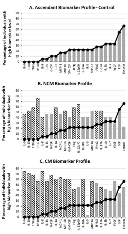
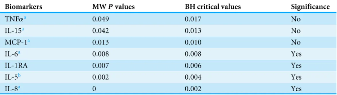
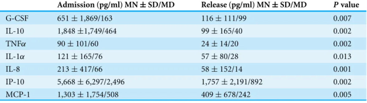
in the CT, NCM and CM groups. An ascendant biomarker profile was then constructed in the CT group by assembling the biomarkers from the one having the smallest percentage of high producers to the one having the largest. The resulting ascendant curve was used as a reference to visualize the variation of the percentage of high producers of biomarkers in the other groups (Fig. 1). In addition to showing the differences in the percentage of high producers (descriptive statistics), the biomarker profiles also indicate the analytes (hatched bars) for which there was a significant difference in adjusted Mann–Whitney U pairwise comparison (inferential statistics).
RESULTS
Study population and clinical data
We performed a retrospective, nonprobability sampling study using sera samples from healthy individuals and from patients admitted at the Hôpital Principal de Dakar, Sénégal. The cohort included 17 and 27 subjects diagnosed with NCM and CM respectively, and 18 healthy controls (CT) (Table 1). The three groups of individuals were comparable in age and gender, and were mainly composed of adults (Table 1). Subjects below 15 years of age included six children aged 5–13 diagnosed with NCM. Several clinical and blood parameters existed but were not recorded for all the individuals preventing reliable statistical analyses. Available data showed, as expected, hemoglobin levels comparable in the CT and NCM individuals while significantly lower in the CM patients (Table 1). Additionally, parasitemia was comparable between the NCM and CM groups (Table 1). Regarding organ defect in the CM group, kidney failure was the most frequent (13/27) followed by liver, hematologic and respiratory malfunction, while hemodynamic failure was rare (Table 1). All the NCM patients were successfully treated, while 9/27 CM subjects died.
Levels of inflammatory but not of anti-inflammatory biomarkers were higher in CM than in NCM patients
Table 1 Demographic, clinical, and disease outcome data.
CT NCM CM Total
Male 11 11 22 44
Gender
Female 7 6 5 17
Range 23–57 5–74 15–80 5–80
Age
Median 28.5 18 26 26
Normal 18 17 11 46
HB
Low 0 0 16 16
Median 0 520 1,452 1,022a
Parasitemia
IQR 0 2,552 9,981 7,654a
Survived 18 17 18 53
Outcome
Deceased 0 0 9 9
Neurological 0 0 27 27
Respiratory 0 0 6 6
Kidney 0 0 13 13
Liver 0 0 8 8
Hematologic 0 0 7 7
Organ failure
Hemodynamic 0 0 2 2
Notes.
CT, Control Individuals; NCM, Non-cerebral Malaria Patients; CM, Cerebral Malaria Patients; HB, Hemoglobin (Low, <100 g/L; Normal, >100 g/L); IQR, Inter Quartile Range.
aValue for NCM+CM.
Figure 2 Serum biomarker levels in control individuals and in non-cerebral and cerebral malaria patients.Biomarkers that significantly differed across the three groups in Kruskal–Wallis test after Benjamini–Hochberg adjustment are shown. Box plots represent medians with 25th and 75th percentiles, bars 10th and 90th percentiles, and dots outliers for biomarker concentrations. P, p[i] values in
Kruskal–Wallis tests. C, critical values in Benjamini–Hochberg correction.(continued on next page. . . )
Levels of inflammatory biomarkers were lower in survivors than in deceased of CM
Figure 2 (. . . continued)
Figure 2 (. . . continued)
Figure 3 Levels of inflammatory immune mediators are higher in cerebral than in non-cerebral malaria patients.The ascendant biomarker profile curve of the NCM (line) was plotted on the CM graph (bars) to visualize the difference in the proportion of high biomarker producers. Hatched bars represent biomarkers for which there is a significant difference in Mann–Whitney U pairwise comparison between the two groups after Benjamini–Hochberg multiple test adjustment.
Table 2 Biomarkers significantly differing between non-cerebral and cerebral malaria patients after multiple testing adjustment.
Biomarkers MWPvalues BH critical values Significance
TNFαa 0.049 0.017 No
IL-15a 0.042 0.013 No
MCP-1a 0.013 0.010 No
IL-6a 0.008 0.008 Yes
IL-1RA 0.007 0.006 Yes
IL-5b 0.002 0.004 Yes
IL-8a 0 0.002 Yes
Notes.
MW, Mann–Whitney U comparison; BH, Benjamini–Hochberg correction.
aIncreased in CM. bDecreased in CM.
Kidney failure showed significant moderate to strong positive correlations with several chemokines and pro-inflammatory cytokines (Table 4), while respiratory, hematological and liver failure displayed weak positive correlations with 5, 5 and 1 biomarkers respectively (not shown).
Variation of biomarker levels before and after cerebral malaria treatment
Figure 4 Levels of inflammatory immune mediators are higher in deceased than in survivors of cere-bral malaria.The ascendant biomarker profile curve of the survivors (line) was plotted on the deceased graph (bars) to visualize the difference in the proportion of high biomarker producers. Hatched bars rep-resent biomarkers for which there is a significant difference in Mann–Whitney U pairwise comparison be-tween the two groups after Benjamini–Hochberg multiple test adjustment.
Table 3 Biomarkers significantly increased in deceased of cerebral malaria compared to survivors.
Biomarkers MWPalues BH critical values Significance
IL-6 0.027 0.015 No
IL-8 0.017 0.010 No
Eotaxin 0.007 0.008 Yes
TNFα 0.003 0.006 Yes
IL-15 0.002 0.004 Yes
MCP-1 0.001 0.002 Yes
Notes.
MW, Mann–Whitney U comparison; BH, Benjamini–Hochberg correction.
Table 4 Biomarkers correlated with kidney failure in cerebral malaria patients.
ρ(pvalue) ρ(pvalue) ρ(pvalue)
Eotaxin 0.514 (0.006) IL-10 0.457 (0.017) IL-1α 0.593 (0.001) G-CSF 0.500 (0.008) IL-12p70 0.445 (0.020) IP-10 0.533 (0.004) GM-CSF 0.714 (<0.001) IL-15 0.621 (0.001) MCP-1 0.581 (0.002) IFNα2 0.525 (0.005) IL-17A 0.401 (0.038) TNFα 0.542 (0.003) IFNγ 0.529 (0.005) IL-1RA 0.390 (0.044)
Notes.
Table 5 Variation of biomarker levels between admission and release from hospital in cerebral malaria patients.
Admission (pg/ml) MN±SD/MD Release (pg/ml) MN±SD/MD Pvalue
G-CSF 651±1,869/163 116±111/99 0.007
IL-10 1,848±1,749/464 99±165/40 0.002
TNFα 90±101/60 24±14/20 0.002
IL-1α 121±165/76 57±80/28 0.013
IL-8 213±417/66 58±152/14 0.001
IP-10 5,668±6,297/2,496 1,757±2,191/892 0.002 MCP-1 1,303±1,754/508 409±678/242 0.005
Notes.
Admission, biomarker levels at the time of hospital admission of CM patients; Release, biomarker levels at the time of release of CM patients from hospital;Pvalue, two-tailedpvalue of a Wilcoxon Rank test; MN, mean; SD, standard deviation; MD, median.
14 CM patients (Table 5). Wilcoxon rank test showed 7 biomarkers (G-CSF, IL-10, IL-1α, IL-8, IP-10, MCP-1, TNFα) that were significantly different between the two time points after Benjamini–Hochberg adjustment. All these biomarkers were lower in the second samples confirming the induction of different types of immune mediators including growth factor (G-CSF), inflammatory (TNFα, IL-1α, IP-10), anti-inflammatory (IL-10) and chemokines (IL-8, MCP-1) during immune response to malaria (Table 5).
DISCUSSION
Inflammation and outcome of cerebral malaria
In this study, we performed a retrospective analysis of 18 controls, and of 17 and 27 NCM and CM patients respectively. The CM patients included 18 survivors and nine (30%) deceased subjects, a proportion similar to the highest mortality rates reported for CM. Beside neurological defect, kidney failure was the most frequent organ malfunction in CM patients and was correlated with death. Analysis of the cytokine response showed a strong induction of pro- and anti-inflammatory biomarkers in malaria patients. However, the magnitude of this response was significantly higher in CM than in NCM patients for inflammatory biomarkers while it was comparable in the two groups for the anti-inflammatory cytokines. Additionally, comparison of the biomarkers in the survivors versus the deceased of CM showed four pro-inflammatory analytes that were significantly higher in the deceased patients. Altogether, our results suggest a scenario in which a strong inflammatory response that was not properly contained led to organ failure and death during CM.
of these cytokines and chemokines with malaria severity and/or poor outcome have been described before (Clark et al., 2008). TNFαis one of the first recognized pro-inflammatory biomarkers that play important role during malaria. With other Th1 type cytokines IL-1, IL-12 and IFNγ it contributes to the control of the infection (Schofield & Grau, 2005). However, elevated levels of TNFα were associated with disease severity in both children and adults (Prakash et al., 2006;Thuma et al., 2011) and can discriminate between SM and UM (Mahanta et al., 2015). Similarly, IL-8 (Berg et al., 2014;Lyke et al., 2004), IL-6 (Jakobsen et al., 1994;Lyke et al., 2004), IL-15 (Hu, 2013;Ong’echa et al., 2011) and MCP-1 (MacMullin et al., 2012;Quelhas et al., 2012) were reported as increased during malaria.
In contrast to TNFα, IL-6, IL-8, IL-15 and MCP-1, eotaxin was not often mentioned in previous malaria studies. Interestingly, the level of eotaxin was higher in the CT individuals than in the malaria patients but the difference lost statistical significance after multiple testing adjustment. A significantly lower level of eotaxin was reported by Requena et al.
(2014)in pregnant women exposed to malaria when compared to controls residing in
malaria-free areas. Additionally, eotaxin was found as a negative predictor of hemoglobin level in children with SMA (Ong’echa et al., 2011). These observations suggest that eotaxin is dowregulated during malaria and that it could be involved in pathogenesis. Eotaxin is a Th2-type chemokine that mediates eosinophil development and recruitment in host tissues (Pope et al., 2001;Queto et al., 2010). Eotaxin is an important biomarker of allergic diseases (Pope et al., 2001) and polymorphism of its encoding gene influence total serum IgE level (Batra et al., 2007;Wang et al., 2007). The role played by IgE in response to malaria infection is controversial with some studies claiming a protective function (Bereczky et al., 2004;Farouk et al., 2005) while other associating IgE with disease severity (Perlmann et al., 1994;Perlmann et al., 1997;Seka-Seka et al., 2004). However, a recent study in a mouse model of experimental CM showed that animals genetically deficient for IgE or for the high affinity receptor for IgE were less susceptible to CM (Porcherie et al., 2011) supporting a role of IgE in the development of CM. The same study showed that CM pathogenesis was mediated by neutrophils expressing the high affinity receptor for IgE that homed to the brain and locally induced high levels of pro-inflammatory cytokines (Porcherie et al., 2011). Whether this function could be translated to human is unknown. However, a recent study reported an elevated neutrophil count that correlated with expression levels of the pro-inflammatory mediators IL-1β and IL-8 in human severe malaria (Mahanta et al., 2015). Altogether, these observations support the hypothesis that elevated levels of eotaxin result in higher production of IgE and deleterious effects during human malaria. If this is the case, the downregulation of eotaxin observed previously (Requena et al., 2014) and in this study might be a mechanism that protects against the damages caused by IgE during malaria. This hypothesis is consistent with higher levels of eotaxin observed in deceased compared to survivors of CM. However it needs to be tested in other studies.
role of this cytokine. This hypothesis is consistent with a recent report of a mouse study demonstrating a protection of rodent against experimental CM by IL-33 treatment (Besnard et al., 2015). The protection against CM was mediated by IL-5 independently of eosinophils, implying a mechanism that does not involve eotaxin.
In conclusion, our study confirms previously reported inflammatory response during malaria. Our findings support the idea of a strong induction of pro-inflammatory immune mediators that was not matched by the production of regulatory, anti-inflammatory biomarkers as the cause of death during CM. Additionally, our results suggests the involvement of eotaxin and of IL-5 in CM development and outcome.
ACKNOWLEDGEMENTS
We would like to thank Theresa Wan-Chen Yap for her help in the dosage of the biomarkers. We are grateful to Dr Becaye Fall, Dr Pape Samba Fall and Dr Ronald Perraut for their constructive suggestions and for stimulating discussions.
ADDITIONAL INFORMATION AND DECLARATIONS
Funding
This study was supported by University of Malaya-Ministry of Education (UM-MoE) High Impact Research (HIR) Grant UM.C/625/1/HIR/MoE/CHAN/13/6 (account no. H-50001-A000034). The funders had no role in study design, data collection and analysis, decision to publish, or preparation of the manuscript.
Grant Disclosures
The following grant information was disclosed by the authors:
University of Malaya-Ministry of Education (UM-MoE) High Impact Research (HIR) Grant: UM.C/625/1/HIR/MoE/CHAN/13/6.
Competing Interests
The authors declare there are no competing interests.
Author Contributions
• Yakhya Dieye conceived and designed the experiments, performed the experiments, analyzed the data, wrote the paper, prepared figures and/or tables.
• Babacar Mbengue conceived and designed the experiments, performed the experiments, analyzed the data, reviewed drafts of the paper.
• Shobha Dagamajalu performed the experiments.
• Mouhamadou Mansour Fall diagnosis of malaria, determination of blood parameters, treatment of patients.
• Mun Fai Loke contributed reagents/materials/analysis tools.
• Cheikh Momar Nguer analyzed the data, prepared figures and/or tables.
• Alassane Thiam performed the experiments, prepared figures and/or tables.
• Alioune Dieye conceived and designed the experiments, contributed reagents/material-s/analysis tools, reviewed drafts of the paper.
Human Ethics
The following information was supplied relating to ethical approvals (i.e., approving body and any reference numbers):
Université Cheikh Anta Diop de Dakar’s institutional research ethics committee; Protocol Number 001/2015/CER/UCAD.
Data Availability
The following information was supplied regarding data availability: The raw data has been supplied as a Supplemental Dataset.
Supplemental Information
Supplemental information for this article can be found online athttp://dx.doi.org/10.7717/ peerj.1965#supplemental-information.
REFERENCES
Batra J, Rajpoot R, Ahluwalia J, Devarapu SK, Sharma SK, Dinda AK, Ghosh B. 2007.A
hexanucleotide repeat upstream of eotaxin gene promoter is associated with asthma, serum total IgE and plasma eotaxin levels.Journal of Medical Genetics44:397–403
DOI 10.1136/jmg.2006.046607.
Bereczky S, Montgomery SM, Troye-Blomberg M, Rooth I, Shaw MA, Farnert A.
2004.Elevated anti-malarial IgE in asymptomatic individuals is associated with
reduced risk for subsequent clinical malaria.International Journal for Parasitology
34:935–942DOI 10.1016/j.ijpara.2004.04.007.
Berg A, Patel S, Gonca M, David C, Otterdal K, Ueland T, Dalen I, Kvaloy JT, Mollnes
TE, Aukrust P, Langeland N. 2014.Cytokine network in adults with falciparum
Malaria and HIV-1: increased IL-8 and IP-10 levels are associated with disease severity.PLoS ONE9:e114480DOI 10.1371/journal.pone.0114480.
Besnard AG, Guabiraba R, Niedbala W, Palomo J, Reverchon F, Shaw TN, Couper KN,
Ryffel B, Liew FY. 2015.IL-33-mediated protection against experimental cerebral
malaria is linked to induction of type 2 innate lymphoid cells, M2 macrophages and regulatory T cells.PLoS Pathog 11:e1004607DOI 10.1371/journal.ppat.1004607.
Bhatt S, Weiss DJ, Cameron E, Bisanzio D, Mappin B, Dalrymple U, Battle KE, Moyes CL, Henry A, Eckhoff PA, Wenger EA, Briet O, Penny MA, Smith TA, Bennett A, Yukich J, Eisele TP, Griffin JT, Fergus CA, Lynch M, Lindgren F, Cohen JM,
Murray CL, Smith DL, Hay SI, Cibulskis RE, Gething PW. 2015.The effect of
malaria control onPlasmodium falciparumin Africa between 2000 and 2015.Nature
526:207–211DOI 10.1038/nature15535.
Clark IA, Alleva LM, Budd AC, Cowden WB. 2008.Understanding the role of
inflamma-tory cytokines in malaria and related diseases.Travel Medicine and Infectious Disease
Crompton PD, Moebius J, Portugal S, Waisberg M, Hart G, Garver LS, Miller LH,
Barillas-Mury C, Pierce SK. 2014.Malaria immunity in man and mosquito: insights
into unsolved mysteries of a deadly infectious disease.Annual Review of Immunology
32:157–187DOI 10.1146/annurev-immunol-032713-120220.
Da Costa AG, Antonelli LR, Costa PA, Pimentel JP, Garcia NP, Tarrago AM, Dos Santos Mdo P, Nogueira PA, Hekcmann MI, Sadahiro A, Teixeira-Carvalho
A, Martins-Filho OA, Malheiro A. 2014.The robust and modulated biomarker
network elicited by thePlasmodiumvivax infection is mainly mediated by the IL-6/IL-10 axis and is associated with the parasite load.Journal of Immunology Research2014: 318250DOI 10.1155/2014/318250.
Deloron P, Roux Lombard P, Ringwald P, Wallon M, Niyongabo T, Aubry P, Dayer
JM, Peyron F. 1994.Plasma levels of TNF-alpha soluble receptors correlate with
outcome in human falciparum malaria.European Cytokine Network5:331–336.
Doolan DL, Dobano C, Baird JK. 2009.Acquired immunity to malaria.Clinical
Microbi-ology Reviews22:13–36, Table of Contents DOI 10.1128/CMR.00025-08.
Espie E, Diene Sarr F, Diop F, Faye J, Richard V, Tall A, Toure Balde A. 2015.
Spatio-temporal variations in malaria incidence in children less than 10 years old, health district of Sokone, Senegal, 2010–2013.PLoS ONE10:e0137737
DOI 10.1371/journal.pone.0137737.
Farouk SE, Dolo A, Bereczky S, Kouriba B, Maiga B, Farnert A, Perlmann H,
Hayano M, Montgomery SM, Doumbo OK, Troye-Blomberg M. 2005.Different
antibody- and cytokine-mediated responses toPlasmodium falciparumparasite in two sympatric ethnic tribes living in Mali.Microbes and Infection7:110–117
DOI 10.1016/j.micinf.2004.09.012.
Frosch AE, John CC. 2012.Immunomodulation inPlasmodium falciparummalaria:
experiments in nature and their conflicting implications for potential therapeutic agents.Expert Review of Anti-infective Therapy 10:1343–1356
DOI 10.1586/eri.12.118.
Hu WC. 2013.Human immune responses toPlasmodium falciparuminfection:
molecular evidence for a suboptimal THalphabeta and TH17 bias over ideal and effective traditional TH1 immune response.Malaria Journal12:Article 392
DOI 10.1186/1475-2875-12-392.
Jakobsen PH, Morris-Jones S, Theander TG, Hviid L, Hansen MB, Bendtzen K, Ridley
RG, Greenwood BM. 1994.Increased plasma levels of soluble IL-2R are associated
with severePlasmodium falciparummalaria.Clinical and Experimental Immunology
96:98–103.
Kurtzhals JA, Adabayeri V, Goka BQ, Akanmori BD, Oliver-Commey JO, Nkrumah
FK, Behr C, Hviid L. 1998.Low plasma concentrations of interleukin 10 in severe
malarial anaemia compared with cerebral and uncomplicated malaria.Lancet
351:1768–1772DOI 10.1016/S0140-6736(97)09439-7.
Lyke KE, Burges R, Cissoko Y, Sangare L, Dao M, Diarra I, Kone A, Harley R, Plowe
CV, Doumbo OK, Sztein MB. 2004.Serum levels of the proinflammatory cytokines
IL-12(p70) in Malian children with severePlasmodium falciparummalaria and matched uncomplicated malaria or healthy controls.Infection and Immunity
72:5630–5637DOI 10.1128/IAI.72.10.5630-5637.2004.
MacMullin G, Mackenzie R, Lau R, Khang J, Zhang H, Rajwans N, Liles WC, Pillai DR.
2012.Host immune response in returning travellers infected with malaria.Malaria
Journal 11:Article 148DOI 10.1186/1475-2875-11-148.
Mahanta A, Kar SK, Kakati S, Baruah S. 2015.Heightened inflammation in severe
malaria is associated with decreased IL-10 expression levels and neutrophils.Innate Immunity 21:546–552DOI 10.1177/1753425914561277.
Nussbaum JC, Van Dyken SJ, Von Moltke J, Cheng LE, Mohapatra A, Molofsky AB,
Thornton EE, Krummel MF, Chawla A, Liang HE, Locksley RM. 2013.Type
2 innate lymphoid cells control eosinophil homeostasis.Nature502:245–248
DOI 10.1038/nature12526.
Olliaro P. 2008.Editorial commentary: mortality associated with severePlasmodium
falciparummalaria increases with age.Clinical Infectious Diseases47:158–160
DOI 10.1086/589288.
Ong’echa JM, Davenport GC, Vulule JM, Hittner JB, Perkins DJ. 2011.Identification of
inflammatory biomarkers for pediatric malarial anemia severity using novel statisti-cal methods.Infection and Immunity79:4674–4680DOI 10.1128/IAI.05161-11.
Perlmann H, Helmby H, Hagstedt M, Carlson J, Larsson PH, Troye-Blomberg M,
Perlmann P. 1994.IgE elevation and IgE anti-malarial antibodies inPlasmodium
falciparummalaria: association of high IgE levels with cerebral malaria.Clinical and Experimental Immunology 97:284–292.
Perlmann P, Perlmann H, Flyg BW, Hagstedt M, Elghazali G, Worku S, Fernandez
V, Rutta AS, Troye-Blomberg M. 1997.Immunoglobulin E, a pathogenic factor in
Plasmodium falciparummalaria.Infection and Immunity65:116–121.
Peyron F, Burdin N, Ringwald P, Vuillez JP, Rousset F, Banchereau J. 1994.High
levels of circulating IL-10 in human malaria.Clinical and Experimental Immunology
95:300–303.
Pope SM, Brandt EB, Mishra A, Hogan SP, Zimmermann N, Matthaei KI, Foster PS,
Rothenberg ME. 2001.IL-13 induces eosinophil recruitment into the lung by an
IL-5- and eotaxin-dependent mechanism.Journal of Allergy and Clinical Immunology
108:594–601DOI 10.1067/mai.2001.118600.
Porcherie A, Mathieu C, Peronet R, Schneider E, Claver J, Commere PH, Kiefer-Biasizzo H, Karasuyama H, Milon G, Dy M, Kinet JP, Louis J, Blank U, Mecheri S. 2011.Critical role of the neutrophil-associated high-affinity receptor for IgE in the pathogenesis of experimental cerebral malaria.Journal of Experimetnal Medicine
208:2225–2236DOI 10.1084/jem.20110845.
Prakash D, Fesel C, Jain R, Cazenave PA, Mishra GC, Pied S. 2006.Clusters of cytokines
determine malaria severity inPlasmodium falciparum-infectedpatients from endemic areas of Central India.Journal of Infectious Diseases194:198–207
Quelhas D, Puyol L, Quinto L, Nhampossa T, Serra-Casas E, Macete E, Aide P, Sanz S, Aponte JJ, Doolan DL, Alonso PL, Menendez C, Dobano C. 2012.
Intermittent preventive treatment with sulfadoxine-pyrimethamine does not modify plasma cytokines and chemokines or intracellular cytokine responses toPlasmodium falciparumin Mozambican children.BMC Immunology13:5
DOI 10.1186/1471-2172-13-5.
Queto T, Gaspar-Elsas MI, Masid-de-Brito D, Vasconcelos ZF, Ferraris FK, Penido C,
Cunha FQ, Kanaoka Y, Lam BK, Xavier-Elsas P. 2010.Cysteinyl-leukotriene type
1 receptors transduce a critical signal for the up-regulation of eosinophilopoiesis by interleukin-13 and eotaxin in murine bone marrow.Journal of Leukocyte Biology
87:885–893DOI 10.1189/jlb.1108709.
Requena P, Campo JJ, Umbers AJ, Ome M, Wangnapi R, Barrios D, Robinson LJ, Samol P, Rosanas-Urgell A, Ubillos I, Mayor A, Lopez M, De Lazzari E, Arevalo-Herrera M, Fernandez-Becerra C, Del Portillo H, Chitnis CE, Siba PM, Bardaji
A, Mueller I, Rogerson S, Menendez C, Dobano C. 2014.Pregnancy and malaria
exposure are associated with changes in the B cell pool and in plasma eotaxin levels.
Journal of Immunology 193:2971–2983DOI 10.4049/jimmunol.1401037.
Sarthou JL, Angel G, Aribot G, Rogier C, Dieye A, Toure Balde A, Diatta B, Seignot
P, Roussilhon C. 1997.Prognostic value of anti-Plasmodium falciparum-specific
immunoglobulin G3, cytokines, and their soluble receptors in West African patients with severe malaria.Infection and Immunity65:3271–3276.
Schofield L, Grau GE. 2005.Immunological processes in malaria pathogenesis.Nature
Reviews Immunology 5:722–735DOI 10.1038/nri1686.
Seka-Seka J, Brouh Y, Yapo-Crezoit AC, Atseye NH. 2004.The role of serum
immunoglobulin E in the pathogenesis ofPlasmodium falciparummalaria in Ivorian children.Scandinavian Journal of Immunology59:228–230
DOI 10.1111/j.0300-9475.2004.01337.x.
Storm J, Craig AG. 2014.Pathogenesis of cerebral malaria–inflammation and
cy-toadherence.Frontiers in Cellular and Infection Microbiology 4:Article 100
DOI 10.3389/fcimb.2014.00100.
Thuma PE, Van Dijk J, Bucala R, Debebe Z, Nekhai S, Kuddo T, Nouraie M, Weiss G,
Gordeuk VR. 2011.Distinct clinical and immunologic profiles in severe malarial
anemia and cerebral malaria in Zambia.Journal of Infectious Diseases203:211–219
DOI 10.1093/infdis/jiq041.
Torrentino-Madamet M, Fall B, Benoit N, Camara C, Amalvict R, Fall M, Dionne P, Ba Fall K, Nakoulima A, Diatta B, Dieme Y, Menard D, Wade B, Pradines
B. 2014.Limited polymorphisms in k13 gene inPlasmodium falciparum
isolates from Dakar, Senegal in 2012-2013.Malaria Journal13:Article 472
DOI 10.1186/1475-2875-13-472.
Trape JF, Tall A, Sokhna C, Ly AB, Diagne N, Ndiath O, Mazenot C, Richard V, Badiane A, Dieye-Ba F, Faye J, Ndiaye G, Diene Sarr F, Roucher C, Bouganali C, Bassene H, Toure-Balde A, Roussilhon C, Perraut R, Spiegel A, Sarthou JL,
fall of malaria in a West African rural community, Dielmo, Senegal, from 1990 to 2012: a 22 year longitudinal study.The Lancet Infectious Diseases14:476–488
DOI 10.1016/S1473-3099(14)70712-1.
Walther M, Tongren JE, Andrews L, Korbel D, King E, Fletcher H, Andersen RF, Bejon P, Thompson F, Dunachie SJ, Edele F, De Souza JB, Sinden RE, Gilbert SC,
Riley EM, Hill AV. 2005.Upregulation of TGF-beta, FOXP3, and CD4+CD25+
regulatory T cells correlates with more rapid parasite growth in human malaria infection.Immunity 23:287–296DOI 10.1016/j.immuni.2005.08.006.
Wang TN, Chiang W, Tseng HI, Chu YT, Chen WY, Shih NH, Ko YC. 2007.The
polymorphisms of Eotaxin 1 and CCR3 genes influence on serum IgE, Eo-taxin levels and mild asthmatic children in Taiwan.Allergy62:1125–1130
DOI 10.1111/j.1398-9995.2007.01485.x.
White NJ, Pukrittayakamee S, Hien TT, Faiz MA, Mokuolu OA, Dondorp AM. 2014.
Malaria.Lancet 383:723–735DOI 10.1016/S0140-6736(13)60024-0.
WHO. 2000.Severe falciparum malaria.Transactions of the Royal Society of Tropical
Medicine and Hygiene94(Suppl1):1–90.