636
DOI:
10.1590/0004-282X20150063
IMAGES IN NEUROLOGY
Primary meningeal melanoma with
cerebrospinal fluid dissemination mimicking
neurofibromatosis type 2
Melanoma meníngeo primário com disseminação liquórica mimetizando neurofibromatose
tipo 2
Marcos Rosa Júnior
1, Luciene Lage da Motta
2, Fabrizio Scardino
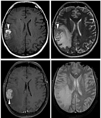
3A 38-year-old man admitted with headache, left paresis and
bilateral sensorineural hearing loss. Neuroimaging showed a
pe-ripheral frontal tumor with hyperintensity on T1WI and
bilater-al internbilater-al auditory canbilater-al (IAC) lesions. (Figures 1, 2 and 3). he
presence of hyperintensity on T1WI, without fat or hemorrhage
should direct for lesions containing melanin
1. Resection of the
frontal tumor diagnosed a primary malignant meningeal
mela-noma with cerebrospinal luid dissemination once the patient
has no melanocytic lesions outside the CNS. he melanocytic
lesions ranges from melanocytoma to melanoma
2,3,4,5.
Malignant
melanoma should be included in the diferential diagnosis of
neoplastic CSF dissemination with bilateral IAC lesions
mim-icking schwannomas in NF2.
1Universidade Federal do Espírito Santo, Seção de Radiologia, Vitoria ES, Brazil;
2Laboratório de Cito e Histopatologia Virchow, Vitoria ES, Brazil;
3Hospital Estadual Jayme Santos Neves, Seção de Neurocirurgia, Serra ES, Brazil.
Correspondence: Marcos Rosa Júnior; Centro de Ciências da Saúde, UFES; Avenida Marechal Campos, 1468; 29043-900 Vitória ES, Brasil; E-mail: [email protected]
Conflict of interest: There is no conflict of interest to declare.
Received 26 December 2014; Received in final form 19 February 2015; Accepted 13 March 2015.
Figure 1.
Non-contrast CT showed a right frontal hyperdense
tumor (arrowhead).
Figure 3.
FLAIR showed bilateral IAC lesions (arrowheads)
(A), which increased over the following 30 days (arrowheads)
(B) mimicking bilateral acoustic schwannoma in
neurofibromatosis type 2.
637
Marcos Rosa Júnior et al. Primary meningeal melanomaReferences
1. Ginat DT, Meyers SP. Intracranial lesions with high signal intensity
on T1-weighted MR images: differential diagnosis. Radiographics. 2012;32(2):499-516. http://dx.doi.org/10.1148/rg.322105761
2. Brat DJ, Giannini C, Scheithauer BW, Burger PC. Primary melanocytic
neoplasms of the central nervous systems. Am J Surg Pathol. 1999;23(7):745-54.
3. Roser F, Nakamura M, Brandis A, Hans V, Vorkapic P, Samii M.
Transition from meningeal melanocytoma to primary cerebral
melanoma: case report. J Neurosurg. 2004;101(3):528-31. http://dx.doi.org/10.3171/jns.2004.101.3.0528
4. Bydon A, Gutierrez JA, Mahmood A. Meningeal melanocytoma:
an aggressive course for a benign tumor. J Neurooncol. 2003;64(3):259-63. http://dx.doi.org/10.1023/A:1025628802228
5. Xie ZY, Hsieh KLC, Tsang YM, Cheung WK, Hsieh CH. Primary