Cardiac Autonomic and Ventricular Mechanical Functions in
Asymptomatic Chronic Chagasic Cardiomyopathy
Daniel França Vasconcelos
1,2and Luiz Fernando Junqueira Junior
1,2Laboratório Cardiovascular da Faculdade de Medicina da Universidade de Brasília1; Serviço de Cardiologia do Hospital Universitário de
Brasília2, DF, Brazil
Abstract
Background: The association of variably altered cardiac autonomic and ventricular systolic and diastolic functions is still controversial and little explored in chronic Chagas’ disease.
Objective: To evaluate the extent to which cardiac autonomic and mechanical ventricular functions are altered and whether they are associated in asymptomatic chagasic cardiomyopathy.
Methods: A total of 13 patients with asymptomatic chagasic cardiomyopathy and 15 normal subjects (control group) were evaluated and the autonomic modulation of heart rate variability for five minutes, in the temporal and spectral domains, in the supine and orthostatic positions, as well as ventricular function based on morphological-functional variables obtained by Doppler echocardiography were correlated. Statistical analysis used the Mann-Whitney test and Spearman’s correlation.
Results: In both positions, the temporal indexes (p = 0.0004 to 0.01) and total (p = 0.0007-0.005) and absolute spectral areas, of low and high frequencies (p = 0.0001 to 0.002), were lower in the chagasic group. The vagal-sympathetic balance was similar in the two groups in both positions (p = 0.43 to 0.89). The echocardiographic variables did not differ between groups (p = 0.13 to 0.82), except the left ventricular end-systolic diameter, which was larger (p = 0.04) and correlated directly with reduced rates of global (p = 0.01 to 0.04) and parasympathetic (p = 0.002 to 0.01) autonomic modulation in patients with Chagas disease in the orthostatic position.
Conclusion: The sympathetic and parasympathetic depressions with preserved balance were associated with only one ventricular dysfunction indicator. This suggests that cardiac autonomic dysfunction may precede and be independently more severe than ventricular dysfunction, with no causal association between both disorders in chronic chagasic cardiomyopathy. (Arq Bras Cardiol 2012;98(2):111-119)
Keywords: Chagas cardiomyopathy; ventricular function Doppler echocardiography; echocardiography, doppler, cardiac autonomic function; heart rate variability.
Mailing Address: Daniel França Vasconcelos •
SHIS, QI 5, Chácara 21, Unidade C , Setor de Habita - 71600-540 - Lago Sul, DF, Brasília, Brazil
E-mail: [email protected], [email protected]
Manuscript received 25/05/11; revised mansucript received 27/08/11; accepted 30/08/11.
the intrinsic autonomic innervation, mainly parasympathetic. Among its manifestations, isolated or combined, are ventricular systolic or diastolic dysfunction, which may culminate in heart failure, various arrhythmias, cardiac autonomic dysfunction, thromboembolism and sudden death3-5.
The primary variable parasympathetic and sympathetic cardiac dysfunction has been thoroughly demonstrated based on poor responses of heart rate to different acute stimuli5-14, as well as in the reduction of the spontaneous variability of heart rate15-18 in chagasic patients with isolated or combined heart disease. Regarding the morphological and functional systolic and diastolic ventricular variables, some studies that used conventional and tissue Doppler echocardiography have shown conflicting results, with no defined morphofunctional standards in chagasic patients with the cardiac and indeterminate forms of the disease. Some alterations were identified19-25, but the preserved morphology and ventricular function have also been described13,26-28.
A question that remains to be clarified is whether the autonomic dysfunction correlates with ventricular morphofunctional alterations or if these disorders are
Introduction
In its different chronic clinical forms, Chagas disease persists as an important endemic disease that affects about 8-11 million individuals in the South American continent and also occurs in countries in Europe and North America1,2. The chronic disease includes the indeterminate form, which exhibits only seropositivity with no overt organ involvement, and the cardiac and digestive forms, either isolated or combined, with or without symptoms3.
independent phenomena with no causal association5,29,30. Some studies have shown that both disorders are independent phenomena and that parasympathetic dysfunction may occur exclusively, precede mechanical alteration or exist in association with the latter, with no causal association5,9,12-14,28.
The present study aimed at assessing to what extent the morphological and ventricular systolic and diastolic functional variables at Doppler echocardiography and the tonic autonomic modulation of the heart assessed by analysis of heart rate variability are altered and whether they are related in asymptomatic chronic chagasic cardiomyopathy.
Methods
Study groups
Two outpatient groups were compared: (a) a control group consisting of 15 healthy subjects (nine men and six women) aged 37 to 54 years (median 43 years) of diverse origin, and (b) a group of 13 chagasic patients with cardiomyopathy (seven men and six women), aged 28 to 62 years (median 44 years). These patients were recruited from the Cardiology Service at the Hospital Universitário de Brasília, DF, where they underwent regular clinical follow-up.
T h e d i a g n o s i s o f C h a g a s ’ d i s e a s e w a s b a s e d on epidemiological history and at least two positive serological tests, including indirect immunofluorescence, hemagglutination and ELISA. Chagasic cardiomyopathy was diagnosed by the presence of isolated or combined specific electrocardiographic alterations at rest: first-degree atrioventricular block (8%), complete left bundle branch block of the His bundle (8%), complete right bundle branch block of the His bundle (23%), right bundle branch block associated with left anterior hemiblock (15%), left anterior hemiblock (23%) and diffuse ventricular repolarization alterations (23%). Conventional chest X-ray was normal in all chagasic patients. Additional inclusion criteria were absence of previous or current manifestations of heart failure and other cardiovascular or systemic condition.
Ten chagasic patients had only the cardiac form of the disease; three had specific symptoms, which were identified with megaesophagus or megacolon, characterizing the cardio-digestive form. No Doppler echocardiography measurement was considered as exclusion or inclusion criterion. All patients in the control and chagasic groups were in perfect physical and mental state and performed regular daily activities, and did not use any drugs.
This study was performed in accordance with the Declaration of Helsinki and the rules of the Ministry of Health and was approved by the Ethics Committee in Human Research, of Faculdade de Medicina da Universidade de Brasília. All subjects signed an informed consent form prior to study enrollment. The authors declare no conflict of interest.
Experimental protocol
Individuals in the chagasic and control groups were submitted to an experimental session between 8 and 11 am, after routine activities, one to four hours after breakfast.
All individuals were instructed to abstain from stimulating beverages as well as exercising, smoking and taking any drugs, at least 24 hours prior to examination. The clinical and anthropometric data, life habits and the conventional electrocardiogram (ECG) were initially obtained in an appropriate examination room, at room temperature (22-28ºC). As previously established in our laboratory18,31-34, after ten subsequent minutes of supine rest, a continuous five-minute ECG recording in DII lead was automatically recorded in real time in a computer system at a velocity of 25 mm/s and at a sampling frequency of 250 Hz
For this purpose, an analog-digital converter was used, as well as dedicated software to capture signal, while viewing occurred in a monitor. Immediately after, the individuals actively adopted the orthostatic posture at the bedside, and after two minutes in this position, a new five-minute ECG recording was stored as previously described.
Individuals breathed spontaneously and regularly, and the respiratory rate was visually monitored and recorded. Those with frequencies below 9 cpm (0.15 Hz) were excluded from the sample, considering that, in this case, the spectral bands of low and high frequencies overlap in the spectral domain of heart rate variability. About an hour after obtaining the RR interval series, the subjects underwent comprehensive Doppler echocardiography assessment. The clinical and autonomic evaluations and echocardiograms were performed by the same author (DFV).
Analysis of R-R interval variability
This analysis was performed as previously described18,31 and according to recommendations32. Each recorded series of RR intervals of the ECG was processed and analyzed off-line, using another specific software program. The software for signal capture, processing and analysis have been developed and validated in our laboratory and the Department of Electrical Engineering of Universidade de Brasilia31.
The periodograms of the RR interval series were initially validated visually to verify the stationarity, sinus rhythm confirmation and the detection of non-sinus and ectopic beats and artifacts, as well as the validation of each R wave. Any spurious beats were eliminated from the series, together with the preceding and subsequent intervals, as well as extreme intervals. The series that needed editing represented less than 1% of the series of chagasic individuals and about 0.1% of those in the control group. The qualified series showed good stationarity, estimated by the percentage differences between the mean and standard deviation of each pair of three segments in which the series were fragmented. After validation and editing, the series were processed and analyzed for variability in the time (temporal analysis) and frequency (spectral analysis) domains18,31-34.
(rMSSD), which estimate the beat to beat parasympathetic modulation associated with respiratory sinus arrhythmia.
For the spectral analysis, the series were initially normalized and resampled to 4 Hz using the cubic polynomial interpolation method (cubic spline), in order to obtain continuous and equidistant intervals. They were subsequently filtered by Hanning windowing to attenuate the effects of discontinuity, and then were processed by the autoregressive model of order 16, for the conversion of wave components in a spectrum of frequencies31,35, which comprises: a very low frequency band (0 to 0.04 Hz), low frequency (0.04 to 0.15 Hz), which reflects slow autonomic modulation and is a marker of combined sympathetic and parasympathetic activity, and high frequency (0.15 to 0.50 Hz), which represents the rapid autonomic modulation, being indicative of almost exclusively parasympathetic sinus influence.
The spectral indices expressed by the areas of frequency bands (ms2/Hz) included: (a) total spectral area (0 to 0.50 Hz), which indicates the degree of global autonomic modulation; (b) absolute and relative spectral area of each of the three frequency bands; (c) normalized spectral area of the low and high frequency bands, calculated as a percentage of the absolute area of each of these bands in relation to the sum of both; (d) ratio of the absolute area of the low and high frequency bands (ratio < 1 indicates predominant parasympathetic modulation; and a ratio > 1 demonstrates a relatively predominant combined vagosympathetic modulation). Thus, the normalized areas and ratio of absolute areas of low and high spectral frequencies reflect the relative sympathetic and parasympathetic modulation, i.e., the vagosympathetic balance.
Echocardiographic Doppler examination
Morphological and functional systolic and diastolic variables were obtained by two-dimensional Doppler echocardiography using an ATL HDI 3500 ultrasound equipment with a 2.5 to 4.0 MHz transducer. During examination, the individual remained in the left supine position, according with the guidelines of the American Society of Echocardiography (ASE)36. The mean values of three consecutive beats were considered, adjusted for body surface area, obtained from the left parasternal view (long and short axis) and apical (four-chamber, two-chamber and apical long axis). The M-mode recording was made by viewing the short axis at the level of the papillary muscles. The examinations were performed blindly, by only one of the authors (DVF).
The evaluated morphological variables included: diameter of the aorta (Ao) and antero-posterior diameter of the left atrium (LA); left ventricular-end systolic (LVESD) and diastolic diameters (LVEDD); left ventricular septal and posterior wall thickness; left ventricular-end diastolic volume (LVEDV), left ventricular mass (M) calculated according to the ASE; LVEDV/M ratio; and cardiac mass index (mass / body surface area - M index).
The left ventricular systolic function was estimated by the ejection fraction (EF) according to Teichholz’s formula, percentage of left ventricular circumferential shortening
(ΔD%), calculated using the formula [(LVEDD - LVESD) /
LVEDD] x 100, and the velocity of circumferential fiber shortening (Vcf)
The measurements of diastolic function included: the transmitral flow velocity, with the sample volume taken at the extremity of the mitral valve leaflets in the apical four-chamber view; the ratio of the early wave velocity of the mitral valve opening (E-wave) and the wave velocity of the maximum atrial contraction (A wave) (E/A ratio); the velocity of the mitral deceleration time in early filling (EF slope); and the ratio of the early wave velocity of the mitral valve opening (E-wave) and the wave of the septal mitral annulus velocity at the tissue Doppler (e ‘) (E/e’ ratio).
The myocardial performance index, or Tei index was also estimated as a measurement of global ventricular function or combined systolic-diastolic function.
Statistical Analysis
As most of the variables showed non-normal distribution at the Kolmogorov-Smirnov, D’Agostino-Pearson and Shapiro-Wilks tests, each one was described by the median, the interquartile range and extreme values , and the groups were compared by Mann –Whitney test. Spearman’s test was used to assess the correlation between the indices of heart rate variability and echocardiographic variables. Categorical variables were compared between groups by Chi-square or Fisher’s exact test. The two-tailed level of statistical significance for differences and correlations was
set at p ≤ 0.05. We used the SigmaStat®
3.11/SigmaPlot 9.01 (Systat Software, Inc., USA, 2004) and Prism®
4 (GraphPad Software, Inc., USA, 2005) software for the statistical analyses.
Results
Table 1 summarizes the general data of groups of individuals, which differed only in terms of alcohol consumption, more frequent in the control group (p = 0.0001). Systolic pressure (p = 0.03) and respiratory rate (p = 0.02) were higher in the chagasic group, but within normal values.
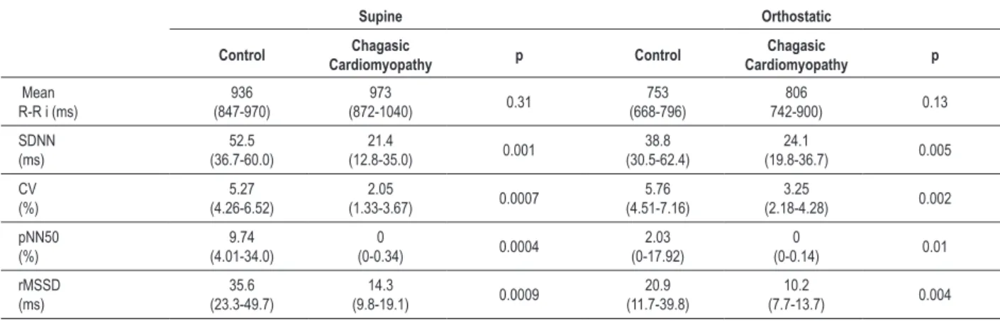
The data of the RR interval variability analysis are shown in Tables 2 and 3. The mean RR intervals showed similar medians for both groups in both positions (p = 0.13 - 0.31). The other temporal indices showed significantly lower medians in the chagasic group, both in the supine (p = 0.0004 to 0.001) and in orthostatic (p = 0.002 - 0.01) position. The absolute total spectral area and areas of low and high frequency spectra also showed lower medians in the chagasic group in both positions (p = 0.000 to 0.002). As for the normalized areas and the low/high spectral frequency ratio, median values were similar in both groups, regardless of the position (p = 0.43-0.89).
A significant negative correlation was observed between LVESD and indices that express the global autonomic modulation – coefficient of variation (rs = -0.58, p = 0.04) and total spectral area (rs = -0.68, p = 0.01) – as well as between those indicators of parasympathetic modulation – rMSSD (rs = -0.77, p = 0.002) – and high-frequency spectral area (rs = -0.68, p = 0.01), only in the orthostatic position. Figure 1 illustrates these correlations.
Discussion
Cardiac autonomic dysfunction, characterized mainly by parasympathetic depression, is a remarkable aspect of human and experimental Chagas disease6,10,29,37-39. Normal, mild to moderate depression or exacerbation of cardiac autonomic function occur in the indeterminate form of the disease and in chagasic patients with doubtful ECG alterations4,7,9,12,13,15,17,18,28,34,40. The more severe autonomic depression is found in the cardio-digestive and isolated digestive forms, while moderate to severe disorder is observed in the cardiac form, exclusively, without heart failure6-12,14-18,29,30,37,38.
The present study reinforces these previous observations with the finding that all the absolute temporal and spectral indices are decreased in the chagasic group, both in the supine and in the orthostatic positions. Although the absolute sympathetic and parasympathetic modulations were depressed, the vagosympathetic balance did not change in
the chagasic group in both positions. This finding, however, does not mean that the depression of sympathetic modulation was similar to the parasympathetic one, considering that the vagosympathetic balance depends on the relative basal level for each autonomic component.
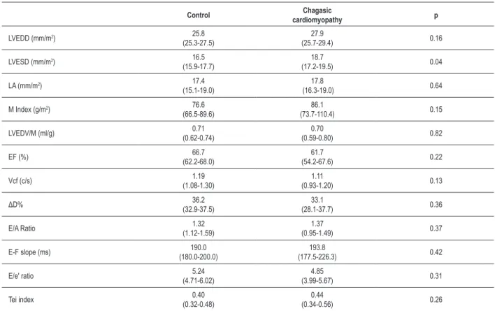
We also observed that chagasic patients exhibited morphological and functional ventricular echocardiographic Doppler measures similar to those of normal individuals, except by the increased LVESD. This alteration alone would represent an incipient morphological alteration, which could precede the established systolic or diastolic dysfunction. Furthermore, it is likely that this finding is only a casual variant of normality, with no morphofunctional significance and so, it should be disregarded. Our results are in agreement with the lack of alterations or mild alterations in ventricular function observed by other authors in chagasic patients with cardiomyopathy12-14,25. On the other hand, they disagree with those that showed left ventricular diastolic dysfunction in this form of the disease, using the same evaluation technique20-22,24, which can be explained by differences in the functional pattern of cardiac involvement of the heart or differences related to different stages of cardiomyopathy evolution in chagasic patients.
As for the possible correlation between ventricular function and cardiac autonomic modulation in patients with Chagas disease, a marked autonomic depression was observed in association with normal systolic and diastolic
Table 1 – Anthropometric and clinical characteristics and life habits of the control group (n = 15) and the group with com chagasic
cardiomyopathy (n = 13)
Control Chagasic
cardiomyopathy p
Age (years)
43 (40-46)
44
(35-49) 0.98
Sex
male/female (%) 60/40 54/46 1.00
Body mass index (kg/m2) 24.4
(20.5-26.4)
23.4
(20.8-25.8) 0.78 Body surface area (m2) 1.74
(1.59-1.85)
1.72
(1.57-1.84) 0.58 Systolic blood pressure
(mmHg)
110 (100-118)
120
(112-129) 0.03 Diastolic blood pressure
(mmHg)
76 (70-84)
80
(78-88) 0.10
Heart rate (bpm)
61 (56-70)
58
(51-68) 0.21
Respiratory rate (irpm)
14 (12-16)
18
(16-20) 0.02
Physical exercise (%) 20 53.8 0.37
Smoking (%) 0 23.1 0.15
Alcohol consumption (%) 100 7.7 0.0001
Stimulating beverages (%) 33.3 53.8 0.28
Table 2 - Median (interquartile interval) of variability indices of R-R intervals in the time domain in the control group (n = 15) and the group with chronic chagasic cardiomyopathy (n = 13), in the supine and active orthostatic positions
Supine Orthostatic
Control Chagasic
Cardiomyopathy p Control
Chagasic
Cardiomyopathy p
Mean R-R i (ms)
936 (847-970)
973
(872-1040) 0.31
753 (668-796)
806
742-900) 0.13 SDNN
(ms)
52.5 (36.7-60.0)
21.4
(12.8-35.0) 0.001
38.8 (30.5-62.4)
24.1
(19.8-36.7) 0.005 CV
(%)
5.27 (4.26-6.52)
2.05
(1.33-3.67) 0.0007
5.76 (4.51-7.16)
3.25
(2.18-4.28) 0.002 pNN50
(%)
9.74 (4.01-34.0)
0
(0-0.34) 0.0004
2.03 (0-17.92)
0
(0-0.14) 0.01 rMSSD
(ms)
35.6 (23.3-49.7)
14.3
(9.8-19.1) 0.0009
20.9 (11.7-39.8)
10.2
(7.7-13.7) 0.004
The groups were compared, in each position, by Mann-Whitney test; see Methods for abbreviations and deinition of indices.
Figure 1 – Spearman’s correlation of heart rate variability indices, indicators of global and parasympathetic autonomic modulation, with left ventricular-end systolic diameter (LVESD) in chagasic patients with cardiomyopathy, in the orthostatic position. rs – Spearman’s correlation coeficient.
L
V
E
S
D
(m
m
/m
m
2)
L
V
E
S
D
(m
m
/m
m
2)
L
V
E
S
D
(m
m
/m
m
2)
L
V
E
S
D
(m
m
/m
m
2)
Total spectral area (ms2) High-frequency spectral area (ms2)
Coeficient of variation (%) rMSSD (ms)
N = 13
rs = 0.58
p = 0.04
N = 13
rs = 0.68
p = 0.01
N = 13
rs = –0.77
p = 0.002
N = 13
rs = –0.68
Table 3 - Median (interquartile interval) of R-R interval variability indices in the frequency domain in the control group (n = 15) and in the group with chronic chagasic cardiomyopathy (n = 13), in supine and active orthostatic positions
Supine Orthostatic
Control Chagasic
cardiomyopathy p Control
Chagasic
cardiomyopathy p
Total spectral area (ms2)
551 (234-761)
101
(34-230) 0.0007
368 (147-770)
82
(46-237) 0.005 Low-frequency spectral area
(ms2)
183 (83-326)
16
(5-60) 0.0001
176 (47-481)
26
(9-69) 0.002 High-frequency spectral area
(ms2)
111 (40-261)
13
(6-29) 0.0007
46 (8-77)
9
(3-11) 0.004 Low-frequency normalized area
(%)
67 (35.2-82.0)
60.3
(38.4-78.1) 0.89
86.2 (67-91)
75
(67.2-88.8) 0.46 High-frequency normalized area
(%)
33 (18-64.8)
39.7
(21.8-61.6) 0.89
13.8 (9-33)
25
(11.2-32.8) 0.46 Ratio between areas of low and high
frequencies
2.02 (0.54-4.45)
1.52
(0.62-3.57) 0.89
6.27 (2.04-10.7)
2.95
(2.06-7.93) 0.43
The groups were compared, in each position, by Mann-Whitney test; see Methods for abbreviations and deinition of indices.
Table 4 - Median (interquartile interval) of the Doppler Echocardiography variables corrected by the body surface area in the control group (n = 15) and the group with chronic chagasic cardiomyopathy (n = 13)
Control Chagasic
cardiomyopathy p
LVEDD (mm/m2) 25.8
(25.3-27.5)
27.9
(25.7-29.4) 0.16 LVESD (mm/m2) 16.5
(15.9-17.7)
18.7
(17.2-19.5) 0.04
LA (mm/m2) 17.4
(15.1-19.0)
17.8
(16.3-19.0) 0.64 M Index (g/m2) 76.6
(66.5-89.6)
86.1
(73.7-110.4) 0.15 LVEDV/M (ml/g) 0.71
(0.62-0.74)
0.70
(0.59-0.80) 0.82
EF (%) 66.7
(62.2-68.0)
61.7
(54.2-67.6) 0.22
Vcf (c/s) 1.19
(1.08-1.30)
1.11
(0.93-1.20) 0.13
ΔD% 36.2
(32.9-37.5)
33.1
(28.1-37.7) 0.36
E/A Ratio 1.32
(1.12-1.59)
1.37
(0.95-1.49) 0.37 E-F slope (ms) 190.0
(180.0-200.0)
193.8
(177.5-226.3) 0.42
E/e' ratio 5.24
(4.71-6.02)
4.85
(3.99-5.67) 0.31
Tei index 0.40
(0.32-0.48)
0.44
(0.34-0.56) 0.26
echocardiographic variables, except for increased LVESD. This ventricular alteration was directly correlated with reduced rates of global and parasympathetic autonomic modulation, but only in the orthostatic position, suggesting that in a situation of cardiovascular functional stress, represented by the postural load, autonomic and ventricular alterations may be slightly correlated. This means that the two disorders are not associated with each other to a great extent, at least at some point in the course of the disease.
Based on these observations, it is unlikely that the marked cardiac autonomic dysfunction may be merely a phenomenon secondary to a ventricular mechanical disorder. Thus, our observations show that autonomic dysfunction is not causally associated with ventricular dysfunction. Cardiac dysautonomia seems to be a primary phenomenon and can occur in isolation or precede ventricular mechanical disorders. That is, the autonomic dysfunction is not a consequence of progressive chronic contractile alteration in chagasic cardiomyopathy.
In contrast, an alternative hypothesis suggests that cardiac autonomic dysfunction is a phenomenon secondary to ventricular dysfunction, based on studies of chagasic patients who simultaneously showed both disorders or ventricular alteration alone29, which does not allow establishing any causal relationship between these disorders. On the other hand, one cannot rule out the possibility that autonomic and contractile dysfunctions may influence each other, considering the important interrelationship between the functions5.
In fact, in a previous correlative study, we observed that the disorders of contractile, electrical and autonomic functions occurred separately or associated in all forms of the disease and that some patients with Chagas disease showed no disorders5. Furthermore, chagasic patients with the indeterminate form showed right ventricular contractile alteration, regardless of autonomic dysfunction9, parasympathetic dysfunction preceding the left ventricular systolic alteration12, deficient vagal responses to orthostatic stress and depressed respiratory arrhythmia, associated with preserved global left ventricular systolic function13.
Moreover, the chronotropic deficiency at the stress test appears to be an early sign of cardiac autonomic dysfunction, regardless of ventricular contractile function in chagasic patients with the cardiac and indeterminate forms14.
In another study, chagasic patients with the indeterminate form of the disease showed preserved right ventricular function in association with reduced heart rate variability and left ventricular diastolic dysfunction as independent phenomena28.
The present study was not longitudinal, so it is impossible to determine any evolution of the association between the autonomic and mechanical functions of the heart, to determine whether one influences the other and to what extent, during the course of the disease. The relatively small number of individuals assessed in each group does not seem to have affected the results, considering that the statistical analysis employed was the nonparametric one and the differences and correlations found were reliably significant or nonsignificant. An additional question is the possible low sensitivity of conventional Doppler echocardiography to detect subtle ventricular abnormalities, in comparison to tissue Doppler26,27, and considering that evaluation under stress not carried out. Still, the limited evaluation of diastolic function based on only a few echocardiographic measurements may not have been enough to detect alterations. However, Doppler echocardiographic assessment shows good accuracy for the purposes of the present study9,12,13,19-25.
Conclusion
The marked decrease in the autonomic modulation of heart rate variability, albeit with preserved vagosympathetic balance, was observed in patients with chagasic cardiomyopathy in the supine and orthostatic positions. A higher LVESD was the only ventricular alteration observed, which was inversely correlated with overall and vagal indices of autonomic modulation.
The observation of marked autonomic dysfunction in association with the normality of most ventricular echocardiographic variables suggests that there is no causal association between the ventricular and autonomic functions, as well as the fact that dysautonomia is actually a primary phenomenon and can precede the ventricular mechanical alterations throughout the evolution of chronic Chagas disease.
Potential Conflict of Interest
No potential conflict of interest relevant to this article was reported.
Sources of Funding
There were no external funding sources for this study.
Study Association
This article is part of the thesis of doctoral submitted by Daniel França Vasconcelos, from Universidade de Brasília.
References
1. Dias JCP, Silveira AC, Schofield CJ. The impact of Chagas’ disease control in Latin America: a review. Mem Inst Oswaldo Cruz. 2002; 97(5):603-12. 2. Guerri- Guttenberg RA, Grana DR, Ambrosio G, Milei J. Chagas
cardiomyopathy: Europe is not spared! Eur Heart J.2008;29(21):2587-91. 3. Prata A. Clinical and epidemiological aspects of Chagas’ disease. Lancet
Infect Dis. 2001;1(2):92-100.
4. Junqueira Jr LF, Soares JD. Impaired autonomic control of heart interval changes to Valsalva manoeuvre in Chagas’ disease without overt manifestation. Auton Neurosci. 2002;97(1):59-67.
6. Amorim DS, Manço JC, Gallo L Jr, Marin-Neto JA. Chagas’ heart disease as an experimental model for studies of cardiac autonomic function in man. Mayo Clin Proc.1982;57(Suppl):48-60.
7. Junqueira LFJr., Gallo Junior L, Manço JC, Marin-Neto JA, Amorim DS. Subtle cardiac autonomic impairment in Chagas’ disease detected by baroreflex sensitivity testing. Braz J Med Biol Res. 1985;18(2):171-8.
8. Marin-Neto JA, Maciel BC, Gallo LJr., Junqueira LFJr., Amorim DS. Effect of parasympathetic impairment on the haemodynamic responses to handgrip in Chagas’ heart disease. Br Heart J. 1986;55(2):204-10.
9. Marin-Neto JA, Bromberg-Marin G, Pazin-Filho A, Simões MV, Maciel BC. Cardiac autonomic impairment and early myocardial damage involving the right ventricle are independent phenomena in Chagas’ disease. Int J Cardiol. 1998;65(3):261-9.
10. Gallo LJr., Morelo Filho J, Maciel BC, Marin-Neto JA, Martins LE, Lima Filho EC. Functional evaluation of sympathetic and parasympathetic system in Chagas’ disease using dynamic exercise. Cardiovasc Res. 1987;21(12):922-7.
11. Junqueira LFJr.. Ambulatory assessment of cardiac autonomic function in Chagas’ heart disease patients based on indexes of R-R interval variation in the Valsalva maneuver. Braz J Med Biol Res. 1990;23(11):1091-102. 12. Ribeiro AL, Moraes RS, Ribeiro JP, Ferlin EL, Torres RM, Oliveira E, et al.
Parasympathetic dysautonomia precedes left ventricular systolic dysfunction in Chagas’ disease. Am Heart J. 2001;141(2):260-5.
13. Ribeiro AL, Ferreira LM, Oliveira E, Cruzeiro PC, Torres RM, Rocha MO. Active orthostatic stress and respiratory sinus arrhythmia in patients with Chagas’ disease with preserved left ventricular global systolic function. Arq Bras Cardiol. 2004;83(1):40-4;35-9.
14. Rocha AL, Lombardi F, da Costa Rocha MO, Barros MV, Val Barros V da C, Reis AM, et al. Chronotropic incompetence and abnormal autonomic modulation in ambulatory Chagas’ disease patients. Ann Noninvasive Electrocardiol. 2006;11(1):3-11.
15. Guzzetti S, Iosa D, Pecis M, Bonura L, Prosdocimi M, Malliani A. Impaired heart rate variability in patients with chronic Chagas’ disease. Am Heart J. 1991;121(6 Pt 1):1727-34.
16. Emdin M, Marin-Neto JA, Carpeggiani C, Maciel BC, Macerata A, Pyntia A, et al. Heart rate variability and cardiac denervation in Chagas’ disease. J Amb Monit. 1992;5:251-7.
17. Ribeiro AL, Lombardi F, Sousa MR, Lins Barros MV, Porta A, Costa Val Barros V, et al. Power-law behavior of heart rate variability in Chagas’ disease. Am J Cardiol. 2002;89(4):414-8.
18. Vasconcelos DF, Junqueira LF Jr. Distinctive impaired cardiac autonomic modulation of heart rate variability in chronic Chagas’ indeterminate and heart disease. J Electrocardiol. 2009;42(3):281-9.
19. Acquatella H, Schiller NB, Puigbó JJ, Giordano H, Suarez JA, Casal H, et al. M-mode and two-dimensional echocardiography in chronic Chagas’ heart disease: a clinical and pathologic study. Circulation. 1980;62(4):787-99. 20. Caeiro T, Amuchastegui LM, Moreyra E, Gibson DG. Abnormal left ventricular
diastolic function in chronic Chagas’ disease: an echocardiographic study. Int J Cardiol.1985;9(4):417-24.
21. Combellas I, Puigbó JJ, Acquatella H, Tortoledo F, Gomez J. Echocardiographic features of impaired left ventricular diastolic function in Chagas’ heart disease. Brit Heart J. 1985;53(3):298-309.
22. Barros MV, Machado FS, Ribeiro AL, Rocha MO. Diastolic function in Chagas’ disease: an echo and tissue Doppler imaging study. Eur J Echocardiogr. 2004;5(3):182-8.
23. Viotti RJ, Vigliano C, Laucella S, Rococo B, Petti M, Bertocchi M, et al. Value of echocardiography for diagnosis and prognosis of chronic Chagas’ disease cardiomyopathy without heart failure. Heart.2004;90(6):655-60.
24. Cianciulli TF, Lax JA, Saccheri MC, Papantoniou A, Morita LA, Prado NG, et al. Early detection of left ventricular diastolic dysfunction in Chagas’ disease. Cardiovasc Ultrasound. 2006;4:18.
25. Pazin-Filho A, Romano MM, Almeida-Filho OC, Furuta MS, Viviani LF, Schmidt A, et al. Minor segmental wall motion abnormalities detected in patients with Chagas’ disease have adverse prognostic implications. Braz J Med Biol Res. 2006;39(4):483-7.
26. Barros MVL, Rocha MOC, Ribeiro ALP, Machado FS. Doppler tissue imaging to evaluate early myocardium damage in patients with undeterminated form of Chagas’ disease and normal echocardiogram. Echocardiography. 2001;18(2):131-6.
27. Barros MVL, Ribeiro ALP, Machado FS, Rocha MOC. Doppler tissue imaging to assess systolic function in Chagas’ disease. Arq Bras Cardiol. 2003;80(1):36-40, 31-5.
28. Molina RB, Matsubara BB, Hueb JC, Zanati SG, Meira DA, Cassolato JL, et al. Dysautonomia and ventricular dysfunction in the indeterminate form of Chagas’ disease. Int J Cardiol. 2006;113(2):188-93.
29. Dávila DF, Inglessis G, Mazzei de Dávila CA. Chagas’ heart disease and the autonomic nervous system. Int J Cardiol. 1998;66(2):123-7.
30. Marin-Neto JA. Cardiac dysautonomia and pathogenesis of Chagas’ heart disease. Int J Cardiol. 1998;66(2):129-31.
31. Carvalho JLA, Rocha AF, Nascimento FAO, Souza-Neto J, Junqueira Jr LF. Development of a Matlab software for analysis of heart rate variability. In: Baozong Y, Xiaofang T. (eds). In:Proceedings of the VI International Conference on Signal Processing. Beijing: IEEE Press; 2002. p. 1488-91. 32. Task Force of the European Society of Cardiology and the North American
Society of Pacing and Electrophysiology. Heart rate variability: Standards of measurement, physiological interpretation and clinical use. Circulation. 1996;93(5):1043-65.
33. Kleiger RE, Stein PK, Bigger JT Jr. Heart rate variability: measurement and clinical utility. Ann Noninvasive Electrocardiol. 2005;10(1):88-101. 34. Correia D, Junqueira LFJr., Molina RJ, Prata A. Cardiac autonomic
modulation evaluated by heart interval variability is unaltered but subtly widespread in the indeterminate Chagas’ disease. Pacing Clin Electrophysiol. 2007;30(6):772-80.
35. Carvalho JLA, Rocha AF, Santos I, Itiki C, Junqueira LF Jr., Nascimento FAO. Study on the optimal order for the auto-regressive time-frequency analysis of heart rate variability. In: 25th Annual International Conference of the IEEE Engineering in Medicine and Biology Society. Cancun: IEEE Press; 2003. p. 2621-4.
36. Lang RM, Bierig M, Devereux RB, Flachskamopf FA, Foster E, Pellikka PA, et al. Recommendations for Chamber Quantification: a Report from the American Society of Echocardiography’s Guidelines and Standards Committee and the Chamber Quantification Writing Group, Developed in Conjunction with the European Association of Echocardiography, a Branch of the European Society of Cardiology. J Am Soc Echocardiogr. 2005;18(12):1440-63.
37. Lopes ER, Tafuri WL. Involvement of the autonomic nervous system in Chagas’ heart disease. Rev Soc Bras Med Trop. 1983;16:206-12. 38. Oliveira JSM. A natural human model of intrinsic heart nervous system
denervation: Chagas’ cardiopathy. Am Heart J. 1985;110(5):1092-8. 39. Junqueira LF Jr, Beraldo PSS, Chapadeiro E, Jesus PC. Cardiac autonomic
dysfunction and neuroganglionitis in a rat model of chronic Chagas’ disease. Cardiovasc Res. 1992; 26(4):314-9.