Hospital Universitário Pedro Ernesto - UERJ
Mailing address: Maria de Fátima M. P. Leite – Rua Magalhães Couto, 784/34 Cep 20735-180 – Rio de Janeiro, RJ, Brazil – E-mail: [email protected] Received 10/27/02
Accepted 1/14/03
Englis version by Stela Maris C. e Gandour
Arq Bras Cardiol, volume 81 (nº 5), 489-93, 2003
Maria de Fátima Monteitro Pereira Leite, Roger Aaron Levy, Mônica Scott Borges,
Eduardo Correa Barbosa, Paulo Ginefra, Paulo Roberto Barbosa Benchimol,
Nadia Barreto Tenório Aoun, Luiz Alberto Christiani, Francisco Manes Albanesi Filho
Rio de Janeiro, RJ - Brazil
Noninvasive Cardiac Assessment in Children of Women with
Systemic Lupus Erythematosus
Neonatal lupus is an autoimmune disease due to passi-ve acquisition of maternal antibodies during pregnancy 1,2.
These antibodies remain in the fetal and newborn circula-tion causing skin, hematological, and cardiologic abnorma-lities, and other conditions that imitate adult systemic lupus erythematosus 1,3,4. These abnormalities are transient if in
tissues capable of continuous regeneration, such as the skin and the hematopoietic system, and irreversible in the cases of impairment of the cardiac conduction system 1.
In healthy pregnant women, the frequency of anti-Ro/ SSA antibodies through immunodiffusion or contraimmu-noelectrophoresis varies around 0.4 and 1%, respectively, and of anti-La/SSB antibodies around 0.7% 5. In patients
with systemic lupus erythematosus, the frequency of Ro/SSA antibodies is approximately 40% and that of anti-La/SSB antibodies is 15 to 20% 5. In patients with Sjögren’s
syndrome, the frequency of anti-Ro/SSA antibodies is 40 to 45% and that of anti-La/SSB antibodies is 15 to 20% 5.
The general prevalence of neonatal lupus in newborns of mothers with systemic lupus erythematosus ranges from 1 to 3% 5, and the general estimate of a child with congenital
total atrioventricular block is 1:60, which increases to 1:20 9
if the mother has circulating anti-Ro/SSA antibodies 5-8.
In 1993, Goble et al 8 reported that newborns from
mothers with systemic lupus erythematosus who develo-ped the disease before or during pregnancy had an increase in heart rate (corrected for age) and in the PR interval in the electrocardiogram corrected for heart rate. Other disorders of cardiac electrical conduction in children of women with anti-Ro/SSA antibodies have been reported, and the classi-fication of these disorders as manifestations of neonatal lupus is still controversial 9,10.
This study aimed at comparing, from the cardiovascu-lar point of view, children of women with systemic lupus erythematosus and children of healthy women through physical examination, electrocardiography, 2-dimensional Doppler echocardiography, color flow Doppler, and signal-averaged electrocardiography.
Objective - Noninvasive cardiac assessment of new-borns and infants of women with systemic lupus erythema-tosus. The children had no congenital total atrioventricu-lar block and were compared with the children of healthy women.
Methods - We prospectively assessed 13 newborns and infants aged 1 to 60 days, children of women with sys-temic lupus erythematosus and without congenital total atrioventricular block. These children were compared with 30 children of women who had no lupus or anti-Ro/ SSA antibodies, and no risk factors for congenital heart disease either. Their age groups matched. The following examinations were performed: cardiological physical examination, electrocardiography, echocardiography, and signal-averaged electrocardiography.
Results - The statistical analysis showed no signifi-cant difference in ventricular function or in the cardiac conduction system between the groups.
Conclusion - In regard to the conduction system and ventricular function in the absence of total atrioventricu-lar block, no statistically significant difference was obser-ved between the children of women with systemic lupus erythematosus and children of healthy women.
Methods
From March 1995 to July 1997, 13 newborns and in-fants from women with systemic lupus erythematosus (group I) were prospectively studied. Their ages ranged from 1 to 60 days, and 8 children were males. All mothers diagnosed with the rheumatologic disease had it confirmed through clinical and laboratory means, and 9 were positive for the anti-Ro/SSA antibodies according to the guidelines of the American Society of Rheumatology 11. The control
group (group II) was composed of healthy children in the same age bracket whose mothers neither had nor were sus-pected of having systemic lupus erythematosus or Sjö-gren’s syndrome, with confirmation of this through clinical and laboratory means. The mothers of group II children we-re negative for anti-Ro/SSA and anti-La/SSB antibodies in a blood sample collected when their children were assessed. The procedures involved in the study were explained to all mothers, who agreed with them through written infor-med consent.
The children studied underwent a physical examination especially directed at the cardiovascular system and the follo-wing complementary tests: conventional 12-lead electrocardio-graphy, 2-dimensional Doppler echocardioelectrocardio-graphy, color flow Doppler, and signal-averaged electrocardiography. The Capurro score was not considered a limitation for inclusion in the control group, in which premature babies as young as 35 gestational weeks were accepted as long as neonatal complica-tions were not present, for matching the gestational age of group I. A Capurro score lower than 38 weeks was considered a prematurity criterion. The criterion for asphyxia was an Apgar score below 7 in the fifth minute of life. During the study, 3 ca-ses of congenital total atrioventricular block were observed and excluded from the statistical analysis.
The electrocardiogram was obtained at rest and without sedatives. The following parameters were analyzed: rhythm, heart rate, electric axis (ÂQRS), amplitude of the R wave in V1 (RV1), and the QT interval corrected for heart rate (QTc).
In the echocardiogram, the following parameters were measured in millimeters (mm): the diameters of the aorta (AO), of the left atrium (LA), of the left ventricle at the end of diastole (EDLVD) and systole (SLVD), of the right ventricle at the end of diastole (RV), diastolic thickness of the inter-ventricular septum (IVS) and of the left inter-ventricular posterior wall (LVPW). Left ventricular ejection fraction (EF) and sys-tolic shortening fraction (SF), used to assess ventricular function, were analyzed. All measures were taken according to the recommendations of the American Society of Echo-cardiography 12. Based on the values obtained on
echocar-diography, left ventricular mass (LVM) was determined in grams (g) with the formula LVM = 0.832 [(IVS + LVPW + EDLVD)3 - (EDLVD)3] + 0.6 13.
The signal-averaged electrocardiogram was obtained using the Predictor IIc system (ART-Austin, Texas, USA). Medi-Trace silver electrodes of the pediatric size or modeled to adapt to the child’s thoracic surface were used and pla-ced in the modified Frank XYZ leads, according to the
follo-wing criteria: X lead – 4th intercostal space, right and left midaxillary lines, positive electrode to the left; Y lead – left midclavicular line, infraclavicular, and left costal margin, the positive electrode being that in the costal margin; Z lead – at the level of the V2 lead of the conventional electrocardio-gram, with its representation in the left paravertebral line, the anterior electrode being the positive.
The signal was considered adequate when the noise at the beginning of the capturing process was below 5 mV in all leads. In children with an initial noise greater than 5 mV, the skin was prepared again at the site of the impaired lead. Considering the arithmetic mean of the 3 leads, the noise was reduced to a level ≤ 0.5 mV, which was automati-cally obtained by the capturing system. The signals were captured in the sampling frequency of 2000 Hz, with the fidu-cial point shifted to the 400 position. The QRS complex was maintained as the region for comparison with the default pa-rameters of the equipment.
The noise window was positioned in a region of the signal away from the QRS and P-wave deflections, usually in the region of the ST segment.
The analysis of the signals was divided into the follo-wing 2 steps: time domain and frequency domain.
Time domain – To assess ventricular activation, each lead underwent a filtering process with the Butterworth bi-directional 4-pole filter with a high-pass cut-off frequency of 40 Hz and a low-pass cut-off frequency of 250 Hz. The 3 leads filtered were combined in a vectorial resultant, the magnitude vector (MV), using the formula: VM = √ X2 + Y2 +
Z2. The following variables were studied: total duration of
the filtered QRS (DQRS); mean square root of tension of the final 40 ms (MSR40) and 20 ms (MSR20) of the filtered QRS; and total duration of the potentials with amplitude ≤ 40 mV in the terminal region of the filtered QRS (LAS40), whose be-ginning and end were visually established after the automa-tic detection performed by the equipment.
The PR interval was measured in each nonfiltered X, Y, and Z lead through visual identification of the beginning of the P and Q or R waves of the QRS complex. The point of the lesser visual curvature at the moment of junction of the baseline with the ascension of the P-wave deflection, in which the tension of the base line did not vary in the prece-ding 2 ms, was considered the beginning of the P wave. The beginning of the Q or R waves was identified as the point of lesser visual curvature at the moment of junction of the ba-seline with the first deflection of the QRS complex. Because of the rapid transition of the PR segment to ventricular acti-vation (QRS complex), the point that was visually preceded by a region of little inclination and whose amplitude remai-ned stable in the 2 preceding points was used. Of the 3 leads, the one whose PR interval was longer was used.
segment being multiplied by a compensation window of the Blackman Harris type to avoid discontinuity in the extremes of the interval. The point preceding the beginning of the fil-tered QRS (initial point of the map) in 25 ms was used. For calculating the spectral components, the window was shif-ted every 2 ms within a region of 200 ms from the initial point in the map. The 25-ms segment FFT was added to values ranging from zero (zero padding) to 64 points for the cons-truction of the range of each window.
The variables used in the analysis of the time-frequen-cy map were as follows: mean (Cem) and standard deviation (Cesd) of the intersegmentary spectral correlation of the electric signal throughout ventricular activation; mean (Bdm) and standard deviation (Bdsd) of the frequency band delimitating the signal energy. The beginning and end of the QRS in the time-frequency map for analysis of the varia-bles were visually detected. The other parameters of analy-sis were maintained in the default mode of the device.
Laboratory tests – Blood samples of all patients under-went the 2 following laboratory tests: FANA (fluorescent antinuclear antibody) (Biorad), and anti-Ro/SSA antibody by hemagglutination (Hemagen). Values greater than 1/50 were considered positive.
In regard to the statistical analysis, the nonparametric Mann-Whitney test (2-tailed) was used. The statistical cal-culation was performed with the aid of electronic computing with SPSS software for Windows. The significance level adopted was P < 0.05.
Results
The patients in both groups studied had a normal phy-sical cardiac examination. Their ages and anthropometric measures showed no statistically significant differences.
The 12-lead surface electrocardiogram showed a signi-ficantly more elevated heart rate in group I as compared with that in group II. The other parameters studied were very similar (tab. I), heart rate being the only significantly different variable between the 2 groups, being greater in the children of women with systemic lupus erythematosus. The results showed no difference, even when considering the group of women with anti-Ro/SSA antibodies and the group of healthy women (tab. II).
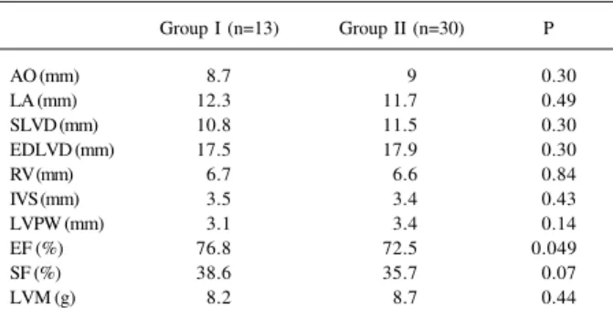
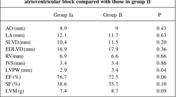
No statistically significant difference was observed in regard to the dimensions of the cavities (tab. III). Analyzing left ventricular function, group I had significantly greater ejection fraction values (76.77 x 72.53; P=0.049) than group II did (tab. III). No difference was found between the left ven-tricular masses in the 2 groups. When the subgroup compri-sed of women positive for anti-Ro/SSA antibodies was se-parated, the difference in the ejection fraction disappeared, and only the difference in the thickness of the left ventricu-lar posterior wall was significant (tab. IV).
The values obtained with the signal-averaged electro-cardiogram showed no significant difference in the QRS analysis and in the time and frequency domains (tab. V). The result did not change when only the subgroup of wo-men with anti-Ro/SSA antibodies was assessed (tab. VI).
The PR interval showed no significant difference bet-ween the 2 groups, even when considering the subgroup of women positive for anti-Ro/SSA antibodies alone.
Discussion
One of the most severe forms of presentation of neo-natal lupus is the one that includes congenital total atrio-ventricularblock 14. Several attempts have been made to
de-monstrate that, even in the absence of that block, cardiolo-gical manifestations of lesser clinical significance exist, indi-cating some degree of myocardial impairment in these pa-tients 8,10. Our study showed some differences between
groups I and II; however, those with statistical significance revealed only small differences between the groups and did
Table I - Assessment of the electrocardiogram of the groups studied
Group I (n=13) Group II (n=30) P
HR 137.6 125.9 0.04
ÂQRS 120.4 130.7 0.20
RV1 20 21.7 0.75
QTc (ms) 40 39 0.20
HR - heart rate; ÂQRS - electrical axis; RV1 - amplitude of the R wave in V1; QTc - corrected QT interval.
Table II - Assessment of the electrocardiograms of the children of anti-Ro/SSA-positive women and healthy women
Group Ia Group II P
HR 132.60 125.20 0.36
ÂQRS 127.20 130.70 0.81
RV1 27.20 21.70 0.79
QTc 0.40 0.39 0.72
HR - heart rate; ÂQRS - electrical axis; RV1 - amplitude of the R wave in V1; QTc - corrected QT interval.
Table III - Values of the measures obtained on the echocardiogram (means of the diameters and mass, and assessment of ventricular
function) in the groups studied
Group I (n=13) Group II (n=30) P
AO (mm) 8.7 9 0.30
LA (mm) 12.3 11.7 0.49
SLVD (mm) 10.8 11.5 0.30
EDLVD (mm) 17.5 17.9 0.30
RV (mm) 6.7 6.6 0.84
IVS (mm) 3.5 3.4 0.43
LVPW (mm) 3.1 3.4 0.14
EF (%) 76.8 72.5 0.049
SF (%) 38.6 35.7 0.07
LVM (g) 8.2 8.7 0.44
difference disappeared, showing once again that the pre-sence of anti-Ro/SSA antibodies was not the factor determi-ning the greater heart rate found in the subgroup of women with those antibodies.
The echocardiographic measures, as expected, were similar, because the patients had neither signals nor symp-toms of fetal or neonatal heart failure. However, a significan-tly greater ejection fraction was found in group I. The sta-tistically significant differences related to ejection fraction may be justified by the fact that that parameter is a mathe-matical formula in which the systolic and diastolic left ventri-cular diameters are cubed. Thus, when cubed, small nonsig-nificant differences in ventricular diameters become statis-tically significant without denoting change in the real func-tion. In addition, the ejection fraction was greater in the group studied, and no scientific explanation exists for the better ventricular function in the children of patients with systemic lupus erythematosus. Moreover, the shortening fraction, another parameter used to assess ventricular function, was similar between the groups, emphasizing once again the possibility that a greater ejection fraction is a bias due to the use of a mathematical resource that increases the difference between the values of left ventricular cavity diameters. Still considering the echocardiographic values, the differences disappeared when only the children of anti-Ro/SSA-positive women were assessed.
Although children of women with systemic lupus ery-thematosus have not been reported as candidates for ven-tricular arrhythmia, the use of signal-averaged electrocar-diography is justified when evidence of myocardial inflam-matory disease is searched for in these patients. Therefore, we could find changes in the spectral turbulence similar to those found in patients in the acute and subacute phases of myocarditis with other causes, in which signal-averaged electrocardiography was more effective than other conven-tional diagnostic methods to detect evidence for a myocar-dial inflammatoryprocess 16. Reports on signal-averaged
Table VI – Assessment of signal-averaged electrocardiogram in the Ia (Anti-Ro/SSA +) and II groups
Group Ia Group II P
DQRS (ms) 54.80 560 0.48
MSR20 (mv) 1760 139.50 0.41
MSR40 (mv) 524.70 464.40 0.32
LAS40 (ms) 9.60 10.20 0.53
Cem 0.96 0.96 0.58
Cesd 0.05 0.05 0.91
Bdm 59.70 62.30 0.09
Bdsd 26.40 27.90 0.39
PR 159.20 158.30 0.76
DQRS - total duration of the filtered QRS; MSR20 - mean square root of the tension of the final 20 ms of the filtered QRS; MSR40 - mean square root of the tension of the final 40 ms of the filtered QRS; LAS40 - total duration of the potentials with amplitude ≤ 40 mV in the terminal region of the filtered QRS; Cem - mean of the intersegmentary spectral correlation of the electric signal throughout ventricular activation; Cesd - standard deviation of the interseg-mentary spectral correlation of the electric signal throughout ventricular ac-tivation; Bdm - mean of the frequency band delimitating the signal energy; Bdsd - standard deviation of the frequency band delimitating the signal energy; PR - PR interval measured on signal-averaged electrocardiogram. Table IV - Measures obtained on the echocardiogram (means of the
diameters and mass, and assessment of ventricular function) in the children of anti-Ro/SSA-positive women without congenital total
atrioventricular block compared with those in group II
Group Ia Group II P
AO (mm) 8.9 9 0.43
LA (mm) 12.1 11.7 0.63
SLVD (mm) 10.4 11.5 0.20
EDLVD (mm) 16.9 17.9 0.36
RV (mm) 6.9 6.6 0.66
IVS (mm) 3.4 3.4 0.86
LVPW (mm) 2.9 3.4 0.04
EF (%) 76.7 72.5 0.06
SF (%) 38.6 35.7 0.10
LVM (g) 7.4 8.7 0.09
Ao- diameter of the aorta; LA- diameter of the left atrium; EDLVD- end-diastolic left ventricular diameter; SLVD- systolic left ventricular diameter; RV- end-diastolic right ventricular diameter; IVS- end-diastolic thickness of the interven-tricular septum; LVPW- thickness of the left veninterven-tricular posterior wall during diastole; EF- left ventricular ejection fraction; SF- systolic shortening fraction; LVM- left ventricular mass.
Table V - Comparison of the means of QRS in the time and frequency domains
Group I (n=13) Group II (n=30) P
DQRS (ms) 550 560 0.57
MSR20 (mv) 177.80 139.50 0.41
MSR40 (mV) 5500 464.40 0.11
LAS40 (ms) 9.70 10.20 0.39
Cem 0.96 0.96 0.17
Cesd 0.06 0.05 0.31
Bdm 60.70 62.30 0.19
Bdsd 26.80 27.90 0.50
PR 187.50 120.70 0.84
DQRS - total duration of the filtered QRS; MSR20 - mean square root of the tension of the final 20 ms of the filtered QRS; MSR40 - mean square root of the tension of the final 40 ms of the filtered QRS; LAS40 - total duration of the potentials with amplitude ≤ 40 mV in the terminal region of the filtered QRS; Cem - mean of the intersegmentary spectral correlation of the electric signal throughout ventricular activation; Cesd - standard deviation of the interseg-mentary spectral correlation of the electric signal throughout ventricular ac-tivation; Bdm - mean of the frequency band delimitating the signal energy; Bdsd - standard deviation of the frequency band delimitating the signal energy; PR - PR interval measured on signal-averaged electrocardiogram.
not suggest cardiological impairment of the children due to maternal systemic lupus erythematosus. No impairment in ventricular function or electrocardiographic change that could confirm cardiac damage was observed in the neonatal period until the 60th day of life.
The 12-lead conventional electrocardiogram showed differences between the groups in regard to heart rate values, which were greater in group I (tab. I). Goble et al 8
electrocardiography in newborns and infants are scarce, ours being one of the first studies on this subject in our country 16. The normal values of the signal-averaged
elec-trocardiogram in the age bracket of the patients studied have not yet been standardized. That was the reason why the control group was carefully selected to include healthy and nonasphyxial newborns and infants, and mothers with normal prenatal follow-up and no risk factors for myocardial damage. The QRS assessment revealed very similar values in the 2 groups, and no pattern suggestive of spectral tur-bulence was found in the time and frequency domains.
The results obtained in the signal-averaged electro-cardiogram suggest that the children of women with syste-mic lupus erythematosus have no changes in intramyocar-dial ventricular conduction. Although this does not defini-tively rule out the presence of myocarditis, it suggests at least that no detectable damage to ventricular intramyocar-dial conduction occurred during intrauterine life.
The PR interval was assessed on the signal-averaged electrocardiogram, and, although it was greater in absolute numbers in the children of women with systemic lupus ery-thematosus, these differences were not statistically signifi-cant and were not the same as those reported by Goble et al8.
This may be because signal-averaged electrocardiography has a greater precision than conventional electrocardiogra-phy with an estimated error of a few milliseconds.
Analyzing the signal-averaged electrocardiogram of the subgroup of women with anti-Ro/SSA antibodies, we also did not find differences in regard to the healthy control group.
When the subject of pregnancy is considered for wo-men with systemic lupus erythematosus, the importance of the prenatal follow-up is worth noting, because women with
severe diseases as such are considered high risk. Therefo-re, they require special attention, such as serial ultrasounds and fetal echocardiography, especially those women positi-ve for anti-Ro/SSA antibodies after the 16th gestational week, when the risks of neonatal lupus are greater 15.
Curren-tly, no doubts exist that a well-conducted prenatal follow-up largely reduces fetal and perinatal mortality. It is also impor-tant to inform the pediatrician about possible delivery com-plications, allowing him to prepare appropriate material for assisting the newborn still in the delivery room. This is a de-termining factor for the good evolution of patients at risk, such as those with congenital total atrioventricular block. From the moment when this study was carried out until the beginning of 2002, 6 cases of congenital total atrioventricu-lar block were diagnosed in the prenatal follow-up, al-though they were not included in this study. The early diag-nosis allowed the successful birth of 4 children, 2 of whom underwent definitive pacemaker implantation within their first 48 hours of life.
In the sample studied, no change in ventricular func-tion or in conducfunc-tion of the cardiac stimulus was observed in children of women with systemic lupus erythematosus. Therefore, no evidence existed confirming lesions in the conduction system of these patients or factors interfering with the ventricular function in the absence of congenital total atrioventricular block, even when assessing the group of women with anti-Ro/SSA antibodies alone. Patients with no congenital total atrioventricular block may not have un-dergone intrauterine myocardial injury, or if they had an in-flammatory process, this may have been successfully resol-ved without any sequelae still during the intrauterine life.
1. Buyon JP, Winchester R. Congenital complete heart block. A human model of passively acquired autoimmune injury. Arthritis Rheum 1990;5:33:609-14. 2. Carrera PE, Gutierrez-Larraya F, Gomez-Reino JJ. Successful intrauterine
thera-py with dexamethasone for fetal myocarditis and heart block in a woman with sys-temic lupus erythematosus. J Rheumatol 1993; 20:1204-7.
3. Tseng CE, Buyon JP. Neonatal lupus syndromes. Rheum Dis Clin North Am 1997; 23:31-53.
4. Alexander E, Buyon JP, Provost TT, et al. Anti-Ro/SSA antibodies in the patho-physiology of congenital heart block in neonatal lupus syndrome, an experi-mental model. In Vitro electrophysiological and immunocytochemical studies. Arthritis Rheum 1992;35:176-89.
5. Talbott JH, Historical background of discoid and systemic lupus erythematosus. In: Dubois’, Lupus Erythematosus. 3rd ed. Philadelphia. London: Lea &
Febiger, 1987; 3-11.
6. Mintz G, Niz J, Gutierrez G, et al. Prospective study of pregnancy in systemic lu-pus erythematosus: results of a multidisciplinary approach. J Rheumatol 1986;13:732-9.
7. Frohn-Mulder IM, Meilof JF, Szatmari A, et al. Clinical significance of maternal anti-Ro/SSA antibodies in children with isolated heart block. J Am Coll Cardiol 1994; 23:1677-81.
8. Goble MM, Dick II M, McCune WJ, et al. Atrioventricular conduction in chil-dren of women with systemic lupus erythematosus. Am J Cardiol 1993;71:94-8. 9. Gordon PA, Khamashta MA, Hughes GR, Rosenthal E. Increase in the heart
rate-References
corrected QT interval in children of anti-Ro positive mothers, with a further in-crease in those with siblings with congenital heart block: comment on the article by Cimaz et al. Arthritis Rheum 2001; 44: 242-3.
10. Cimaz R, Stramba-Badiale M, Bucrato A, Catelli L, Panceri P, Meroni PL. QT in-terval prolongation in asymptomatic anti-SSA/Ro – positive infants without congenital heart block. Arthritis Rheum 2000; 43:1049-53.
11. Tan EM, Cohen AS, Fries JF, et al. The 1982 revised criteria for the classification of systemic lupus erythematosus. Arthritis Rheum 1982; 25: 1271-7. 12. Sahn DJ, DeMaria A, Kisslo J, Weyman AL. The Committee on M-mode
standar-dization of the American Society of echocardiography. Recommendations regar-ding quantitation in M-mode echocardiography: Results of survey on echocar-diographic measurements. Circulation 1978; 58:1072-83.
13. Devereux RBH, Alfonso DT, Lutas EM, et al. Echocardiographic assessment of left ventricular hypertrophy comparison to necropsy findings. Am J Cardiol 1986; 57:450-8.
14. Waltuch J,Buyon JP. Autoantibody-associated congenital heart block: outcome in mothers and children. Ann Intern Med 1994;120:544-51.
15. Kuramochi Y, Takechi N, Ohkubo T, Ogawa S. Longitudinal estimation of signal-ave-raged electorcardiogram in patiens with Kawasaki disease. Pediatr Int 2002; 44: 12-7. 16. Leite MFMP, Barbosa EC, Barbosa P, et al. O eletrocardiograma de alta resolução