Session editor: Alfredo José Mansur ([email protected])
Associate editors: Desiderio Favarato ([email protected]) Vera Demarchi Aiello ([email protected])
Mailing address: Alfredo José Mansur InCor Av. Dr. Enéas C. Aguiar, 44 -05403-000 - São Paulo, SP, Brazil
English version by Stela Maris C. Gandour
Ca se 3 / 2 0 0 1 – Tw enty-yea r-old w oma n w ith pulmona r y hypertension, syncope, a nd severe hea da che (Instituto do Cora çã o do Hospita l da s Clínica s – FM USP - Sã o Pa ulo)
Clinicopathologic Session
A 20-year-old woman sought medical care due to hea-dache and vomiting following syncope (9/22/95).
The patient was born from an uneventful normal deli-very at the end of a full-term pregnancy. Up to the age of 1 year and 6 months, the patient had fatigue when being fed. At the age of 7 years, she began to have fatigue on strenu-ous exertion and palpitations during tense situations. At the age of 8 years, she had 2 episodes of cyanosis and dys-pnea on exertion accompanied by the sensation of imminent loss of consciousness. She also complained of arthralgia in her knees and ankles, and pain in her thoracic and lumbar spine. At the age of 9 years, the patient was referred to our hospital (11/26/84).
The physical examination at that time (11/26/84) sho-wed an eupneic and acyanotic child, with a regular and sym-metric pulse. The heart rate was 72bpm and blood pressure was 120/90mmHg. The lung examination was normal. The heart examination showed an increased intensity of the se-cond cardiac sound, which was palpable in the pulmonary area, where a systolic murmur could also be heard. The liver was palpated in the right costal margin, and no edema of the lower limbs was observed.
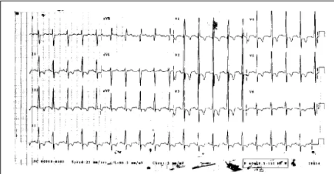
The electrocardiogram (11/23/84) showed sinus rhy-thm, a heart rate of 79bpm, QRS axis of +110° forward, and right ventricular hypertrophy (fig. 1). The chest X-ray sho-wed enlargement of the right ventricle and bulging of the pulmonary trunk. Laboratory tests are shown in table I. The search for lupus erythematosus cells, antinuclear factor, and rheumatoid factor was negative.
The echocardiogram (10/3/85) disclosed an aneurysm
of the membranous part of the ventricular septum and a ventricular septal defect of small hemodynamic repercussi-on. The heart had normal dimensions and contractility (Table II).
The patient underwent hemodynamic and angiogra-phic studies (November ’85) which evidenced severe pul-monary hypertension, a mild increase in the end-diastolic volume, normal left ventricular contractility, and aneurysm of the membranous septum through which blood flow into the left ventricle was detected (Table III). New studies car-ried out in October ’86 showed severe pulmonary hyperten-sion (Table III).
The radionuclide ventriculography (May ’86) showed a mild global reduction in left ventricular function and bulging of the pulmonary trunk (Table IV).
On the exercise test (November ’87), the patient sho-wed a low physical capacity and no signs of myocardial is-chemia (Table V). New assessments were performed on 8/18/ 88 and 5/23/91 (Table V).
The definitive diagnoses were primary pulmonary hy-pertension, and, after rheumatologic assessment, juvenile rheumatoid arthritis. Nifedipine, prednisone, and chloroqui-ne were then started.
Cardiac scintigraphy with gallium-67 (November ’93) showed no concentration of the radiotracer in the cardiac area not sugestive of myocardial inflammation.
The patient evolved with fatigue on major exertion up to September ’95.
A new echocardiogram (April ’94) showed severe pul-monary hypertension and mild tricuspid insufficiency (Table II). The radionuclide ventriculography (April ’94) was almost unaltered with dilation of the pulmonary trunk and mild dilation of the ventricles (Table IV).
In March ’94, the patient had syncope after emotional stress.
In April ’95, the patient underwent a left tympanoplas-ty due to repetitive episodes of otitis.
In September ’95, a new episode of syncope occurred
Paulo Bocayuva Cauduro, Moacyr Roberto Cucê N obre, Edmar Atik, Léa M. M. F. Demarchi
São Paulo, SP - Brazil
with falling and facial trauma. Four days later, the patient had intense pulsatile headache accompanied by nausea, vomiting, and dizziness, but with no eyesight changes.
On physical examination (September ’95), the patient had a regular pulse, a heart rate of 100 bpm, and a blood pressure of 140/95mmHg. Her lung examination was normal. Her heart examination showed increased intensity of the
second cardiac sound in the pulmonary area, a systolic murmur (++/4) in the tricuspid area, and an aspirating dias-tolic murmur (+/4) in the high left sternal margin. The abdo-minal examination was normal. The neurological examina-tion showed an oriented patient with mild neck stiffness and no motor deficit. On ophthalmoscopy, bilateral papillary edema was present.
The diagnosis of endocranial hypertension seconda-ry to an expansive process in the posterior cranial fossa was established.
Fig. 1 – Electrocardiogram – Sinus rhythm and right ventricular hypertrophy.
Table I – Laboratory test results
Tests/dates April ‘85 May ‘88 Dec ‘89 Sept ‘95
Red blood cells/mm3 5.200.000 4.200.000 4.800.000
Hemoglobin (g/dL) 14.5 12.4 16.4
Hematocrit 0.42 0.38 0.48
MCV (m3) 81 90 100
MCHC (g/dL) 35 33 34
Leukocytes/mm3 5.800 6.200 14.900
Band neutrophils (%) 1 6 3
Segmented neutrophils 49 65 84
Eosinophils (%) 4 2 0
Lymphocytes (%) 42 25 10
Monocytes (%) 3 2 3
Platelets/mm3 228.000 237.000
Platelet aggregation Normal
Prothrombin time (s) 14 13.4
Observed/control ratio 1.01 1.05
APTT (s) 50.4 21.8
Observed/control ratio 1.01 0.72
Thrombin time (s) 11.3 (10.5)
-Urea (mg/dL) 49
Creatinine (mg/dL) 1
Glycemia (mg/dL) 97
Sodium (mEq/L) 137
Potassium (mEq/L) 4.3
Table II – Echocardiographic evolution
Measures/dates October ‘85 April ‘94
Ventricular septal thickness (mm) 6 8 Posterior wall thickness (mm) 6 8
Aortic diameter (mm) 26 27
Left atrial diameter (mm) 26 28 Left ventricle
Diastolic diameter (mm) 40 36
Systolic diameter (mm) 23 21
Ejection fraction - 0.42
Right ventricular diastolic diameter (mm) 14 28 Ventricular septal defect (mm) 8 Absente Mean pulmonary artery pressure (mmHg) Normal 77
Table IV – Evolution on roentgenography and radionuclide ventriculography
Measures/dates 9/5/86 18/4/94
Radiocardiography
Pulmonary transit time (s) 6 7
Pulmonary trunk Dilated Dilated
Ventriculographies Left ventricle
Volumes Mild increase Mild increase
Kinetics Septal and apical hypokinesia Normal
Ejection fraction 0.49 0.62
Right ventricle
Volumes Mild/moderate increase Mild increase
Kinetics Normal Normal
Ejection fraction - 0.60
Table III – Hemodynamic data
Measures/dates November ‘85 October ‘86
Pressures (mm Hg) Basal Nifedipina
Right atrial mean 9 6 2
Right V. 128/15/15 94/7/7 67/3/4
(syst/initial diast/end-diast)
Pulmonary artery 127/96/111 95/68/81 67/49/59 (syst/diast/mean)
Pulmonary wedge (mean) 12 8 11
Left V. 136/0/14 120/0/17 136/5/12
(syst/initial diast/end-diast)
Aorta 135/96/114 118/75/94 130/95/111
(systolic/diastolic/mean) Vascular resistance (dyn.seg.cm-5)
Pulmonary 587 481
-Systemic 1624 1455
-Cardiac index (L/min/m2) 2.3 3.9
The test for detecting the IgG class antibody to the he-patitis A virus was positive. Blood cultures showed no growth of microorganisms.
The electrocardiography (10/9/95) showed sinus rhy-thm, a heart rate of 125bpm, QRS axis of +150° forward, and right ventricular hypertrophy (fig. 2).
The cranial radiography was normal.
The computerized tomography revealed a lesion with reduced density and a ring contrast in the right cerebellar hemisphere, with no fourth ventricular deviation and no ventricular dilation. The image was considered suggestive of an abscess.
The patient was prescribed 2g of vancomycin, 1,500mg of metronidazole, 4g of ceftriaxone, and 12mg of dexametha-sone, daily. In the following days, the patient remained on ceftriaxone, metronidazole, and dexamethasone. On the 6th day of hospitalization, the dose of ceftriaxone was reduced to 2g daily, vancomycin and metronidazole were suspen-ded, and 2,400mg of clindamycin were introduced.
The patient’s neurological condition and the papillary edema improved. On the 18th day of hospitalization, she complained of pain and paresthesia in the lower limbs with no signs of inflammation or blood hypoperfusion. The neu-rological examination showed no reduction in strength or abnormal reflexes. A global reduction in neuromuscular re-flexes existed.
On the 19th day of hospitalization, the patient under-went left suboccipital trepanation for stereotaxic drainage of a cerebellar abscess. A dense and chocolate-like material suggestive of an abscess collection was drained.
Five hours after the procedure, the patient had intense cyanosis and prolonged hypotension, followed by cardio-pulmonary arrest and death (10/10/95).
Discussion
Clinical considerations – Our patient is a young
wo-man with antecedents suggesting pneumopathy since her birth and rheumatologic findings during childhood. During the clinical investigation, the diagnostic hypotheses of pri-mary pulmonary hypertension and juvenile rheumatoid arth-ritis were formulated. During a hemodynamic study at the age of 10 years, a ventricular septal defect was observed, but it was no longer reported at the age of 20 years. After re-petitive episodes of otitis, the patient underwent
tympano-plasty. Five months after the procedure, a diagnostic hypo-thesis of cerebellar abscess was made and the patient was treated with antibiotics and corticoids. On the 19th day of hospitalization, the patient underwent trepanation and stereotaxic drainage, evolving to death in 5 hours. We will focus on the diagnoses of pulmonary hypertension and ce-rebral abscess.
Pulmonary hypertension is defined as systolic pulmo-nary pressure above 30mmHg and mean pulmopulmo-nary pressu-re above 20mmHg. The diagnosis of primary pulmonary hy-pertension is established only when, after appropriate investigation, no secondary cause is found 1-3.
On anamnesis, the patient complained of fatigue, dysp-nea and cyanosis on exertion, presyncope, and syncope. On physical examination, the patient had an increased in-tensity of the second cardiac sound, in pulmonary area. The electrocardiogram showed deviation of the axis to the right and right ventricular hypertrophy. The chest X-ray showed enlargement of the right ventricle and bulging of the pulmonary trunk. These findings are very suggestive of pulmonary hypertension.
Rheumatoid disease may be a cause of pulmonary hy-pertension. If the diagnosis of juvenile rheumatoid arthritis made during the rheumatologic investigation of our patient is considered true, it may constitute a possible secondary cause of pulmonary hypertension. Juvenile rheumatoid ar-thritis may be classified into 3 types depending on the de-gree of involvement observed: systemic, in many joints, or in few joints. Systemic involvement manifests as high fever, rash, general lymphadenopathy, hepatosplenomegaly, and cardiopulmonary involvement. Articular involvement mani-festation varies. Regarding pulmonary involvement, the most common finding is pleuritis, followed by pulmonary nodules, and, more rarely, by pulmonary fibrosis 4,5. In some cases of pulmonary fibrosis, the chest X-ray shows a diffuse reticulonodular infiltrate. However, it is worth noting that even in patients with a normal chest X-ray, alterations in pulmonary function have been found with a reduction in vi-tal capacity, which suggests the existence of a higher inci-dence than expected of cases of pulmonary fibrosis with no radiographic alterations. Some patients with pulmonary fi-brosis will develop pulmonary hypertension 6-8. We conclu-de that regarding the clinical case in question, the hypothe-sis of pulmonary hypertension secondary to rheumatoid disease may not be eliminated; however, examinations such as pulmonary function tests and pulmonary biopsy, which
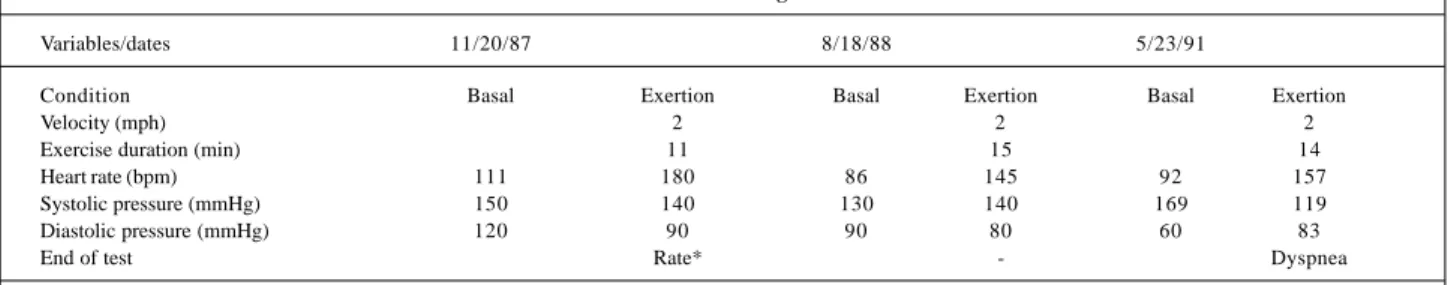
Table V – Assessment through exercise test
Variables/dates 11/20/87 8/18/88 5/23/91
Condition Basal Exertion Basal Exertion Basal Exertion
Velocity (mph) 2 2 2
Exercise duration (min) 11 15 14
Heart rate (bpm) 111 180 86 145 92 157
Systolic pressure (mmHg) 150 140 130 140 169 119
Diastolic pressure (mmHg) 120 90 90 80 60 83
End of test Rate* - Dyspnea
were not performed, would be important for establishing the diagnosis.
The echocardiogram, which is an important comple-mentary test in investigating pulmonary hypertension, showed ventricular septal defect and eliminated mitral valve disease or cardiomyopathy, which are other possible cau-ses of pulmonary hypertension 9.
The hemodynamic and angiographic studies perfor-med in November ’85 confirperfor-med the severe pulmonary hy-pertension, increased vascular pulmonary resistance, and ventricular septal defect, leading to right-to-left reverse shunt, which are findings reported in Eisenmenger’s syn-drome 8. Data reported in the anamnesis, physical examina-tion, electrocardiogram, and chest X-ray are compatible with this hypothesis supported by the fact that patients with Eisenmenger’s syndrome may have transient bacteremia, which may enter the cerebral circulation through the shunt without being filtered in the pulmonary circulation, leading to a cerebral abscess 10,11. The hypothesis of Eisenmenger’s syndrome can be questioned due to the presence of the small size of the ventricular septal defect (8mm in diameter), which, even though present, may not have been the cause of pulmonary hypertension. Other discordant points are the absence of an elevated hematocrit in our patient and the fact that on the echocardiogram of November ’94 the ventricular septal defect was no longer detected.
Regarding the investigation of pulmonary hyperten-sion, we have to consider the possibility of pulmonary thromboembolism as an important cause of pulmonary hy-pertension. Therefore, ventilation-perfusion mapping or pulmonary angiography, which were not performed, is re-quired so the diagnosis of primary pulmonary hypertension may be established 12,13.
Regarding the hypothesis of pulmonary thrombo-embolism, we could argue that no clinical data suggestive of this diagnosis exist, and if no cause for pulmonary hyperten-sion is identified, it should be considered primary. The cur-rent theories about the causes of primary pulmonary hyper-tension are as follows: 1) recurring occult systemic venous thrombosis with thromboembolism: patients with recurring thromboembolism develop pulmonary hypertension and cor pulmonale slowly, with no clinical manifestations of thromboembolism, and characteristic angiographic fin-dings may only be present in an early phase, disappearing later 14; 2) development of pulmonary hypertension due to in situ thromboses in smaller pulmonary arteries resulting in diffuse vascular pulmonary obstruction. This theory is ba-sed on the existence of defects in coagulation, including ab-normalities in platelet function and defective fibrinolysis 12. Other less probable causes of pulmonary hyperten-sion in our case are the following: chronic obstructive pul-monary disease, to be investigated with a pulpul-monary func-tion test; Takayasu’s disease, which involves the peripheral pulmonary arteries (our patient, however, showed no invol-vement of the large arteries, as would be expected in this di-sease); thrombosis of the portal vein, in which the hepatic function tests would be very useful; and sickle cell disease,
which could be confirmed with erythrocytic morphology and electrophoresis of hemoglobin.
In the hemodynamic study in ’85, a good response to nifedipine occurred with a reduction in the pressure in the pulmonary artery and in the right atrium, an increase in car-diac output, and a slight change in systemic blood pressure. This test is very important to recognize which patients would benefit from the prolonged use of nifedipine to con-trol pulmonary hypertension.
Some authors recommend the prophylactic use of anti-coagulant agents in pulmonary hypertension, even if no thromboembolism has been evidenced on complementary tests during the investigation of the cause of the disease. They state that patients have a high risk for pulmonary thromboembolism due to their sedentary lifestyle, venous insufficiency, and dilation of the right cardiac chambers. This medicamentous option is being studied to determine its efficacy.
In 1995, the patient being discussed was hospitalized and the diagnostic hypothesis of left cerebellar abscess was formulated.
According to the literature, in approximately 75% of the patients with cerebral abscess, the duration of the symp-toms equals or is less than 2 weeks. Only 50% of the pati-ents have the classic triad of fever, headache, and focal neu-rological deficit. Headache is usually moderate to intense, and it may be hemicrania or generalized headache. Fever oc-curs in 45% to 50% of the patients. Changes in mental state ranging from lethargy to coma may occur. Focal neurolo-gical findings are present in 50% of the patients and de-pend on the localization of the lesion and adjacent edema. Nausea and vomiting affect half the patients, probably due to intracranial hypertension. Convulsions occur in 25% to 35% of the patients, usually when the frontal lobe is impai-red. Stiffness of the neck and papilledema are present in 25% of the cases each 15-17. In regard to complementary tests, mo-derate leukocytosis may be found in the hemogram; howe-ver, 40% of the patients have a normal white blood cell co-unt. The sedimentation rate is increased up to around 45-50mm/h. C-reactive protein is increased and may be used in the differential diagnosis between neoplasia and cerebral abscess. Lumbar puncture is contraindicated in cases sus-pected of having cerebral abscess because of the risk of herniation and because of the low specificity of this type of examination. Cranial X-ray is normal in most cases.
may use scintigraphy with leukocytes marked with indium-111, which will accumulate in the inflammatory foci. The cli-nical case in question is highly suggestive of cerebellar abs-cess, considering the data presented.
A cerebral abscess may develop in the following condi-tions: 1) associated with a continuous suppurative focus (47% of the cases of cerebral abscess) such as in: a) otitis media 17: the patient had repetitive episodes of otitis, which ended in a tympanoplasty. Most cerebral abscesses caused by otitis media are located in the temporal lobe, followed by the cerebellar location (it is worth noting that 85% to 89% of the cerebellar abscesses are secondary to otitis media). The-se procesThe-ses occur more frequently when the tympanic membrane is damaged as in the case of our patient; b) sinusi-tis: most cases involve the frontal lobe and not the cerebel-lum, as is the case of our patient; c) mastoiditis: no alteration in the mastoid process was observed on cranial computed to-mography; therefore, this possibility does not match our case 18; d) trauma: the patient had facial trauma; however, cra-nial radiography and tomography showed no signs of frac-ture; 2) through hematogenous via: a distant infectious fo-cus is characteristically present, usually in the thorax; abs-cesses occurring through hematogenous via are usually multiple. They may be associated with pulmonary abscess or bronchiectasis, which is not the case of our patient, or with localized infectious foci in other parts of the body.
It should be noted that cerebral abscess complicating bacterial endocarditis is a rare occurrence when the blood-brain barrier is intact 18. Among the cases of endocarditis, the acute form predominates, which in our case is not sug-gested by the clinical findings, over the subacute form. The cases of endocarditis and cerebral abscess are more fre-quent among patients with cyanotic congenital heart disea-ses, as in Eisenmenger’s syndrome. These cases are clinical-ly evidenced in the pediatric age bracket. Our patient had the cerebellar abscess at the age of 20 years, and the echo-cardiography performed 1 year earlier did not show a ventri-cular septal defect; these facts are contrary to hematogenic dissemination.
Five hours after trepanation and stereotaxic drainage, the patient had intense cyanosis, hemodynamic shock, cardiopul-monary arrest, and died. In cases of pulcardiopul-monary hypertension, the most common causes of death are the following: right heart fai-lure (64%), pneumonia (7%), sudden death (7%), and other cau-ses (7%), among which we can cite pulmonary thromboembo-lism, malignant arrhythmias, massive pulmonary hemorrhage, and sudden ischemia of the right ventricle 19.
(Dr. Paulo Bocayuva Cauduro)
Diagnostic hypotheses – Eisenmenger’s syndrome;
pulmonary hypertension related to repetitive pulmonary thromboembolism, primary pulmonary hypertension, cere-bral abscess, juvenile rheumatoid arthritis.
(Dr. Paulo Bocayuva Cauduro)
Rheumatoid arthritis that evolves with systemic vas-culitis is usually of the classical type with impairment of the bone-joint function, joint deformities, and high levels of rheumatoid factor. It usually has rheumatoid nodules, which are the clinical expression of cutaneous vasculitis. The cli-nical findings of our patient comprising arthralgia, absence of cutaneous nodules and of other manifestations such as peripheral neuritis and episcleritis are very different from those expected in the vasculitis of rheumatoid arthritis.
The usual pulmonary manifestation of rheumatoid ar-thritis is pleural effusion, which is followed in order of fre-quency by pulmonary interstitial disease, characteristically non-hypertensive. Another classical pulmonary lesion of rheumatoid arthritis is the presence of a nodule in the pul-monary parenchyma similar to the subcutaneous nodule, being considered an expression of vasculitis in the lungs.
The image of selective pulmonary arteriography, whi-ch shows a reduction in the lumen of the first branwhi-ch of the right pulmonary artery, as well as a reduction in the branch of the arterial tree, suggests involvement of medium- and lar-ge-caliber vessels. These findings differ from the typical im-pairment of the vasculitis of diffuse connective tissue di-seases, which is restricted to small vessels and arterioles. The pathognomonic lesion of rheumatoid vasculitis is a pe-rivascular inflammatory infiltrate, with a granuloma sur-rounded by palisaded histiocytes and a central focus of fibrinoid necrosis.
The diffuse connective tissue diseases that more commonly lead to vascular and interstitial pulmonary di-sease accompanied by hypertension are progressive syste-mic sclerosis and systesyste-mic lupus erythematosus. These diseases could be hardly considered as diagnostic hypo-theses in the present case due to the lack of clinical manifes-tations of these diseases and the lack of antinuclear factor.
(Dr. Moacyr Roberto Cucê Nobre)
Diagnostic and evolutionary characteristics of pulmo-nary vascular disease are evident in the present case.
Worthy of note are the symptoms and signs of pulmona-ry hypertension and right heart failure with a greater expression of temporal evolution, including the electrocardiographic alterations with progressive right ventricular hypertrophy.
In regard to the major cardiovascular causes of the syndrome, there are heart diseases with left-to-right shunt and increased pulmonary flow of the ventricular septal de-fect type, the obstructive lesions of the left side of the heart, and primary pulmonary hypertension.
Left obstructive defects usually cause severe condi-tions with a rapid fatal outcome in the first months of life, and the heart diseases of the ventricular septal defect type are initially present as pulmonary venocapillary congestion. Neither situation existed in our case, despite the anatomical demonstration of the latter anomaly.
The above question may be appropriate to ask about other similar cases, in which signs of primary pulmonary hypertension do not depend on heart defects that are even-tually discovered in the follow-up. The patient had clinical findings of pulmonary hypertension in the absence of any manifestation of ventricular septal defect. This fact may not have accounted for the alterations in the pulmonary arterial tree, which were so expressive of pulmonary hypertension. In addition, one may consider the possibility of the pulmonary vascular alteration that may occur in rheumatoid arthritis, despite its rarity, with findings similar to those of primary pulmonary hypertension.
Angiographic images and epidemiological data exclu-de other causes, such as chronic pulmonary thromboembo-lism, Takayasu’s disease, and pulmonary parenchyma le-sions, among which is schistosomiasis mansoni, which, even though rare, should be remembered in discussing this syndrome.
(Dr. Edmar Atik)
Autopsy
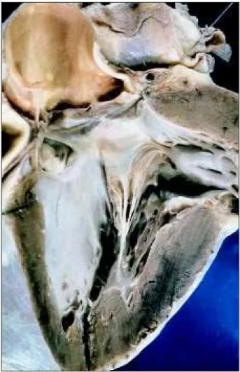
The heart weighed 400g and grossly showed an in-creased volume with predominance of the right chambers. On the sections, a perimembranous ventricular septal de-fect in the ventricular inlet was found, measuring approxi-mately 1.5cm in diameter. The defect was totally closed due to fibrosis of the anterior and septal leaflets of the tricuspid valve and their adhesion to the margins of the septal defect orifice (fig. 3). The tricuspid valve also had dilation of the ring and rolling of the free margin of the leaflets. The right atrium was very dilated and intense hypertrophy of the right ventricular wall existed. No intracavitary thrombi or other cardiac alterations were found.
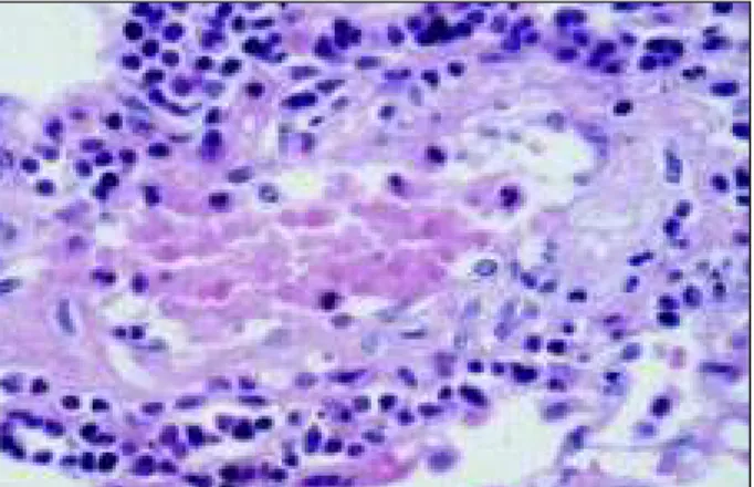
Microscopically, plexiform lesions in pulmonary arte-rioles compatible with Heath-Edwards grade IV pulmonary hypertension were seen (figs. 4 and 5). A few arterioles had multiple fibrin thrombi in their lumens, and multiple small and focal recent pulmonary infarcts were seen. A moderate lymphomononuclear inflammatory infiltrate was observed around rare pulmonary arterioles, but no histological altera-tions compatible with active vasculitis existed. Focal and sparse areas of mild pleural fibrosis with no inflammatory in-filtrate were present.
A large anemic cavitated infarct was observed in the ri-ght cerebellar hemisphere measuring 4cm of diameter, with approximately 3 weeks of evolution and no liquid content. In-side the cavitation, a drain with patent lumen and no obstruc-tion could be seen. The histochemical search for bacteria, fungi, and alcohol-acid resistant bacilli in the fragments of the cerebellar infarct was negative. The brain had an intense and diffuse edema with herniation of the cerebellar amygdalae.
No relevant morphological alterations were observed in the remaining organs.
(Dr. Léa M. M. F. Demarchi)
Anatomicopathological diagnoses – 1)
Perimembra-nous ventricular septal defect in the ventricular inlet, which was naturally closed by the anterior and septal leaflets of the tricuspid valve; 2) Pulmonary vascular alterations cor-responding to Heath-Edwards grade IV pulmonary hyper-Fig. 3 – Section of the left ventricular outflow tract showing the defect of the perimembranous ventricular septum, which is totally occluded due to the adhesion and fibrosis of the anterior and septal leaflets of the tricuspid valve to the margins of the septal orifice.
tension; 3) Surgically drained anemic and cavitated cerebel-lar infarct; 4) Cerebral edema.
(Dr. Léa M. M. F. Demarchi)
Comments
We have a case of pulmonary hypertension in a young woman with juvenile rheumatoid arthritis and naturally clo-sed ventricular septal defect. The patient died because of alterations related to intracranial hypertension secondary to an anemic cerebellar infarct with approximately 3 weeks of evolution.
Despite the clinical suspicion of abscess, the histolo-gical cerebellar alterations are more consistent with cavita-ted anemic infarct. Cerebellar anemic infarcts are caused by a deficiency in irrigation of the superior or inferior cerebellar arteries, which are part of the vertebro-basilar territory. The most common causes are thrombosis, vasospasm, or arterial hypotension, and, more rarely, congenital malformations, such as the presence of rings in the walls of the cerebellar arteries, which reduce even more the lumen of these small-caliber arteries. However, on autopsy, we could not find morphological changes of cerebellar ischemia.
The major reason for discussion in this case is pulmo-nary hypertension, because the patient had 2 diseases that could lead to pulmonary vascular alterations: juvenile rheu-matoid arthritis and ventricular septal defect. Therefore, we can not say that the patient had primary pulmonary hyper-tension.
Pulmonary involvement is one of the extraarticular ma-nifestations of juvenile rheumatoid arthritis 2. Different pleural lesions may be found, pleural effusion being the most common. Parenchymal lesions, such as interstitial pneumonia, pulmonary fibrosis, rheumatoid nodules, and, more rarely, vasculitis and pulmonary hypertension, may also be found 20. In the lungs, we could not find other mor-phological alterations in addition to the vascular lesions of pulmonary hypertension, the moderate
lymphomononu-clear inflammatory infiltrate, and the focal areas of mild pleu-ral fibrosis suggesting impairment due to rheumatoid arthri-tis. However, this does not allow us to completely eliminate the possibility of juvenile rheumatoid arthritis being the cause of or contributing to the development of pulmonary hypertension in our patient.
Ventricular septal defects represent the most frequent congenital cardiac anomaly. They are usually found in isola-tion, but may also be present in association with other car-diac malformations 21.
In the case being discussed, likewise with most ventri-cular septal defects 22, the defect was of the perimembra-nous type. It was located in the right ventricular inlet and was totally and naturally closed by the anterior and septal leaflets of the tricuspid valve.
Ventricular septal defects may be naturally closed by 2 mechanisms 21: 1) by formation of fibrous tissue beginning at the fibrous or valvar structures, usually of the tricuspid val-ve, located in the proximities of the defect 21. In these cases, the diagnosis of aneurysm of the membranous septum is made, which is a wrong determination, because only rarely is the fibrous tissue that closes the defect derived from the membranous septum or its remnants 23,24; 2) by adhesion of the septal leaflet of the tricuspid valve, which occurs only if the leaflet is adjacent to the communication orifice, ie, when the defect is located in the right ventricular inlet 21, as is the case of our patient.
Hemodynamic alterations of a patient with ventricular septal defect are determined more by the size of the defect than by its location 22. Pulmonary hypertension develops in patients with nonrestrictive defects, which comprise those defects large enough to cause no difference in pressure between the 2 sides of communication 25. Therefore, the flow through the defect is not determined by a pressure gra-dient, but by the relative resistance between the pulmonary and systemic circulations 25. At birth, the pulmonary vascu-lar resistance of an individual usually decreases suddenly in regard to systemic resistance 25. In the presence of a non-restrictive ventricular septal defect, blood flows from the left to the right ventricle through the ventricular communica-tion, increasing pulmonary flow 25. Therefore, if the defect is not surgically corrected, the patient will develop pulmonary hypertension 26.
In the case being discussed, even though the assess-ment of the size of communication has been impaired by the natural closure, the diameter of the defect seemed to mea-sure around 1.5cm, with a great possibility of being nonres-trictive. A few authors consider that ventricular septal de-fects with a diameter equal to or higher than 1.5cm are non-restrictive 27.
With elevation of pulmonary resistance, blood flow through the defect becomes minimal, and reversion in the flow direction may occur. Therefore, a right-to-left ventri-cular blood flow may result, characterizing Eisenmenger’s complex.
In most congenital cardiac anomalies, Eisenmenger’s com-plex manifests in the first year of life, and is well established by the Fig. 5 - Histologic section of the lung showing moderate lymphomononuclear
age of 2 to 3 years, being observed in around 5% to 10% of the patients with large nonrestrictive ventricular septal defect 26.
Indications for surgical treatment comprise the follo-wing: left-to-right ventricular blood flow higher than 2:1, re-curring endocarditis, and progressive aortic insufficiency. Once established, Eisenmenger’s complex contraindicates the surgical repair of any intracardiac communication 26.
Therefore, we could prevent the pulmonary complica-tion of pulmonary hypertension secondary to a large ventri-cular septal defect through surgical repair before the age of
2 years 26. In the case being discussed, we believe that, des-pite the natural closure of the ventricular septal defect, pul-monary hypertension could have resulted from an increase in pulmonary blood flow, which the patient underwent when the defect was still patent. Once more we emphasize that we have not found enough morphological data to eliminate the possibility that juvenile rheumatoid arthritis may have also contributed to the appearance of pulmonary hypertension.
(Dr. Léa M. M. F. Demarchi)
References
1. Rich S, Dantzker DR, Ayres SM et al. Primary pulmonary hypertension: a national prospective study. Ann Intern Med 1987; 107: 216-23.
2. Rubin LJ. Primary pulmonary hypertension - ACCP Consensus Statement. Chest 1993; 104: 236-50.
3 Vender R. Chronic hypoxic pulmonary hypertension. Chest 1994; 106: 236-43. 4. Calabro JJ, Holgerson WB, Sonpal GM, Khoury MI. Juvenile rheumatoid arthritis: a general review and report of 100 patients observed for 15 years Semin. Arthritis Rheum 1976; 5 : 257-98.
5. Gordon DA, Stein JL, Broder IThe extra-articular features of rheumatoid arthritis: a systematic analysis of 127 cases. Am J Med 1973; 54: 445-52.
6. Davidson C, Brooks AG, Bacon PA. Lung function in rheumatoid arthritis. A clinic survey. Ann Rheum Dis 1974; 33: 293-7.
7. Cervantes-Perez P, Toro-Perez AH, Rodriguez-Jurado P. Pulmonary involve-ment in rheumatoid arthritis. JAMA 1980; 243: 1715-9.
8. Kanemoto N. Electrocardiogram in primary pulmonary hypertension Eur J Cardiol 1981; 12 : 181-93.
9. Goodmann J, Harrison DC, Popp RL Echocardographic features of primary pul-monary hypertension Am J Cardiol 1974; 33: 438-43.
10. Fischbein CA, Rosenthal A, Fischer EG, Nadas AS, Welch K. Risk factors of brain abscess in patients with congenital heart disease. Am J Cardiol 1974; 34: 97-102. 11. Park SC, Neches WH. Neurologic complications of congenital heart disease.
Neurol Clin 1993; 11: 441-62.
12. Rubin LJ. Pathology and pathophisiology of primary pulmonary hypertension. Am.J.Cardiol 1995; 75: 51A-54A.
13. Pietra GG, Edwards WD, Kay JM, et al. Histopathology of primary pulmonary hypertension: a qualitative and quantitaive study of pulmonary blood vessels from 58 patients in the National Heart, Lung, Nd Blood Institute. Circulation 1989, 80: 1198-206.
14. Moser KM, Page GT, Ashburn WL, Fedullo PF. Perfusion lung scans provide a guide twich patients with apparent primary pulmonary hypertension merit angiography. West J Med 1988; 148: 167-70.
15. Carey ME, Yang SH. Brain abscess: A review of 400 cases J Neurosurg 1981; 55: 794-9. 16. Heineman HS, Braude AI, Osterholm JL. Intracranial suppurative disease. JAMA
1971; 218 1542-7.
17. Shaw MDM, Russel JA Cerebellar abscess - a review of 47 cases J. Neurol Neuro-surg Psychiatry 1975; 38: 429-35.
18. Pritt AA, Rubin RHJ, Karchmor AW, et al Neurological complications of bacterial endocarditis. Medicine 1978; 57: 329-43.
19. Sandoval J, Bauerle O, Palomar A, et al. Survival in primary pulmonary hyper-tension. Circulation 1994; 89: 1733-44.
20 Anaya JM, Diethelm L, Ortiz LA, et al. Pulmonary involvement in rheumatoid arthritis. Semin Arthritis Rheum 1995; 24: 242-54.
21. Anderson RH, Becker AE. In: Pathology of congenital heart disease. 2nd edition.
London: Butterworths & Co. Ltd, 1981.
22. Engle MA. The adult whith congenital heart disease. Br Heart J 1981; 27: 1-59. 23. Chesler E, Korns ME, Edwards E. Anomalies of the tricuspid valve, including pouches, resembling aneurysms of the membranous ventricular septum. Am J Cardiol, 1968; 21: 661-8.
24. Anderson RH, Kenox CC, Zuberbuhler JR. Mechanisms of closure of perimem-branous ventricular septal defects. Am J Cardiol 1983; 52: 341-5.
25 Hopkins WE. Severe pulmonary hypertension in congenital heart disease: a re-view of Eisenmenger syndrome. Curr Opin Cardiol 1995; 10: 517-23. 26. MacNamara DG. The adult with congenital heart disease. Curr Probl Cardiol
1989; 14: 57-114.