Cinecor (IV Centenário e Pró-Cardíaco) Rio de Janeiro e Hospital Universitário Antônio Pedro - UFF, Niterói
Correspondência: Edison C. Sandoval Peixoto - Av. Epitácio Pessoa, 4986/301 - 22471-001 - Rio de Janeiro, RJ
Objective - To evaluate prior mitral surgical commis-surotomy and echocardiographic score influence on the outcomes and complications of percutaneous mitral balloon valvuloplasty.
Methods - We performed 459 complete mitral valvu-loplasty procedures. Four hundred thirteen were primary valvuloplasty and 46 were in patients who had undergone prior surgical commissurotomy. The prior commissuro-tomy group was older, had higher echo scores, and a tendency toward a higher percentage of atrial fibrillation.
Results - When the groups were compared with each other, no differences were found in pre- and postprocedure mean pulmonary artery pressure, mean mitral gradient, mi-tral valve area, and mimi-tral regurgitation . Because we found no significant differences, we subdivided the entire group based on echo scores, those with echo scores ≤ 8 and those with echo scores >8 the mitral valve area being higher in the ≤ 8 echo score group 2.06±0.42 versus 1.90±0.40cm2
(p=0.0090) in the >8 echo score group.
Conclusion - Dividing the groups based on echo sco-re sco-revealed that the higher echo scosco-re group had smaller mitral valve areas postvalvuloplasty.
Key words: percutaneous mitral balloon valvuloplasty, prior mitral commissurotomy, mitral stenosis.
Arq Bras Cardiol, volume 76 (nº 6), 478-82, 2001
Edison C. Sandoval Peixoto, Rodrigo T. Sandoval Peixoto, Ivana Picone Borges, Paulo Sérgio de O liveira, Marta Labrunie, Mário Salles N etto, Ronaldo A. Villela, Pierre Labrunie, Guilherme A. Xavier de Brito,
Ricardo T. Sandoval Peixoto
Rio de Janeiro - Niterói, RJ - Brazil
Influence of the Echocardiographic Score and Not of the
Previous Surgical Mitral Commissurotomy on the Outcome of
Percutaneous Mitral Balloon Valvuloplasty
In 1984, Inoue at al.1 reported on the percutaneous mitral valve balloon dilatation technique, using the balloon subsequently named after Inoue.
We began percutaneous mitral balloon valvuloplasty in 1987 using a single balloon, and soon after we used the double balloon technique, Inoue’s balloon technique, and the low profile single balloon technique 2-5.
Percutaneous mitral balloon valvuloplasty has beco-me the treatbeco-ment of choice for symptomatic severe mitral stenosis without great damage to the subvalvular mitral apparel or great calcification or thickening of the mitral valve leaflets. It has had consistent results, and is currently preferred over surgical mitral commissurotomy in the type of patients mentioned above.
We have been using the Inoue balloon and single balloon techniques above all others 2-5. We use the double balloon technique less frequently. The literature shows in general similar results with these techniques 3-8.
We present here our experience with primary mitral balloon valvuloplasty compared with mitral balloon valvuloplasty following restenosis after surgical mitral commissurotomy, which we have been studying for some time 9. The efficacy of percutaneous mitral balloon valvu-loplasty for treating restenosis postsurgical mitral commis-surotomy has been demonstrated by us and others.
Methods
restenosis after they have already undergone surgical commissurotomy formed the prior surgical commissuro-tomy group. Seventeen procedures were either not comple-ted or had complications that impeded their performance, so were excluded from the study.
Of the 46 patients in the prior surgical commissuro-tomy group, 39 were female and 7 were male, but in the primary valvuloplasty group 339 were female and 74 were male (p=0.6486). No significant statistical difference existed. Age in the prior surgical commissurotomy group was 42.7±12.4 years and 36.9±12.5 years in the primary valvulo-plasty group (p=0.0030), the prior surgical commissurotomy group being older.
Ten patients were in NYHA functional class II, 32 were in functional class III, and 4 were in functional class IV in the prior surgical commissurotomy group. In the primary valvuloplasty group, 6 patients were in functional class I, 91 were in functional class II, 272 were in functional class III, and 44 were in functional class IV (p=0.8236), with no significant differences existing among the groups.
Heart rhythm was sinusal in 35 patients in the prior surgical commissurotomy group, and atrial fibrillation was present in 11. In the primary valvuloplasty group, heart rhythm was sinusal in 358, and atrial fibrillation was present in 55 (p=0.0520), with a tendency toward a greater percen-tage of atrial fibrillation in the prior surgical commissuro-tomy group.
Nine patients underwent mitral balloon valvuloplasty, developed restenosis during the follow-up period, and had to undergo a new mitral balloon valvuloplasty.
Echocardiographic mitral area determined by the half pressure time method before mitral balloon valvuloplasty
was 1.00±0.22cm2 in the prior surgical commissurotomy
group and 0.94±0.21cm2 in the primary valvuloplasty
group, (p=0.0699). The echo score 10 was 7.7±1.5 points in the prior surgical commissurotomy group and 7.2±1.4 in the primary valvuloplasty group (p=0.0158). It was significantly higher in the prior surgical commissurotomy group.
We used Inoue’s balloon in 65 procedures (maximum inflations of 24, 26, and 27mm diameter were used once, 25mm was used twice, and 28mm was used 60 times), the low profile single balloon in 385 (25mm was used 12 times, 25mm followed by 30mm was used 6 times and, finally, 30mm alone was used 367), the conventional single balloon was used 3 times (20mm diameter), and the double balloon in 6 (20+15mm in 3 and 20+18mm in 3), being a total of 459 procedures with those techniques.
After mitral dilation, left ventriculography in the right anterior oblique view was performed, in addition to new right and left catheterization and a new measure of the left atrium-left ventricle gradient. Measurement was always made of the simultaneous left atrium-left ventricle gradient immediately before and immediately after the dilation of the mitral valve. Early and middiastolic and presystolic gra-dients were measured, the mean gradient (method of the 3 points) being the arithmetic average of the 3 measures previously reported or, in a second phase, by the automatic
planimetric area of the gradient 11. Mitral valvular area was determined both pre- and postdilation. Cardiac output was determined by thermodilution and, the Gorlin and Gorlin formula 12 was used to calculate the area. The presence of mitral insufficiency was graduated according to the semi-quantitative criterion of Sellers et al. 13.
The following parameters were studied: age, sex, rhythm, preprocedure NYHA functional class, mitral valve, echo score 10, preprocedure echo mitral valve area, pre- and postballoon valvuloplasty mean pulmonary artery pressure, pre- and postprocedure mean mitral gradient, pre- and postprocedure hemodynamic measured mitral valve area, and pre- and postprocedure mitral regurgitation. The data obtained in the 2 groups were compared.
As pre- and postprocedure data studied revealed no significant statistical differences, the entire group of the 459 procedures performed was then subdivided into 2 new groups, the first one with echo scores ≤8 and the second one with echo scores >8. We then looked again at pre- and postballoon valvuloplasty mean pulmonary artery pressure, and postprocedure mean mitral gradient, and the pre-and postprocedure hemodynamically measured mitral valve area.
The statistical analysis was accomplished with the use
of the program “EPI-INFO version 6” 14 to determine the
frequency of the qualitative and numeric variables. The numeric variables were also studied through the analysis of the variance and the qualitative variables with the chi-square test.
Results
Four hundred fifty-nine percutaneous mitral balloon valvuloplasty procedures were performed, 46 in patients with restenosis in the follow-up of previous surgical com-missurotomy and 413 in intervention patients in whom per-cutaneous mitral balloon valvuloplasty was the only inter-ventional treatment.
See Tables I and II for the hemodynamic data obtained before and after mitral balloon valvuloplasty in the prior surgical commissurotomy and primary mitral valvuloplasty groups. No significant statistical differences were found between the two groups.
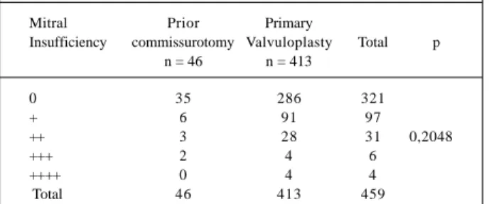
Tables III and IV show the mitral competence observed both pre- and postpercutaneous mitral balloon valvu-loplasty in the prior surgical commissurotomy and primary mitral valvuloplasty groups. Ten cases of serious mitral regurgitation occurred, 2 in the prior surgical commis-surotomy group and 8 in the primary valvuloplasty group (p=0.2048). No significant difference existed in the distri-bution of the serious mitral regurgitation pre- and postper-cutaneous mitral balloon valvuloplasty.
As the hemodynamic data were not significantly different between the groups, the entire group of 459 procedures was then subdivided into two new groups
based on echo scores, one group with echo scores ≤8 and
hemodynamic data for both pre- and post percutaneous mitral balloon valvuloplasty in the two groups. The pre- and postprocudure data was not significantly statistically different. However, the mitral valve area in the post-procedure group was significantly smaller in the >8 echo score group.
Discussion
Introduced by Inoue 1, percutaneous mitral
valvul-plasty quickly became one of the preferred treatments for severe mitral stenosis. It was reported in the beginning that mitral valve area after percutaneous mitral balloon valvu-loplasty was larger when the double balloon technique was used rather than the conventional single balloon 6,15. The efficiency and low level of complications with the Inoue
balloon 16,17 have also been reported. Although values
obtained with the double balloon 18 have been reported, the immediate results with the double balloon and the Inoue
Table I - Comparison of hemodymanic data prepercutaneous mitral balloon valvuloplasty in the prior surgical
commissuroto-my and primary mitral valvuloplasty groups
Prior Primary
Commissurotomy Valvuloplasty p N = 46 n = 413
Mean pulmonary pressure 37.6 ± 14.4 37.8 ± 14.2 0.9515 (mmHg)
Mean mitral gradient 18.3 ± 6.9 19.6 ± 6.9 0.2342 (mmHg)
Mitral valve area (cm²) 0.93 ± 0.19 0.90 ± 0.21 0.4092
Table II - Comparison of hemodymanic data postpercutaneous mitral balloon valvuloplasty in the prior surgical
commissuro-tomy and primary mitral valvuloplasty groups
Prior Primary Commissurotomy Valvuloplasty p
n = 46 n = 413
Mean pulmonary pressure 26.6 ± 10.9 26.8 ± 10.2 0.9062 (mmHg)
Mean mitral gradient 6.3 ± 4.2 5.4 ± 3.5 0.1492 (mmHg)
Mitral valve area (cm²) 1.92 ± 0.41 2.04 ± 0.42 0.0801
Table III – Mitral competence prepercutaneous mitral balloon valvuloplasty in the prior surgical commissurotomy and of
primary mitral valvuloplasty groups
Mitral Prior Primary
Insufficiency commissurotomy Valvuloplasty Total P n = 46 n = 413
0 41 352 393
+ 5 60 65 0.7602
++ - 1 1
Total 46 413 459
Table IV – Mitral competence postpercutaneous mitral balloon valvuloplasty in the prior surgical commissurotomy and of
primary mitral valvuloplasty groups
Mitral Prior Primary
Insufficiency commissurotomy Valvuloplasty Total p n = 46 n = 413
0 35 286 321
+ 6 91 97
++ 3 28 31 0,2048
+++ 2 4 6
++++ 0 4 4
Total 46 413 459
balloon are similar 19. Today, it has been proven that similar mitral valve area after percutaneous mitral valvuloplasty can be obtained with any of the balloon techniques whether the double balloon, Inoue balloon, or low profile single balloon are used if effective balloon dilation areas are comparable 3-5. The most recent studies 4,15 have shown that average mitral valve area equal to or greater than 2cm2 can result in spite of the technique used 4,15.
The incidence of mitral regurgitation is smaller when the balloon dilation area is corrected for the surface corporal area, being less than or equal to 4.0cm² 20. Accor-ding to Roch et al. 21, the only variable that can predict an increase in mitral regurgitation is the relationship between the effective balloon dilation area and the surface corporal area. Aurora et al. 22 showed that the size of the balloon, the degree of compromise of the subvalvar apparel, or the gravity of the mitral stenosis is not related to the appearance of mitral regurgitation.In spite of the high effective balloon dilation area that we used along with our experience, our incidence of significant mitral regurgitation was low 2-5.
It has been shown that percutaneous mitral valvulo-plasty can be accomplished with the same efficiency or at least with good results in groups of patients who undergo the procedure for either postsurgical mitral commissurotomy restenosis 9,23-25 or postpercutaneous mitral valvuloplasty restenosis 26.
In the present study, we evaluated the results obtained in a group of patients who underwent percutaneous mitral balloon valvuloplasty as the only treatment (primary valvu-loplasty) in comparison with the results obtained in a group of patients who underwent percutaneous mitral balloon val-vuloplasty for postsurgical mitral commissurotomy tenosis. The postsurgical mitral commissurotomy res-tenosis group was older, had higher echocardiographic sco-res, and had a tendency toward a higher percentage of atrial fibrillation and a larger mitral valve area before percuta-neous mitral balloon valvuloplasty. Higher echocardiogra-phic scores in this group of patients have been found also by others 27.
in spite of no significant difference existing, a tendency was noted toward smaller mitral valve areas in the postsurgical
mitral commissurotomy restenosis group, 1.92±0.41cm2
versus 2.04±0.42cm2 (p=0.0801), in the primary mitral
valvuloplasty group (tables I and II).
Lee et al.23 also found a smaller increase in the mitral valve area postpercutaneous mitral balloon valvulo-plasty in their postsurgical mitral commissurotomy restenosis group, but the difference was also not significant. In a subsequent report, Lau et al. 25, of the same group, reported mitral gradient, mitral valve area, mitral insufficiency, and restenosis in the follow-up similar to that in the postsurgical mitral commissurotomy restenosis group and in the primary mitral valvuloplasty
group.Although both authors reported smaller valvular
areas after percutaneous mitral balloon valvuloplasty (1.6cm2) than we did. Sharma et al. 27 and Medina et al.28 found similar results in their 2 groups of patients for hemodynamic data and for mitral insufficiency, although the postsurgical mitral commissurotomy restenosis
group of Sharma et al.27 had higher echocardiographic
score.
Like the last authors mentioned above, we also did not find a difference in the presence of mitral insufficiency either before percutaneous mitral balloon valvuloplasty mitral insufficiency (table III) or after percutaneous mitral balloon valvuloplasty (table IV) in the 2 study groups. Results very similar to ours were reported by Ha et al. 29, who obtained larger values for mitral gradient and smaller for mitral valve area, both without statistical differences, and similar variations for mitral insufficiency.
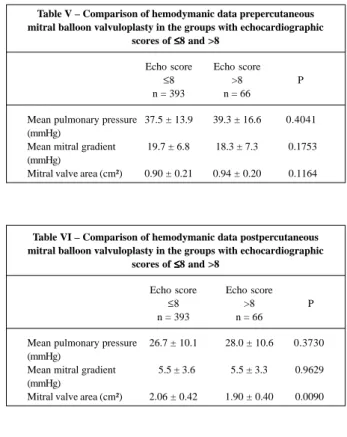
The differences that we obtained were not sig-nificant. The importance of the Wilkins et al. 10 echocar-diographic scores is demonstrated in the results obtained in percutaneous mitral balloon valvuloplasty 30. Because our percutaneous mitral balloon valvuloplasty post-surgical mitral commissurotomy group had higher echo scores than the primary percutaneous mitral balloon valvuloplasty group, 7.7±1.5 versus 7.2±1.4 points (p=0.0158) and a tendency toward slightly better results in the primary percutaneous mitral balloon valvuloplasty group, we redivided the total 459 procedures into a patient group with echo scores ≤8 and a patient group with echo scores >8 to better study the influence of echo-cardiographic scores on the obtained results. The new groups did not have significant differences for mean pulmonary pressure, mean mitral gradient, and mitral valve
area in the prepercutaneous mitral balloon valvuloplasty results (table V), or for mean pulmonary artery pressure and mean mitral gradient in the postpercutaneous mitral balloon valvuloplasty results (table VI). However, mitral valve area postpercutaneous mitral balloon valvuloplasty was significantly smaller in the group with scores >8 when compared with the mitral valve area obtained in the group with scores ≤ 8, respectively, 1.90±0.40 versus 2.06±0.42cm2 (p= 0.0090), (table VI).
Jang et al. 31 also demonstrated the importance of the echocardiographic score when they showed worse imme-diate results and in the follow-up of their postsurgical mitral commissurotomy group; however, they redivided this group into two subgroups with scores ≤ 8 and scores >8. The subgroup with the score ≤8 had results and evolution similar to that in the primary valvuloplasty group.
We conclude that mitral balloon valvuloplasty is an effective treatment for severe mitral stenosis secondary to postsurgical mitral commissurotomy restenosis, similar statistical results being present in the 2 study groups. A tendency existed toward a slightly smaller mitral area in the postsurgical mitral commissurotomy group, influenced by a higher echocardiographic score in this group.
Table V – Comparison of hemodymanic data prepercutaneous mitral balloon valvuloplasty in the groups with echocardiographic
scores of ≤≤≤≤≤8 and >8
Echo score Echo score
≤8 >8 P
n = 393 n = 66
Mean pulmonary pressure 37.5 ± 13.9 39.3 ± 16.6 0.4041 (mmHg)
Mean mitral gradient 19.7 ± 6.8 18.3 ± 7.3 0.1753 (mmHg)
Mitral valve area (cm²) 0.90 ± 0.21 0.94 ± 0.20 0.1164
Table VI – Comparison of hemodymanic data postpercutaneous mitral balloon valvuloplasty in the groups with echocardiographic
scores of ≤≤≤≤≤8 and >8
Echo score Echo score
≤8 >8 P
n = 393 n = 66
Mean pulmonary pressure 26.7 ± 10.1 28.0 ± 10.6 0.3730 (mmHg)
Mean mitral gradient 5.5 ± 3.6 5.5 ± 3.3 0.9629 (mmHg)
References
1. Inoue K, Owki T, Kikamara T, Kitamura F, Miyamoto M. Clinical application of‘ transvenous mitral commissurotomy by a new balloon catheter. J Thorac Cardiovasc Surg 1984; 87: 394-402.
2. Peixoto ECS, Oliveira PS, Salles Netto M, et al. Valvoplastia mitral percutânea por balão. Resultados imediatos, complicações e evolução hospitalar. Arq Bras Cardiol 1995; 64: 109-16.
3. Peixoto ECS, Oliveira PS, Salles Netto M, et al. Valvoplastia mitral percutânea com a técnica do balão único. Resultados imediatos, complicações e evolução intra-hospitalar. Arq Bras Cardiol 1996; 66: 267-73.
4. Peixoto ECS, Oliveira PS, Salles Netto M, et al. Balão único versus balão de Inoue na valvoplastia mitral percutânea por balão. Resultados imediatos e complicações. Arq Bras Cardiol 1998; 71: 59-64.
5. Peixoto ECS, Oliveira PS, Salles Netto M, et al. Comparação dos resultados e complicações das técnicas do balão único e do balão de Inoue na valvoplastia mitral percutânea por balão. Rev Bras Cardiol Invas 1998; 6: 6-12
6. Chen CR, Huang ZD, Lo ZX, Cheng TO. Comparison of single rubber nylon balloon and double polyethylene balloon valvoplasty in 94 patients with rheumatic mitral stenosis. Am Heart J 1990; 119: 102-11.
7. Patel J, Vylhilingum S, Mitha AS. Balloon dilatation of the mitral valve by a single, bifoil (2 x 19mm) or trifoil (3 x 15mm) catheter. Br Heart J 1990; 64: 342-6. 8. Ribeiro PA, Fawzy ME, Arafat MA, et al. Comparison of mitral valve area results of balloon mitral valvotomy using the Inoue and double balloon techniques. Am J Cardiol 1991; 68: 687-8.
9. Peixoto ECS, Burello DM, Borges IP, et al: Prior mitral comissurotomy and echocardiographic score influence in mitral balloon valvuloplasty. Am J Cardiol 1999, 84(supl 6A): 122P-123P.
10. Wilkins GT, Weyman AE, Abascal VM, Block PC, Palacios IF. Percutaneous mitral valvotomy: An analysis of echocardiographic variables related to outcome and the mechanism of dilatation. Br Heart J 1988; 60: 299-308. 11. Yang SS, Bentivoglio L, Maranhão V, Golberg H. From cardiac catherization data
to hemodynamic parameters. F A Davis Company, 2nd. Edition, Philadelphia, 1978, p.1-54.
12. Gorlin R, Gorlin SG. Hydraulic formula for calculation of the area of the stenotic mitral valve, other cardiac values and central circulatory shunts. Am Heart J 1951, 41: 1-29.
13. Sellers RD, Levy MJ, Amplatz K, Lillehei CW. Left retrogade cardioan-giography in acquired cardiac disease. Technic, indication and interpretations in 700 cases. Am J Cardiol 1964; 14: 437-47.
14. Dean AG, Dean JA, Coulombier D, et al. Epi-Info, version 6: A word processing, database and statistic program for public health on IBM-microcomputers. The division of surveillance and epidemiology. Epidemiology program office. Centers for disease control and prevention, Atlanta, 1995.
15. Waller BF, Vantassel JW, McKay C. Anatomic basis for and morfologic results from catheter balloon valvoplasty of stenotic mitral valves. Clin Cardiol 1990; 13; 655-61.
16. Nishimura RA, Holmes Jr J, Rucler GS. Eficacy of percutaneous mitral balloon valvuloplasty with the Inoue balloon. Mayo Clin Proc 1991; 66: 276-82. 17. Feldman T, Carroll JD. Valve deformity and balloon mechanics in percutaneous
transvenous mitral commissurotomy. Am Heart J 1991; 121: 1628-33. 18. Farhat MB, Belbout F, Gamra H, et al. Results of percutaneous double-balloon
mitral commissurotomy in one medical center in Tunisia. Am J Cardiol 1995; 76: 1266-70.
19. Cardoso LF, Grinberg M, Patrício M, et al. Estudo comparativo entre balão único de Inoue e duplo balão na valvoplastia mitral percutânea. Resultados imediatos e após seguimento de um ano. Arq Bras Cardiol 1996; 66: 213-6.
20. Abascal VW, Wilkins GT, Choong CY, Block PC, Palacios IF, Weyman AE. Mitral regurgitation after percutaneous balloon mitral valvuloplasty in adults: Evaluation by pulsed doppler echocardiography. J Am Coll Cardiol 1988; 11: 257-63.
21. Roth RB, Block PC, Palacios IF. Predictor of increased mitral regurgitation after mitral balloon valvotomy. Cathet Cardiovas Diagm 1990; 20: 17-21. 22. Aurora R, Nair M, Kaha GS, et al. Non surgical mitral valvuloplasty for rheumatic
mitral stenosis. Indian Heart J 1990; 42: 329-34.
23. Lee CY, Lau KW, Ding ZP, et al. Percutaneous balloon valvuloplasty in mitral restenosis after previous surgical commissurotomy. Singapore Med J 1995; 36: 474-8.
24. Rediker DE, Block PC, Abascal VM, Palacios IF. Mitral balloon valvuloplasty for mitral restenosis after surgical commissurotomy. J Am Coll Cardiol 1988; 11: 252-6.
25. Lau KW, Ding ZP, Gao W, Koh TH, Johan A. Percutaneous balloon mitral valvuloplasty in patients with mitral restenosis after previous surgical commissurotomy. A matched comparative study. Eur Heart J 1996; 17: 1367-72. 26. Pathan AZ, Mahdi NA, Leon MN, et al. Is redo percutaneous mitral balloon valvuloplasty (PMV) indicated in patients with post-PMV mitral restenosis? J Am Coll Cardiol 1999; 34: 49-54.
27. Sharma S, Loya YS, Desai DM, Pinto RJ. Balloon valvotomy for mitral resteno-sis after open or closed surgical commissurotomy. Int J Cardiol 1993; 39: 103-8. 28. Medina A, Lezo JS, Hernandez E, et al. Balloon valvuloplasty for mitral restenosis after previous surgery: a comparative study. Am Heart J 1990; 120: 568-71.
29. Ha JW, Shim WH, Yoon JH, et al. Percutaneous mitral balloon valvuloplasty in patients with restenosis after surgical commissurotomy: a comparative study. Yonsei Med J 1993; 34: 243-7.
30. Palacios IF, Tuzcu ME, Weyman AE, Newell JB, Block PC. Clinical follow-up of patients undergoing percutaneous mitral balloon valvotomy. Circulation 1995; 91: 671-6.