Article
ISSN 0102-695X
http://dx.doi.org/10.1590/S0102-695X2011005000214 Received 20 Apr 2011 Accepted 1 Sep 2011 Available online 23 Nov 2011
nitidum
with anti-inl ammatory potential
modulate cytokine levels and the expression of
Nuclear Factor kB
Marília S. Nascimento,
1Joedyson E. M. Magalhães,
1Thuane
S. Pinheiro,¹ Thayse Azevedo da Silva,
2Leonam Gomes
Coutinho,
2Iuri G. Baseia,
3Lucymara F. Agnez Lima,
3Edda
Lisboa Leite
*,11Laboratório de Glicobiologia, Departamento de Bioquimica, Universidade Federal
do Rio Grande do Norte, Brazil,
2Laboratório de Biologia e Genômica Molecular, Departamento de Genética e
Biologia Celular, Universidade Federal do Rio Grande do Norte, Brazil,
3Laboratório de Micologia, Departamento de Botânica, Ecologia e Zoologia,
Universidade Federal do Rio Grande do Norte, Brazil.
Abstract: Several pharmacological properties are attributed to polysaccharides and glucans derived from fungi such as tumor, anti-inflammatory, and immunomodulatory activity. In this work, the anti-inflammatory potential of polysaccharides from the fungus Scleroderma nitidum and their possible action mechanism were studied. The effect of these polymers on the inflammatory process was tested using the carrageenan and histamine-induced paw edema model and the sodium thioglycolate and zymosan-induced model. The polysaccharides from S. nitidum were effective in reducing edema (73% at 50 mg/kg) and cell infiltrate (37% at 10 mg/kg) in both inflammation models tested. Nitric oxide, a mediator in the inflammatory process, showed a reduction of around 26% at 10 mg/kg of body weight. Analysis of pro- and anti-inflammatory cytokines showed that in the groups treated with polysaccharides from S. nitidum there was an increase in cytokines such as IL-1ra, IL-10, and MIP-1β concomitant with the decrease in INF-γ (75%) and IL-2 (22%). We observed the influence of polysaccharides on the modulation of the expression of nuclear factor κB. This compound reduced the expression of NF-κB by up to 64%. The results obtained suggest that NF-κB modulation an mechanisms that explain the anti-inflammatory effect of polysaccharides from the fungus S. nitidum.
Keywords:
anti-inflammatory fungus
Immunomodulators nitric oxide NF-κB
Scleroderma nitidum
Introduction
Inl ammation is a complex event that consists
of recognizing the lesion stimulus and the consequent attempt at restoring the damaged tissue (Nathan, 2002). This involves a series of cellular and vascular events
(Rocha & Silva, 1978). In inl ammatory reactions, nitric
oxide derived from cells stimulated by the action of cytokines promotes changes in the vascular permeability
of the inl amed tissue (Barnes & Karin, 1997). The Nuclear κB (NF-κB) factor, an important transcription factor in the progression of inl ammation, is related to
gene activation in many of the mediators involved in
inl ammatory response, such as TNF-α, IL-1β and iNOS (Barnes & Karin, 1997; Ben-Neriah & Schmidt-Supprian,
2007). The biodiversity found in Brazil accounts for 15 to
20% of total biodiversity worldwide and is considered a source of biologically active compounds with enormous potential as a source of new drugs (Barreiro & Fraga, 1999).
A number of mushroom-based compounds with pharmacological potential can be mentioned,
such as triterpenes (Liu et al., 2006), lectins (Koyama
et al., 2002), and polysaccharides. Among the various polysaccharides isolated from basidiomycetes are glucans, heterogalactans, and heteromanans (Schepetkin
& Quinn, 2006). The β-glucans, for example, exert potent
anti-inlammatory potential has been widely studied (Dore et al., 2007; Carbonero et al., 2008; Smiderle et al., 2008).
Several in vitro and in vivo mechanisms have been related
to the anti-inlammatory activity of polysaccharides and
extracts rich in fungal polysaccharides. Among these mechanisms are cyclooxygenase inhibition (Hong et al.,
2004), inhibition, or decrease in cytokine levels (Kim et al., 2003) and inhibition of inlammatory cell recruitment (Dore et al., 2007).
Scleroderma genus Pers.: Fr (Gasteromycetes, Sclerodermatales) has included species commonly known
as "earth balls", worldwide distributed (Kirk et al., 2001).
Although this genus poisonous mushroom contain,
species have been investigated their anti-inlammatory and haemostatic porperties (Guzmán & Ovrebo, 2000;
Liu et al., 1984). Thus, the aim of this study was to verify
the anti-inlammatory potential of polysaccharides from
the fungus Scleroderma nitidum, as well as investigate the possible mechanism involved in this process.
Material and Methods
Fungus
Fresh fruiting bodies of the fungus Scleroderma
nitidum, Phylum Basidiomycota, Class Agaricomycetes,
Order Boletales, Family Sclerodermataceae, were collected in the Dunes State Park in Natal, Brazil, and identiied by Iuri Goulart Baseia from the Department
of Botany, Ecology, and Zoology of UFRN. The species were deposited in the Herbarium at UFRN (UFRN-fungos 688).
Reagents
Sodium thioglycolate, zymosan, carrageenan, histamine, and N-nitro-L-arginine methyl ester (L-NAME) were purchased from Sigma Aldrich, EUA (St Louis,
MO, USA). Diclofenac sodium was purchased from Novartis. Other reagents and their respective sources include the following: polyvinylidene diluoride (PVDF) membrane (Qbiogene); primary monoclonal antibodies against NF-kappa B (Santa Cruz biotechnology, INC);
secondary antibodies conjugated to peroxidase (Santa
Cruz biotechnology, INC); DAB (0.5%) (Across Organics, R&D systems and β-actin antibody (SIGMA).
Animals
The studies were conducted with Wistar rats (150-200 g) and Swiss mice (25-30 g) kept in the vivarium
of the Department of Biochemistry-UFRN. All the
animals used in the experimental phase were submitted to water and diet ad libitum under controlled illumination (12 h light/dark cycle) and constant temperature at 23±2
ºC. The animals were acclimatized in the laboratory for
at least 4 h before the experiments and were used only once. The assays conducted were approved (nº012/2009)
by the Ethics Committee (of the Federal University of
Rio Grande do Norte (UFRN), and followed established norms.
Polysaccharide extraction
The polysaccharides from fruiting bodies of the fungus Scleroderma nitidum were washed and dissected
at 40 ºC and then pulverized. For the extraction of
polysaccharides, 50 g of pulverized fungal tissue was used. Two volumes of 80% acetone were added and the
mixture was kept at 25 °C for 24 h and then iltered.
Extraction with chloroform-methanol (2:1, v/v) was
performed for 2 h at 60 ºC, under relux, followed by
centrifugation at 8000 x g for 10 min. The supernatant was discarded and 500 mL of distilled water was added to the supernatant, and the mixture remained in a double
boiler at 100 ºC for 3 h. The precipitate was treated with
4 volumes of ethanol, resulting in a precipitate that was separated from the supernatant by centrifugation (8000 x
g for 20 min at 4 ºC). Next, the precipitate obtained was
dried and pulverized.
Chemical analyses
The proteins present in the polysaccharide fraction of the mushroom were determined using the Bradford method (Bradford, 1976). Total sugars were
determined by the method proposed by Dubois (1956).
Total sulphate content was determined by turbidimetry
using the gelatin-barium method (Dodgson & Price, 1962) after previous acid hydrolysis with HCl 6 N, for 6 h at 100 ºC.
Monosaccharide composition
The polymers (25 mg) were hydrolyzed (2M
HCl, 100 °C, 2 h) and monosaccharide composition was determined by HPLC with a refractive index detector using a column LiChroCART® (250-4 mm). The
following sugars were used as standard for analysis: glucose, galactose, arabinose, fucose, mannose, rhamnose and xylose.
Carrageenan and histamine-induced paw edema
The carrageenan and histamine-induced paw
edema model was performed with modiications to the method proposed by Winter et al., (1962). The rats
animal weight or L-NAME. In the negative and positive controls, sterile saline solution was injected. After 30 min carrageenan (0.1 mL at 1%) or histamine (0.1 mL at 0.1%) was injected subcutaneously into the right paw of the rats, except the negative control, which received saline solution. Immediately after this procedure a new measurement of right paw thickness was taken in all the groups and this was repeated after 1, 2, 3, and 4 h.
Histological examinations
Paw tissue samples were removed after the
second induction of inlammatory reaction in all the
groups of histamine-induced paw edema. The tissue
was ixed in formaldehyde, embedded in parafin and
sectioned. The sections were stained in hematoxylin and eosin (HE).
Sodium thioglycolate and zymosan-induced peritonitis
The animals were treated subcutaneously with polysaccharides at different doses. The positive (zymosan) and negative (saline) control groups were submitted to the same procedure, but received 500 µL of PBS buffer. After 30 min the animals received an intraperitoneal injection containing 1 mL of 3% sodium thioglycolate or zymosan, according to methodology proposed by Xie et al., (2000). The negative control group received an injection of 1 mL of PBS buffer. Three hours after the application of 3% sodium thioglycolate (1 mL) or zymosan (1 mg/600 µL), we anesthetized and sacrificed the animals and washed the peritoneal cavity with 5 mL of a PBS buffer solution. The peritoneal liquid of each animal was collected
and placed into hemolysis tubes containing EDTA.
Next, the leukocytes present in the liquid collected in the peritoneal cavity of the animals were stained with Türck solution and counted in a Neubauer counter.
Drug potentiation
The animals were subcutaneously treated with the concomitant application of sodium diclofenac and polysaccharides, both at 10 mg/kg, and L-NAME and polysaccharides also at 10 mg/kg each.
Nitric oxide
The nitrite-nitrate concentration was measured using the Griess reaction and the addition of 100 µL of peritoneal wash obtained in item 2.8. Absorbance at 540 nm was measured using the ELISA reader.
Ex vivo determination of cell viability
Cell viability was determined using the trypan
blue exclusion method described by Phillips, (1973).
Different doses of polysaccharides were inoculated and after 4 and 24 h the animals were sacriiced. The control
group received only PBS (pH 7.4). After peritoneal wash collection, as previously described, an aliquot of 20 µL of the cell suspension was removed and diluted in 20 µL of trypan blue. The percentage of viable cells was determined after differential counting in Neubauer chamber, with the help of a light microscope. The viable cells, which excluded this stain, were translucent and the dead cells were purplish.
Cytokine analysis
Concentrations of IL-1ra, IFN-γ, IL-2, MIP-1β, and IL-10 in the peritoneal wash were measured in duplicate by LINCOplex assay (Linco Research, Inc. St Charles Missouri, USA). This assay is based on the use of polystyrene beads marked with luorescent stains, in
which each group of spheres has a distinct color and can be distinguished using a Luminex100 system analyzer
(Luminex Corp, Austin, TX). Each bead was coated with a speciic antibody for the cytokine of interest. Each speciic cytokine-antibody pair in this multiplex assay
did not react with the other analyses on the panel.
Dot-blot assay for NF-kappa B
Proteins were obtained with 5 µL aliquots
of the peritoneal wash from the mice; these aliquots
were centrifuged at 15000 x g, at 4 °C, for 5 min. The
samples were transferred to an activated membrane of
PolyVinylidene DiFluoride (PVDF) and the non-speciic
ligand was blocked by incubation with blocking buffer (5% powdered milk (non-fat) diluted in TBS) for 1 h. The membranes were then incubated with primary monoclonal antibodies against NF-kappaB. After washing, the membrane was incubated with peroxidase-conjugated secondary antibodies. The protein-antibody reaction was detected by the reaction with diaminobenzidine (0.5%), amonium acetate at 50mM, pH 5.0 and H2O2 (0.06%).
β-actin antibody was used as control of the endogenous
expression. To quantify the expression levels, the membranes were photographed digitally and the images were analyzed using ImageJ software. The NF-kappaB levels were determined from the division of the respective optical density obtained by the optical density of the
β-actin membrane.
Statistical analysis
Values were presented as mean±SEM. Analyses of variance (ANOVA) and Tukey-Kramer were used,
Results
Extraction
Aqueous extraction is one of the most widely
used techniques in scientiic studies (Barbisan et al., 2002; Bellini et al., 2006; Giacomini & Eira, 2003; Lavi et al., 2006). Its eficiency in obtaining polysaccharides
and the low cost may be the main reasons for its wide use.
Chemical analyses
The chemical analyses conducted after obtaining polysaccharides from the fungus S. nitidum showed high carbohydrate content (94.71%), low protein content (5.29%), and no sulphate (Table 1).
Table 1. Percentual chemical components after fractionation of bodies fructiication from Scleroderma nitidum.
Components (%)
Total sugar 94,71
Proteins 5,29
Sulfate 0
Phenolic compounds 0
Monosaccharide composition
The polysaccharide on acid hydrolysis by 2M
HCl showed the presence of glucose, galactose, mannose and xylose that were detected by HPLC analysis and
found to be present in a molar ratio of 1 :0.4:0.6 :0.2 respectively.
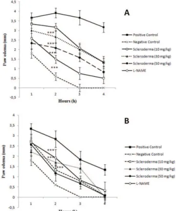
Carrageenan and histamine-induced paw edema
To determine the effect of polysaccharides on
the inlammatory process, we performed carrageenan
and histamine-induced paw edema assays in Wistar rats. Glucans from the fungus S. nitidum reduced
carrageenan-induced paw edema by up to 41±7% at the inlammatory
peak (2rd h) and by approximately 73% at the end of the
4th h at a dose of 50 mg/kg (Figure 1A). In the
histamine-induced paw edema, all the doses of polysaccharides (10,
30 and 50 mg/kg) were eficient in reducing the edema.
The greatest reduction observed occurred in the second h, where the reduction obtained using a dose of 50 mg/ kg of polysaccharides exceeded the effect obtained with L-NAME by 26.5%. At the end of the fourth h, all the groups treated with the different doses of polysaccharides exhibited no edema (Figure 1B).
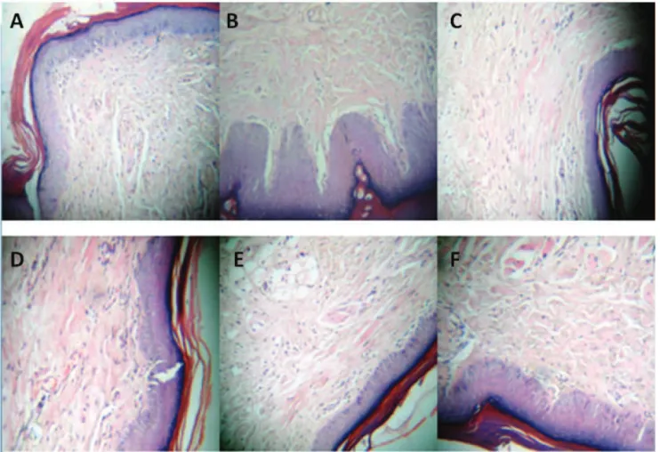
Histological examinations
Histological examinations demonstrated that the animals sensitized only with histamine (positive
control) exhibited intense cell iniltrate, characteristic of the inlammatory reaction (Figure 2A). However, those
that received only saline solution (negative control)
showed an absence of inlammatory reaction (Figure
2B). It was also observed that in the animals treated with polysaccharides from the fungus S. nitidum (10, 30, and
50 mg/kg, respectively) showed a signiicant decrease in PMN cell recruitment at the inlammation site (Figure 2D,E,F). These results were conirmed by histological analysis of the paw treated with L-NAME (Figure 2C).
Figure 1. Anti-inlammatory effect of polysaccharides from
the fungus Scleroderma nitidum in the carrageenan and histamine-induced paw edema model. A. Effect of different doses of polysaccharides and L-NAME on the carrageenan-induced plantar edema model; B. Effect of different doses of polysaccharides and L-NAME on the histamine-induced plantar edema model.
Sodium thioglycolate and zymosan-induced peritonitis
The anti-inlammatory action of polysaccharides
enzymatic activity of the iNOS (Bogle et al., 1992). Figure 3C shows that the dose of the polysaccharides tested
(10 mg/kg), applied jointly with diclofenac, exceeded the action of this drug by around 10±1%. However, the polysaccharides applied in conjunction with the L-NAME
showed no signiicant differences. The data demonstrate
that these fungal polysaccharides may act synergically
with other inlammatory drugs, potentiating their effect.
Nitric oxide
Nitric oxide in most body luids is rapidly
metabolized to stable products such as nitrite and nitrate.
According to the results obtained (Figure 3D) in the
sodium thioglycolate-induced model, the polysaccharides from the fungus Scleroderma nitidum showed a reduction
of NO2/NO3 content of around 32.4±10%. However, in the zymosan-induced peritonitis model, no signiicant reduction in NO2/NO3 levels were observed at any of the
doses tested (Figure 3E). polysaccharides from S. nitidum was administered. The
other polysaccharide concentrations tested (30 and 50
mg/kg) reduced the inlammatory process by 31±1%
and 15±3%, respectively (Figure 3A). In the zymosan-induced peritonitis model, a reduction of around 66±3% was observed in overall leukocytes with 10 mg/kg and 30 mg/kg of polysaccharides from S. nitidum. At polysaccharide concentrations of 50 and 100 mg/kg,
the reduction observed in the inlammatory process was
60±10% and 52±10%, respectively (Figure 3B). Thus, the polysaccharides showed no dose-dependent effect. In
this inlammation model, the 10 mg/kg dose had a better anti-inlammatory effect.
Drug potentiation
To check if the polysaccharides were capable of potentiating drug action, two drugs were used for this
assay: L-NAME and diclofenac. Diclofenac is a COX
1 and 2 inhibitor, whereas L-NAME is a competitive inhibitor of arginine that promotes a decrease in the
Figure 2. Histological analyses (HEX200) of paws from different groups belonging to the histamine-induced paw edema model. A. Positive control (Histamine, without treatment); B. negative control (saline); C: L-NAME (50 mg/kg); D: polysaccharides from
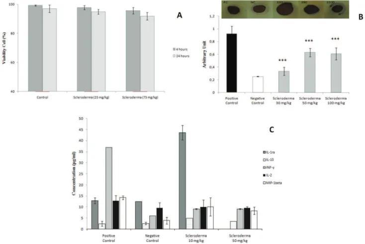
Ex vivo cell viability
These results show that the polysaccharides had no toxic effect at the doses tested. The maximum percentage of non-viable cells was 10% at a dose of 75 mg/kg 24 h after administration. Thus, the polysaccharides
did not reach IC50 at any of the doses tested (Figure
4A).
Cytokine analysis
The glucans from S. nitidum showed an effect
on cytokine expression. We observed a two-fold and three-fold increase in the release of anti-inlammatory
cytokines IL-1ra and IL-10 respectively, in the groups treated with polysaccharides when compared with the
positive control. A signiicant decrease (75%) was also
Figure 3. Anti-inlammatory activity of polysaccharides from Scleroderma nitidum in the sodium thioglycolate and
induced peritonitis model. A. on the sodium thioglycolate-induced peritonitis model; B. on the zymosan-induced peritonitis model. C. Anti-inlammatory effect of polysaccharides from Scleroderma nitidum (10 mg/kg) on
drug potentiation in the zymosan-induced peritonitis model. The factors were expressed as mean±SD. A value of
p<0.001(***) was considered statistically signiicant. D. Effect of different doses of polysaccharides from S. nitidum
in NO (NO2/NO3) content on the sodium thioglycolate-induced peritonitis model; E. Effect of different doses of
polysaccharides in NO content on the zymosan-induced peritonitis model. The values were expressed as mean±SD.
observed in IFN-γ levels as well as a slight reduction in IL-2 and MIP-1β levels of 22% and 29% respectively,
in the groups treated with the different polysaccharide doses. In addition, the effect of polysaccharides was not dose-dependent (Figure 4B).
Dot-blot assay for NF-κB
Nuclear factor kB activation is a critical event
in inlammation. NF-kB activation is related to gene
activation for many cytokines, chemiokines, and adhesion molecules. This experiment was conducted to determine whether the polysaccharides from the fungus Scleroderma nitidum are capable of modulating NF-κB expression.
According to the results obtained, the dose that had the
best effect was 30 mg/kg, reducing NF-κB expression by
63±8%, whereas doses of 50 and 100 mg/kg reduced it by only 30±1%. Thus, it can be observed that all the fungal
polysaccharide doses tested signiicantly reduced nuclear factor NF-kB expression (Figure 4C).
Discussion
Acute inlammation is essentially mediated by
exudate formation and leukocyte migration. Innumerable studies have proven the potential of polysaccharides extracted from fungi in reducing leukocyte migration to
the inlammation sites. To conirm the anti-inlammatory
potential of fungal polysaccharides, the object of this study, we tested their effect in models of plantar edema induced by carrageenan and histamine and of peritonitis induced by sodium thioglycolate and zymosan.
During the inlammatory process, monocytes
are recruited for parenchyma tissue, where they are activated to become cells with a phagocytic function, that
is, macrophages. These cells can release inlammatory
cytokines, free radicals, and nitric oxide, which can
mediate the tissue lesion related to inlammatory response (Almeida at al., 1980; Gordon, 2001; Hermann et al.,
2001). Studies on mushrooms with medicinal potential have been conducted to discover compounds that can
positively or negatively modulate the immunological
system. The signiicant reduction of edema observed in
the rats treated with polysaccharides from the fungus
Scleroderma nitidum can be observed in two plantar
edema models, suggesting their anti-inlammatory
effect.
Macrophage activation via the classic pathway
leads to the secretion of nitric oxide and pro-inlammatory
cytokines, whereas activation via the alternative pathway
leads to the release of anti-inlammatory cytokines
(Gordon, 2003).The polysaccharides from the fungus (glucans) Scleroderma nitidum did reduce INF-γ levels
in the peritoneal wash of the mice. Futhermore, we observed a reduction in the levels of IL-2, a cytokine that stimulates T cell proliferation (Malek & Bayer, 2004).
The macrophage inlammatory protein 1 beta (MIP-1β) is a chemiokine that induces chemiotaxis
and T cell adhesion (Tanaka et al., 1993). The
MIP-1β levels were found to be reduced after treatment
with polysaccharides from Scleroderma nitidum.
IL-1ra can inhibit the pro-inlammatory effects of IL-1 (Dinarello, 1992; 1994). Polysaccharides from the fungus
Scleroderma nitidum caused a three-fold increase in IL-1ra levels in the peritoneal wash of mice with zymosan-induced peritonitis. In the present study a substantial increase in IL-10 levels was also found.
The increase in anti-inlammatory cytokine levels concomitant with the decrease in pro-inlammatory
cytokine release is a plausible explanation for the
anti-inlammatory activity of polysaccharides extracted
from the fungus Scleroderma nitidum. Given that polysaccharides from the fungus Scleroderma nitidum
were capable of decreasing nitric oxide levels and modulating cytokine expression, we decided to assess a possible action mechanism to explain their biological effects. Thus, an assay was conducted to determine the expression of transcription factor NF-kB. In the groups treated with polysaccharides from the fungus Scleroderma nitidum, a signiicant reduction in NF-κB expression
was observed, showing that the polysaccharides could modulate nitric oxide and cytokine levels through the
regulation of NF-κB expression. This fact may explain the anti-inlammatory effect of these polysaccharides.
Acknowledgements
The authors would like to thank the Brazilian
agencies Conselho Nacional de Desenvolvimento Cientíico e Tecnológico and Coordenação de Aperfeiçoamento de Pessoal de Nível Superior for inancial support.
References
Almeida AP, Bayer BM, Horakova Z, Beaven MA 1980. Inluence of indomethacin and other anti-inlammatory drugs on mobilization and production of neutrophils: studies with carrageenan-induced inlammation in rats.
J Pharmacol Exp Ther 214: 74-79.
Barbisan LF, Miyamoto M, Scolastici C, Salvadori DMF, Ribeiro LR, Eira AF 2002. Inluence of aqueous extract of Agaricus blazei on rat liver toxicity induced by different doses of diethylnitrosamine. J Ethnopharmacol 83: 25-32.
Barnes J, Karin M 1997. Nuclear factor-κB-A pivotal transcription factor in chronic inlammatory diseases.
NEJM 336: 1066-1071.
Barreiro EJ, Fraga CAM 1999. A utilização do safrol, principal componente químico do óleo de sassafráz, na síntese de substâncias bioativas na cascata do ácido araquidônico: antiinlamatórios, analgésicos e antitrombóticos. Quim Nova 22: 744-759.
Bellini MF, Angel JPF, Matuo R, Terezan AP, Ribeiro RL, Mantovani MS 2006. Antigenotoxicity of Agaricus blazei mushroom organic and aqueous extracts in chromosomal aberration and cytokinesis block micronucleus assays in CHO-k1 and HTC cells. Toxicol in Vitro 20: 355-360.
Ben-Neriah Y, Schmidt-Supprian M 2007. Epithelial NF-κB maintains host gut microlora homeostasis. Nature Immunol 8: 479-481.
Bogle RG, Baydoun AR, Pearson JD, Moncada S, Mann GE 1992. L-arginine transport is increased in macrophages generating nitric oxide. Biochem J 284: 15-18.
Bradford MM 1976. A rapid and sensitive method for the quantitation of microgram quantities of protein utilizing the principle of protein-dye binding. Anal Biochem 7: 248-254.
Brown GD, Gordon S 2001. Immune recognition: A new receptor for β-glucans. Nature 413: 36-37.
Carbonero ER, Gracher AHP, Komura DL, Marcon R, Freitas CS, Baggio CH, Santos ARS, Torri G, Gorin PAJ, Iacomin M 2008. Lentinus edodes heterogalactan: Antinociceptive and anti-inlammatory effects. Food Chem 111: 531-537.
Dinarello CA 1994. Interleukin-1. In: Thomson A (org) The cytokine handbook, 1st ed.. San Diego: Academic Press.
Dinarello CA 1992. Reduction of inlammation by decreasing production of interleukin-1 or by speciic receptor antagonism. Int J Tissue React 14: 65-75.
Dodgson KS, Price RG 1962. A note on the determination of the ester sulphate content of sulphated polysaccharides.
Biochem J 84: 106-110.
of β-glucan-rich extract from Geastrum saccatum
mushroom. Int Immunopharmacol 7: 1160-1169. Dubois M, Gilles KA, Hamilton JK, Rebers A, Smith F 1956.
Colorimetric method for determination of sugars, and related substances. Anal Chem 28: 350-356.
Giacomini NL, Eira AF 2003. Anticlastogenic effect of aqueous extracts of Agaricus blazei on CHO-k1 cells, studying
different developmental phases of the mushroom.
Toxicol in Vitro 17: 465-469.
Gordon S 2003. Alternative Activation of Macrophages. Nature Rev Immunol 3: 23-35.
Gordon S 2001. Macrophage Function Disorders. ELS 1: 1-11. Guzmán, G, Ovrebo CL 2000. New observations on
sclerodermataceous fungi. Mycologia 92: 174-179. Hermann GE, Rogers RC, Bresnahan JC, Beattie MS 2001.
Tumor necrosis factor-alpha induces Cfos and strongly potentiates glutamate-mediated cell death in the rat spinal cord. Neurobiol Dis 8: 590-599.
Hong KJ, Dunn DM, Shen CL, Pence BC 2004. Effects of
Ganoderma lucidum on apoptotic and anti-inlammatory
function in HT-29 human colonic carcinoma cells.
Phytother Res 18: 768-770.
Kim GY, Roh SI, Park SK, Ahn SC, Oh YH, Lee JD, Park YM 2003. Alleviation of experimental septic shock in mice by acidic polysaccharide isolated from the medicinal mushroom Phellinus linteus. Biol Pharm Bull 26: 1418-1423.
Kirk PM, Cannon PF, David JC, Stalpers JA 2001. Ainsworth & Bisby´s Dictionary of the Fungi. 9th ed., CAB
International, Wallinford.
Koyama Y, Katsuno Y, Miyoshi N, Hayakawa S, Mita T, Muto H, Isemura S, Aoyagi Y, Isemura M 2002. Apoptosis induction by lectin isolated from the mushroom
Boletopsis leucomelas in U937 cells. Biosci Biotechnol Biochem 6: 784-789.
Lavi I, Friesem D, Geresh S, Hadar Y, Schwartz B 2006. An aqueous polysaccharide extract from the edible mushroom Pleurotus ostreatus induces anti-proliferative and pro-apoptotic effects on HT-29 colon cancer cells.
Cancer Lett 244: 61-70.
Liu B 1984. The Gasteromycetes of China. Beihefte zur Nova Hedwigia (Vaduz: J. Cramer) 74: 1-235.
Liu J, Kurashiki K, Shimizu K, Kondo R 2006.
Structure-activity relationship for inhibition of 5a-reductase by triterpenoids isolated from Ganoderma lucidum. Bioorg Med Chem Lett 14: 8654-8660.
Malek TR, Bayer AL 2004. Tolerance, not immunity, crucially depends on IL-2. Nature Rev Immunol 4: 665-674. Nathan C 2002. Points of control in inlammation. Nature 420:
846-852.
Phillips HJ 1973. Dye exclusion test for viability. In: Kruse Jr PF, Patterson Jr MH (org) Tissue Culture: methods and application. New York: Academic Press.
Rocha & Silva MO 1978. A brief history of inlammation. In: Vane JR, Ferreira SH (org) Handbook Exp Pharmacol. Berlin: Springer-Verlag. p. 6-25.
Schepetkin IA, Quinn MT 2006. Botanical polysaccharides: Macrophage immunomodulation and therapeutic potential. Int Immunopharmacol 6: 317-333.
Smiderle FR, Olsen LM, Carbonero ER, Baggio CH, Freitas CS, Marcon R, Santos AR, Gorin PA, Iacomini M 2008. Anti-inlammatory and analgesic properties in a rodent model of a (1-->3), (1-->6)-linked beta-glucan isolated from Pleurotus pulmonarius. Eur J Pharmacol 597: 86-91.
Tanaka Y, Adams DH, Hubscher S, Hirano H, Siebenlist U, Shaw S 1993. T-cell adhesion induced by proteoglycan-immobilized cytokine MIP-lβ. Nature 361: 79-82. Winter CA, Risley EA, Nuss GW 1962. Carrageenin-induced
oedema in hind paw of the rat as an assay for anti-inlammatory drugs. Proc Soc Exp Biol Med 111: 544-547.
Xie X, Rivier A-S, Zakrzewicz A, Bernimoulin M, Zeng X-L, Wessel HP, Schapira M, Spertini O 2000. Inhibition of selectin-mediated cell adhesion and prevention of acute inlammation by nonanticoagulant sulfated saccharides. Studies with carboxyl-reduced and sulfated heparin and trestatin a sulfate. J Biol Chem 275: 34818-34825. *Correspondence
Edda Lisboa Leite
Department of Biochemistry, Bioscience Center, UFRN Av Sen Salgado Filho, 3000, Natal-RN, Brazil