13
Radiol Bras. 2012 Jan/Fev;45(1):13–19
Diagnostic difficulties in osteoid osteoma of the elbow:
clinical, radiological and histopathological study
*
Dificuldades diagnósticas no osteoma osteoide do cotovelo: estudo clínico, radiológico e histopatológico
Ana Cristina Cotta1, Rogério Teles de Melo2, Renato César Rezende de Castro3, Francineide Sadala
de Souza4, Yana Senna Jeronymo Najjar5, Júlia Filardi de Oliveira Paim1, Ricardo Karam Kalil4
Objective: To describe the clinical and imaging findings that may aid in correctly diagnosing osteoid osteoma of the elbow. Materials and Methods: The authors have retrospectively evaluated seven patients diagnosed with osteoid osteoma of the elbow histologically confirmed by bone biopsy. The patients’ medical records and imaging studies were reviewed. Results: Among the 142 patients with osteoid osteoma identified in the archives of the institutions, 4.9% had the lesion in the elbow, predominantly in men (2.5:1), with a mean age of 25 years. Pain and limitation of movement were the most common symptoms. Five of the patients (71.4%) had previously received a different clinical diagnosis. The mean duration of symptoms was 21 months. Radiography did not demonstrate the nidus in 42.8% of cases. Computed tomography and magnetic resonance imaging clearly demonstrated the nidus. Joint effusion was a constant finding. The histological findings corresponded to the ones usually observed in such cases. Surgical resection has brought relief of symptoms and/or functional improvement in all of the cases. Conclusion: It is important to consider the possibility of osteoid osteoma in young adult patients with pain, limitation of movement and signs of synovitis in the elbow refractory to conservative treatment. The detection of bone sclerosis, cortical thickening and/or periosteal reaction at radiography allows directing computed tomography towards the accurate identification of the nidus.
Keywords: Osteoid osteoma; Intra-articular; Elbow; Synovitis; Imaging; Biopsy.
Objetivo: Descrever os aspectos clínicos e de imagem que podem auxiliar no diagnóstico correto do osteoma osteoide no cotovelo. Materiais e Métodos: Realizado estudo retrospectivo de sete pacientes com diagnóstico de osteoma osteoide no cotovelo confirmado histologicamente por biópsia óssea. Eles tiveram seus prontuários médicos e exames de imagem revisados. Resultados: Do total de 142 pacientes identificados em nossos arquivos, 4,9% apresentavam a lesão no cotovelo, com predomínio no sexo masculino (2,5:1) e idade média de 25 anos. Dor e limitação de movi-mento foram os sintomas mais comuns. Cinco pacientes (71,4%) receberam outro diagnóstico clínico prévio. A dura-ção média dos sintomas foi de 21 meses. As radiografias não demonstraram o nidus em 42,8% dos casos. A
tomo-grafia computadorizada e a ressonância magnética mostraram claramente o nidus. Derrame articular foi um achado
constante. O aspecto histológico observado foi o usual. A ressecção cirúrgica promoveu alívio dos sintomas e/ou melhora funcional em todos os casos. Conclusão: É importante considerar a possibilidade de osteoma osteoide em paciente adulto jovem com dor, limitação do movimento e sinais de sinovite no cotovelo, refratária ao tratamento conservador. Esclerose óssea, espessamento cortical e/ou reação periosteal detectados na radiografia permitem direcionar a tomo-grafia computadorizada para a visualização precisa do nidus.
Unitermos: Osteoma osteoide; Intra-articular; Cotovelo; Sinovite; Imagem; Biópsia.
Abstract
Resumo
* Study developed at Departments of Surgical Pathology, Ra-diology and Orthopedics – The Sarah Network of Rehabilitation Hospitals, Hospital Sarah Belo Horizonte, MG, and Hospital Sarah Brasília, DF, Brazil.
1. Specialists in Pathological Anatomy, MDs, Surgical Patholo-gists at Hospital Sarah, Belo Horizonte, MG, Brazil.
2. Titular Member of Colégio Brasileiro de Radiologia e Diagnóstico por Imagem (CBR), MD, Radiologist at Hospital Sarah, Belo Horizonte, MG, Brazil.
3. Specialist in Orthopedics and Traumatology, MD, Orthope-dist at Hospital Sarah, Belo Horizonte, MG, Brazil.
4. Specialists in Pathological Anatomy, MDs, Surgical Patholo-gists at Hospital Sarah, Brasília, DF, Brazil.
5. Titular Member of Colégio Brasileiro de Radiologia e Diag-nóstico por Imagem (CBR), MD, Radiologist at Hospital Sarah, Brasília, DF, Brazil.
Cotta AC, Melo RT, Castro RCR, Souza FS, Najjar YSJ, Paim JFO, Kalil RK. Diagnostic difficulties in osteoid osteoma of the elbow: clinical, radiological and histopathological study. Radiol Bras. 2012 Jan/Fev;45(1):13–19.
Most patients present the classical bone pain symptom that worsens at night and im-proves with the use of aspirin or other non-steroidal anti-inflammatory drugs. Diaphy-sis and metaphyDiaphy-sis of long bones, particu-larly the femur and tibia constitute the most common sites of osteoid osteomas.
In cases of intra-articular involvement, the clinical manifestations are nonspecific and the radiographic findings may be subtle, becoming a diagnostic challenge. Patients with osteoid osteoma of the elbow INTRODUCTION
Osteoid osteoma is a small and rela-tively common benign bone tumor, with well defined clinical, radiological and pathological characteristics(1–3). It is most
prevalent in the 10-25 year old age group.
Mailing Address: Dr. Rogério Teles de Melo. Avenida Amazo-nas, 5953, Gameleira. Belo Horizonte, MG, Brazil, 30510-000. E-mail: [email protected]
frequently present pain, chronic synovitis, joint effusion, limitation in range of motion and flexion contracture simulating inflam-matory arthropathy. At radiography, reac-tive sclerosis may be minimal or absent, leading to a significant delay in the diag-nosis or even to misdiagdiag-nosis(4–6).
The present study is aimed at highlight-ing clinical and radiological findhighlight-ings which allow early diagnosis and appropriate man-agement in cases of osteoid osteoma of the elbow.
MATERIALS AND METHODS
In the databanks of the Sarah hospitals in the cities of Brasília, DF, and Belo Hori-zonte, MG, Brazil, 142 patients with histo-logically confirmed diagnosis of osteoid osteoma were identified. Among them, seven patients presenting elbow lesions
were selected and had their records and imaging studies reviewed.
The following clinical data were col-lected: sex, age, affected side, presence of pain, fever, edema, functional limitation, symptoms duration, previous diagnoses and treatments, and response to use of non-steroidal anti-inflammatory drugs and sur-gical treatment (Table 1).
All the seven patients were submitted to plain radiography and computed tomogra-phy (CT). Three patients were also under-went magnetic resonance imaging (MRI) in a 1.5 T equipment. The radiographs were utilized to evaluate the presence of nidus, sclerotic halo, cortical thickening and pe-riostitis. Appearance and location of the nidus, sclerosis signs, cortical thickening and rupture, besides the presence of joint effusion, were analyzed at CT. MRI was useful in the assessment of changes in
sig-nal intensity in the bone marrow, identifi-cation of the nidus, presence of joint effu-sion, synovitis and abnormalities in adja-cent soft tissues. The imaging findings are described on Table 2.
The study was approved by the Commit-tee for Ethic in Research of the Institution.
RESULTS
Out of a total of 142 patients diagnosed with osteoid osteoma, seven (4.9%) pre-sented lesions in the elbow. Five of these patients were male, and two were female (2.5:1 ratio), with ages ranging between 11 and 47 years (mean = 25 years). The distal humerus was affected in four cases while the proximal ulna was affected in three cases, and four cases involved the left el-bow and three, the right elel-bow. Pain and limitation in the range of motion were the
Table 2 Imaging findings related to osteoid osteoma of the elbow. Case No. 1 2 3 4 5 6 7 Site Distal humerus Distal humerus Distal humerus Distal humerus Proximal ulna Proximal ulna Proximal ulna Nidus identification (Rx) No No Yes Yes No Yes No
Nidus appearance (CT) Partially calcified Densely calcified Partially calcified Partially calcified Two partiallly calcified
nidi Densely calcified Non calcified Medullary sclerosis (CT) Yes Yes Yes No No No Yes Cortical thickening (Rx and CT)
Yes Yes Yes Yes Yes (intracortical) No (intramedullary) Yes Yes Cortex rupture (CT) Yes Yes Yes Yes* Yes* (intracortical) No (intramedullary) Yes* No Joint effusion (CT and MRI)
Mild Severe Severe Severe No Mild Mild Rx, plain radiography; CT, computed tomography; MRI, magnetic resonance imaging. * Extraosseous nidus component.
Table 1 Clinical presentation and follow-up.
Case No. 1 2 3 4 5 6 7 Sex M M F M M M F Age (years) 21 20 15 11 18 47 47 Side L R L L R L R Symptoms Pain and motion loss Pain and extension loss
Difficulty in extension and pain Pain, heat and edema; initial
fever; functional limitation Continuous and incapacitating
pain which worsens at night, irradiating to the wrist Pain; loss of strength in the arm, forearm and fingers; wrist
weakness and extension loss Constant pain during night and
day in the medial epicondyle; nodulation; movement deficit
Duration of symptoms (months) 12 6 36 4 24 48 18 Previous diagnosis Pseudogout — Osteomyelitis Tendinitis Tendinitis Tendinitis — Previous treatment Physiotherapy — Antibiotics Physiotherapy Nonsteroidal anti-inflamma-tory drug, no relief; rest
Nonsteroidal anti-inflamma-tory drug; physiotherapy
Nonsteroidal anti-inflamma-tory drug, no relief
Postoperative follow-up Pain relief Pain relief; functional
improvement Pain relief Pain relief Functional improvement Pain relief Symptoms relief
most common symptoms, found in all ana-lyzed cases. One patient also presented fe-ver, articular heat and joint edema.
Among the seven studied patients, five (71.4%) had previously received other
clinical diagnosis. The previously raised diagnostic hypotheses were the following: tendinitis (three patients), pseudogout (one patient) and osteomyelitis (one patient). The mean duration of symptoms was 21
months. The previous treatments consisted of utilization of non-steroidal anti-inflam-matory drugs for three patients, without symptoms relief in two of them; physio-therapy for two patients; and use of antibi-otics in one case.
The radiographic studies demonstrated periosteal reaction and/or variable cortical thickening in all of the patients (Figure 1) but could not demonstrate the nidus in three of the patients (42.8%). CT clearly demon-strated the nidus and bony reaction in all analyzed cases (Figure 2). Cortical bone rupture was identified in six of the seven patients, and in three of those patients there was protrusion through the cortex with extraosseous component of the nidus (Fig-ure 3). Two nidi were identified in one pa-tient (Figure 4). As regards mineralization pattern within the eight nidi, calcification/ ossification was absent in one case, was partial in five cases and dense in two cases (Figure 5). MRI could demonstrate the ni-dus, joint effusion, synovitis, bone edema and soft tissue edema in the three patients (cases 1, 2, and 4) who underwent such an examination (Figure 6). The effusion present in six patients was considered as mild in three cases and severe in the other three cases (Figure 3).
The histological diagnosis was based on the classical signs of an interwoven net-Figure 1. Case 6. Radiography of left elbow –
ob-lique view demonstrating the presence of periosti-tis (circle and white arrow) adjacent to the nidus (black arrow) located in the proximal ulna.
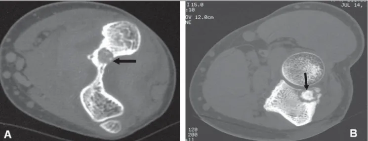
Figure 4. Case 5. Axial tomographic section reveal-ing the presence of two nidi in the proximal ulna, one intramedullary (black arrow) and the other pro-truding through a cortex rupture (white arrow). Note the presence of minute medullary sclerosis adja-cent to the lesion.
Figure 2.Case 1. Axial computed tomography depicting osteolytic nidus (black arrow) including calcification foci, in association with periostitis (white arrow) in the posterolateral region of the distal humerus.
Figure 3. Case 3. A: Radiography of left elbow – anteroposterior view depicting a partially calcified nidus (arrow) in the humerus coronoid fossa. B: Axial tomographic section with bone window showing the nidus (arrow) protruding on the ventral aspect of the humerus. C: Image in the same plane with soft tissue window demonstrating association with significant joint effusion (arrow).
Figure 5. Nidus appearance at tomography. A: Partially calcified osteolytic lesion (arrow), smaller than 1 cm, adjacent to the olecranon fossa. B: Nidus located in the proximal ulna, including a densely calcified central focus surrounded by a hypodense halo (arrow).
work of osteoid trabeculae with variable degrees of mineralization, consistent with nidus, surrounded by osteoblasts and some osteoclasts intermingled with highly vascu-larized mixed connective tissue (Figure 7A). The analysis of the synovial tissue demonstrated diffuse edema and focal
mononuclear perivascular inflammatory cell infiltrate. In some cases, such infiltrate was dense and was associated with the presence of lymphoid follicles (Figure 7B). In one patient, prominent focal nerve fibers were found as part of the adjacent soft tis-sue reaction (Figure 7C).
All the patients were submitted to open surgery with nidus resection, with symptom relief and/or functional improvement in all cases. The patients who presented the best degree of joint motion recovery were those whose symptoms duration was less than one year from admission. However, the patient who experienced symptoms for two years also presented significant improve-ment after the surgical treatimprove-ment.
DISCUSSION
Osteoid osteoma is a relatively common bone lesion, corresponding to 10–12% of the benign bone tumors(7,8). It generally
af-fects young individuals in the age range from 10 to 25 years, with 90% of the cases occurring between 5 and 30 years of age, with a mean of 21 years(9). Men are more frequently affected than women (2:1 to 4:1 ratio).
Although it has been first described by Bergstrand in 1930, the term osteoid os-teoma was introduced in the medical litera-ture in 1935 by Jaffe who, based on the study of five cases, described such condi-tion as a specific pathological entity(10).
The tumor may occur in almost all bones of the body; however the long bones are the site of origin in more than 70% of cases, with the femur and the tibia being most commonly affected (50–60%). In long bones, osteoid osteomas are usually located in the diaphysis or in the metaphy-sis. The epiphyseal and intra-articular lo-cations are rarely observed(11), although some authors estimate their prevalence to be between 10% and 13%(6,9), the hip
be-ing the most common location. Ankle, el-bow, wrist and knee are other less affected locations. The lesion is considered to be intra-articular in cases where it occurs at the extremity of long bones, around or within the joint, in a bone limited by the capsule, very close to it and to the synovia.
The etiology of this condition is un-known. Cytogenetic changes such as chro-mosome 22q deletion have been described in cases of osteoid osteoma(12), but further
studies are necessary to definitively link such an entity to a clonal mutation. Famil-ial occurrence is extremely rare. In a study developed in the authors’ institution(3),
os-teoid osteoma was described in two broth-ers that had the same age when the symp-toms onset led them to seek medical assis-tance. Both of them also had the lesion lo-cated in proximal femoral cortex (the most common site). Such findings suggest the presence of an associated genetic factor.
Immunohistochemical studies of neu-ronal antigens and associated with nerves
Figure 6. Case 2. Sagittal, contrast-enhanced MRI T1-weighted s with fat saturation demonstrating a hypointense nidus (white arrow), medullary edema and edema in the triceps muscle, besides signifi-cant thickening and synovial enhancement. The solid periosteal reaction (black arrow) at the pos-terior margin of the distal metaphysis of the hu-merus occurs far from the nidus since the perios-teum is not present within the articular capsule.
have demonstrated that osteoid osteomas present nervous fibers in the reactive zone. Their innervation seems to be unique among bone tumors(13). The clinical
symp-tom of intense pain in the affected area which worsens at night and in general im-proves after 30 minutes with the use of salicylates is present in 75% of the cases and suggests the diagnosis. The pain seems to be mediated by high levels of E2 pros-taglandins released by the tumor which are inhibited by the use of non-hormonal anti-inflammatory drugs. The prominent focal nerve fibers found in the region of adjacent soft tissue reaction in one patient might be related to the disproportionate pain re-ported by such patient. This is in agreement with the previous description of nerves found in the peritumor tissue in a case of osteoid osteoma. In the intra-articular pre-sentation, the response to utilization of nonsteroidal anti-inflammatory drugs for pain relief seems to be less effective than
in the extra-articular presentation. Such a fact was observed in two of the three pa-tients in the present study who utilized such type of drug.
The tumor presents a limited growth po-tential and a tendency toward spontaneous maturation in two to five years as the pain tends to decrease. The lesion can sponta-neously regress, but the mechanism of such regression is not yet known.
Plain radiography is the imaging method utilized for initial evaluation. In cases of osteoid osteoma, the classical ra-diographic finding consists of a round or ovoid lytic lesion, called nidus (or nest), smaller than 2.0 cm, normally measuring 5 to 10 mm, in association with an area of bone sclerosis and/or reactive cortical thickening(14). Such finding may vary ac-cording to the affected bone, to the tumor site, symptom duration and the patient’s age. The nidus may be uniformly radiolu-cent or including a variable amount of
calcification/ossification. Bone maturation is more pronounced in lesions with a longer evolution.
The diagnosis is not difficult in cases where the typical clinical and radiographic findings are present. However, tumor pre-sentation in unusual locations, like the in-tra-articular presentation, may be associ-ated with atypical manifestations that com-promise the diagnosis.
Osteoid osteoma can be classified into three types as follows: cortical, medullary and subperiosteal. Intra-articular tumors are normally of the medullary and subpe-riosteal types, which contributes to the ab-sence or preab-sence of limited sclerosis around the nidus, making its identification difficult at radiography.
particularly in cases where it is occult at radiography (Figure 8). CT is also very useful in cases where there is a suspicion of residual lesion. However, such a tech-nique has the inconvenience of exposing the patient to ionizing radiation.
The role of MRI in the evaluation of os-teoid osteoma is controversial. The variable appearance of the lesion, extensive reactional changes in the bone marrow and in soft tissues may simulate a more aggres-sive lesion or one of a different nature(15), though such was not the impression of the authors on the three MRI studies. Based on the hypervascularization of the nidus, the dynamic study with gadolinium combined with high resolution images increases the nidus conspicuity, moreover in cases where the lesion is located in an atypical site and in those patients presenting with nonspe-cific clinical symptoms(16–18).
Bone scintigraphy utilizing technetium-99 presents a high sensitivity in the
diag-nosis of osteoid osteomas. The tumor pre-sents a typical pattern known as “double-density” sign. Such a sign is characterized by intense activity/hyper-uptake centrally in the nidus region and less intense accu-mulation of the radioisotope in the periph-ery of the lesion, where the reactive scle-rotic bone and perilesional edema are lo-cated. However, many times such sign is absent in cases of intra-articular lesions, as a generalized increase activity increase within the joint due to the presence of syno-vitis associated with osteoporosis and hy-peremia.
In the literature, there are few reports on osteoid osteoma of the elbow, describing tumors in the head and tuberosity of the radius, in the olecranon and coronoid pro-cess of the ulna, in the capitulum and ole-cranon fossa of the humerus.
The intra-articular lesion is associated with pain, soft tissue edema and joint ef-fusion. Synovitis may be intense and the
patient may present with articular rigidity. Six out of the seven patients presented joint effusion. Osteopenia, uniform reduction of the articular space and osteo-hypertrophic changes may be observed(9). Clinically and
radiologically, the condition may simulate a primary articular process resulting in di-agnosis delay of months or even years. Patients presenting intra-articular lesion may progress with signs of osteoarthrosis secondary to synovitis and synovial prolif-eration with cartilage destruction. Muscle atrophy may affect the limb, particularly in the case of long-lasting tumors.
The differential diagnoses for osteoid osteoma of the elbow include: (inflamma-tory/infectious) monoarthritis, nonspecific synovitis, tendinitis/epicondylitis, osteo-chondritis dissecans, osteomyelitis/ Brodie’s abscess, eosinophilic granuloma, enostosis, stress fracture, chondroblastoma and osteoblastoma. Osteoid osteoma and osteoblastoma have the same histopatho-logical characteristics; but osteoblastoma is larger than 2.0 cm, presents growth pro-gression and tends to be more aggressive. The results of the present study, in agreement with those from previous stud-ies(5), show that the radiographic findings
of sclerosis, cortical thickening and subtle periosteal reaction in elbow bones in asso-ciation with the presence of joint effusion should raise the possibility of osteoid os-teoma.
Complete nidus excision is curative and is the most traditional treatment method. However, minimally invasive techniques have been utilized as equally safe and ef-fective alternatives for the treatment of osteoid osteomas, with low morbidity rates and faster recovery. Radiofrequency abla-tion is the most prominent of such tech-niques; alternating current radiofrequency is transmitted by means of an electrode needle positioned in the nidus, producing heat and causing irreversible cell damage/ necrosis by means of thermal coagula-tion(19). Aiming at symptoms relief and
functional improvement of their patients, the authors of the present study have opted for open surgery. Such an option was based on the difficulties in the preoperative diag-nosis, on the raised hypotheses, as well as on the surgeon’s experience. Additionally, the authors highlight the scarcity of reports Figure 8. Case 1. A,B: Lateral and anteroposterior radiography of left elbow cannot demonstrate the
nidus. C,D: Tomographic reconstructions in the sagittal and coronal planes of a single patient clearly demonstrate the lesion (arrows).
on cases of osteoid osteoma of the elbow in the literature to guide them towards a specific treatment modality that might be adopted as a treatment standard.
CONCLUSION
Osteoid osteoma is a rare cause of el-bow pain. The diagnosis of this tumor is difficult as, generally, the clinical manifes-tations and radiographic findings are not characteristic. It is important to consider the possibility of osteoid osteoma in young adult patients with pain, limitation of move-ment and signs of synovitis in the elbow with unknown etiology, refractory to con-servative treatment and not related to sys-temic disease. CT is the imaging method of choice for identification of the nidus. The early diagnosis allows appropriate surgical treatment, reducing the related morbidity.
Acknowledgements
To Rede Sarah de Hospitais de Reabi-litação, for the institutional support. To Drs. Antônio Lopes da Cunha Júnior, Sandra Alvarenga Coutinho Passos, Maria Henri-queta Freire Lyra, Jorge Henrique Calil and to the histology technician Cleides Campos
de Oliveira, for their contribution in the present study.
REFERENCES
1. Fletcher CDM, Unni KK, Mertens F, editors. World Health Organization Classification of Tumours. Pathology & genetics. Tumours of soft tissue and bone. Lyon, France: IARC Press; 2002. 2. Picci P, Mirra JM. Osteoid osteoma. In: Mirra JM, editor. Bone tumors. Philadelphia, PA: Lea & Febiger; 1989. p. 226–48.
3. Kalil RK, Antunes JS. Familial occurrence of os-teoid osteoma. Skeletal Radiol. 2003;32:416–9. 4. Weber KL, Morrey BF. Osteoid osteoma of the el-bow: a diagnostic challenge. J Bone Joint Surg Am. 1999;81:1111–9.
5. Moser RP Jr, Kransdorf MJ, Brower AC, et al. Os-teoid osteoma of the elbow. A review of six cases. Skeletal Radiol. 1990;19:181–6.
6. Szendroi M, Köllo K, Antal I, et al. Intraarticular osteoid osteoma: clinical features, imaging re-sults, and comparison with extraarticular localiza-tion. J Rheumatol. 2004;31:957–64.
7. White ML, Kandel R. Osteoid-producing tumors of bone. Semin Musculoskelet Radiol. 2000;4: 25–43.
8. Dahlin DC, Unni KK. Bone tumors: general as-pects and data on 8,542 cases. 4th ed. Springfield, IL: Thomas; 1987.
9. Allen SD, Saifuddin A. Imaging of intra-articu-lar osteoid osteoma. Clin Radiol. 2003;58:845–52. 10. Jaffe HL. “Osteoid osteoma”. A benign osteoblas-tic tumor composed of osteoid and atypical bone. Arch Surg. 1935;31:709–28.
11. Resnick D, Kyriakos M, Greenway GD. Tumors
and tumor-like lesions of bone: imaging and pa-thology of specific lesions. In: Resnick D, Kransdorf MJ, editors. Bone and joint imaging. Philadelphia, PA: Elsevier/Saunders; 2005. p. 1120–6.
12. Baruffi MR, Volpon JB, Barbieri Neto J, et al. Osteoid osteomas with chromosome alterations involving 22q. Cancer Genet Cytogenet. 2001; 124:127–31.
13. O’Connell JX, Nanthakumar SS, Nielsen GP, et al. Osteoid osteoma: the uniquely innervated bone tumor. Mod Pathol. 1998;11:175–80. 14. Cassard X, Accadbled F, De Gauzy JS, et al.
Os-teoid osteoma of the elbow in children: a report of three cases and a review of the literature. J Pediatr Orthop B. 2002;11:240–4.
15. Davies M, Cassar-Pullicino VN, Davies AM, et al. The diagnostic accuracy of MR imaging in os-teoid osteoma. Skeletal Radiol. 2002;31:559–69. 16. Liu PT, Chivers FS, Roberts CC, et al. Imaging of osteoid osteoma with dynamic gadolinium-en-hanced MR imaging. Radiology. 2003;227:691– 700.
17. Zampa V, Bargellini I, Ortori S, et al. Osteoid os-teoma in atypical locations: the added value of dynamic gadolinium-enhanced MR imaging. Eur J Radiol. 2009;71:527–35.
18. von Kalle T, Langendörfer M, Fernandez FF, et al. Combined dynamic contrast-enhancement and serial 3D-subtraction analysis in magnetic reso-nance imaging of osteoid osteomas. Eur Radiol. 2009;19:2508–17.