ABSTRACT
http://dx.doi.org/10.1590/1678-775720150125
Serum big endothelin-1 as a biomarker in oral
squamous cell carcinoma patients: an analytical
study
Pritam Kumar MANKAPURE, Suresh Ramchandra BARPANDE, Jyoti Dilip BHAVTHANKAR, Manda MANDALE
Government Dental College and Hospital, Department of Oral Pathology and Microbiology, Aurangabad, India.
Corresponding address: Pritam K. Mankapure, Assistant Professor - Department of Oral Pathology and Microbiology, Government Dental College and Hospital - Dhanvantari Nagar - Aurangabad 431001 - Maharashtra - India - Phone: +91-9011179100 - e-mail: [email protected]
6XEPLWWHG$SULO0RGL¿FDWLRQ-XO\$FFHSWHG$XJXVW
D
etection of abnormally elevated levels of molecules in patients with oral cancer may be useful in early diagnosis. These markers can be included in current Histopathology grading and in TNM staging systems of Oral Squamous Cell Carcinoma (OSCC) to make it more ef¿cient. Several pro-angiogenic molecules have been assessed for the same reason. Endothelin-1 (ET-1) is a vasoactive peptide associated with the development and spread of many solid tumors, including Squamous Cell Carcinoma (SCC), but its utility in OSCC has not been con¿rmed. ObMective: This study aims to evaluate the role of the serum big ET-1 as a biomarker of OSCC, by correlating it with the clinical staging and the histopathological grading. Material and Methods: Serum levels of big ET-1 measured by the sandwich Enzyme-Linked Immunosorbent Assay (ELISA) in 40 OSCC cases were compared with the levels from the control group using independent t-test. Clinical stages and histopathological grades of OSCC cases were compared in relation to their mean levels of serum big ET-1, one using the Analysis of Variance (ANOVA) test and the other the independent t-test, respectively. The signi¿cance of the mean difference between the groups was evaluated by Tukey¶s multiple comparison test. All statistical analyses were performed on GraphPad statistical software version 5.0. Results: By comparing the mean of the big ET-1 concentrations of cases and controls, the independent t-test revealed signi¿cant higher big ET-1 concentration of OSCC cases when compared to controls (p<0.0001). Tukey’s multiple comparison test also revealed statistically signi¿cant difference among all OSCC stages in relation to the mean levels of serum big ET-1. However, the mean of the big ET-1 concentrations of cases of grade I and of grade II did not differ statistically (p=0.729). Conclusion: Serum big ET-1 levels may be useful as a diagnostic tool in OSCC and as an adjunct to OSCC staging. However, its use as a prognostic marker warrants larger prospective studies.Ke yw or ds: Endothelin-1. Oral cancer. Cancer staging. Neoplasm grading.
I N TROD UCTI ON
Oral cancer is the 6th most common cancer in the world. India is the highest ranked country in the world with over 1,00,000 cases per year with mortality rate of 22.9%, which is largely attributed to the late diagnosis of the disease33. However, Histopathology remains the gold standard for the diagnose of oral cancer; the grade of differentiation alone shows poor correlation with the outcome and the response to treatment of an individual
more precisely in terms of its aggressiveness and biology10.
Interest in the role of ET-1 as an angiogenic molecule in cancer has grown over the last two decades, following the demonstration of ET-1 production by several tumor cell lines5,8,14,21.The contribution of ET-1 in tumor growth and progression has been evaluated in prostatic, ovarian, renal, pulmonary, colorectal, cervical, breast, lung, bladder, and endometrial cancer1,4,14,17,29-31. In the context of oral tissues, it has been demonstrated that keratinocytes can translate and secrete ET-1 protein into the keratinocyte conditioned medium and oral epithelial cell line can produce ET-134,35. ET-1 levels from culture supernatants of human oral SCC cell lines were found to be higher when compared to the one of normal human epidermal keratinocytes6. This study also demonstrated that OSCC cells possess the required machinery to synthesize and secrete ET-1. They suggested that ET-1 may act in an autocrine manner on oral SCC cells, promoting their growth, and in paracrine manner on endothelial cells and oral
¿broblasts assisting tumor growth and invasion6. Currently there is evidence that ET-1 contributes to carcinogenesis directly through proliferation, resistance to apoptosis, migration, and invasion while indirectly through angiogenesis, Extracellular Matrix (ECM) breakdown and deposition14,19. Thus, the ET-1 presents itself as a promising biomarker in OSCC. Therefore, this study was designed to assess the use of serum big ET-1 as a biomarker of oral SCC, correlating it with clinical staging and histopathological grading.
M ATERI AL AN D M ETH OD S
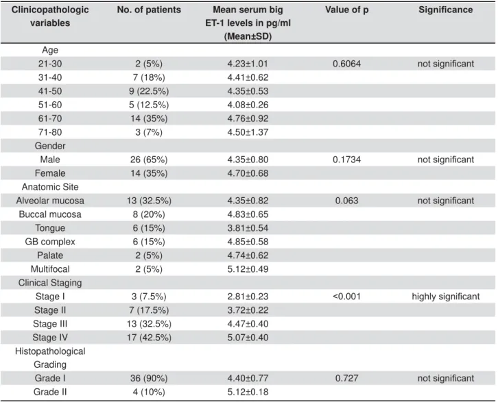
This study was approved by the Institutional Ethics Committee, including a total of 80 subjects and being composed of a study group of 40 patients histopathologically diagnosed with Oral Squamous Cell Carcinoma (OSCC) and a control group of age 40 and with healthy and sex matched subjects. Patients who had received or were receiving any treatment for carcinoma and patients with known systemic illness or with any other oral disease were excluded from the study. Demographic and clinical features of OSCC cases have been given in Table 1. Informed consent was obtained from all patients and control participants.
The diagnosis of squamous cell carcinoma was con¿rmed in each case by the histopathological examination of an incisional biopsy in the representative site of the lesion. The relevant history of each patient was done and recorded through clinical examination to rule out any systemic illness or any other local disease or prior therapy. Clinical staging of patients with
oral squamous cell carcinoma was performed according to the TNM classi¿cation given by the American Joint Committee on Cancer (AJCC) for Staging Atlas15 (2006) and End Results Reporting13. The histopathological grading of oral squamous cell carcinoma was performed according to the malignancy grading system proposed by Anneroth, et al.2 (1987). This grading system is consisted of six parameters: degree of keratinization, nuclear polymorphism, number of mitoses per high power
¿eld, pattern of invasion, stage of invasion, and lympho-plasmacytic in¿ltration. Each parameter was graded from 1 to 4 points. As recommended by Araújo, et al.3 (1997), the parameter “stage of invasion” was omitted as incisional biopsy was used for histopathological grading of OSCC. Total scores for malignancy histological grading were obtained by the sum of the scores given for each parameter, and then divided by the number of parameters used. The ¿nal grading was done as follows: 1.0 to 2.0 – Grade I; 2.1 to 3.0 – Grade II, and 3.1 to 4.0 – Grade III.
From each subject, 5 ml of intravenous blood was collected using an evacuated blood collection system (Becton, Dickinson and Company, Franklin Lakes, New Jersey, USA). Then, the blood was allowed to clot at room temperature for about 2 hours and then centrifuged at 3000 RPM for 10 minutes to separate the serum for the study. The concentrations of serum big ET-1 were estimated in both cases and controls by the solid phase sandwich ELISA (Immuno-Biological Laboratories Co. Ltd., Fujioka-Shi, Gunma, Japan).
St a t ist ica l a n a lysis
The mean levels of serum big ET-1 among the study and the control group was compared by the independent t-test. The ANOVA test and independent t-test were applied respectively to compare clinical stages and histopathological grades of OSCC cases in relation to their mean levels of serum big ET-1. The signi¿cance of the difference between the means of groups was evaluated by Tukey’s multiple comparison test. All statistical analyses were performed using GraphPad Prism v.5.0 software. All p-values of <0.05 were considered statistically signi¿cant.
RESULTS
in the 5th and in the 7th decade of life in males. In the present study, 11 (27.5%) patients were 45 Clinicopathologic
variables
No. of patients Mean serum big ET-1 levels in pg/ml
(Mean±SD)
Value of p 6LJQL¿FDQFH
Age
21-30 2 (5%) 4.23±1.01 0.6064 QRWVLJQL¿FDQW
31-40 7 (18%) 4.41±0.62
41-50 9 (22.5%) 4.35±0.53
51-60 5 (12.5%) 4.08±0.26
61-70 14 (35%) 4.76±0.92
71-80 3 (7%) 4.50±1.37
Gender
Male 26 (65%) 4.35±0.80 0.1734 QRWVLJQL¿FDQW
Female 14 (35%) 4.70±0.68
Anatomic Site
Alveolar mucosa 13 (32.5%) 4.35±0.82 0.063 QRWVLJQL¿FDQW
Buccal mucosa 8 (20%) 4.83±0.65
Tongue 6 (15%) 3.81±0.54
GB complex 6 (15%) 4.85±0.58
Palate 2 (5%) 4.74±0.62
Multifocal 2 (5%) 5.12±0.49
Clinical Staging
Stage I 3 (7.5%) 2.81±0.23 KLJKO\VLJQL¿FDQW
Stage II 7 (17.5%) 3.72±0.22
Stage III 13 (32.5%) 4.47±0.40
Stage IV 17 (42.5%) 5.07±0.40
Histopathological Grading
Grade I 36 (90%) 4.40±0.77 0.727 QRWVLJQL¿FDQW
Grade II 4 (10%) 5.12±0.18
Table 1- Comparison of clinico pathologic variables of OSCC cases with serum big ET-1 levels
Figure 1- Comparison of mean levels of big ET-1 between oral squamous cell carcinoma (OSCC) cases and controls using LQGHSHQGHQWWWHVWPHDQVSLQFRPSDULVRQZLWKKHDOWK\FRQWUROV
years and the remaining 29 (72.5%) were above 45 years. Male: female ratio of the OSCC cases was 1.8:1.There was no signi¿cant difference among groups of different age (p=0.6064 ) and gender (p=0.1734) in relation to the serum big ET-1 values analyzed by the ANOVA test and the independent t-test, respectively (Table 1). The alveolar mucosa was the most common site (32.5%) to be involved in patients with OSCC, followed by the buccal mucosa (20.0%), the tongue (15.0%), and the gingivobuccal complex (15.0%). Comparing the mean of the big ET-1 concentration between the sites, ANOVA revealed no signi¿cant difference (p=0.063) (Table 1). The mean big ET-1 level in multifocal lesions and in the gingivobuccal complex
were the highest, the lowest being the mean for tongue.
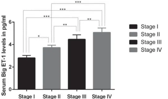
Comparing the mean big ET-1 concentrations of the two groups, the independent t-test revealed a signi¿cantly higher big ET-1 concentration in OSCC cases when compared to controls (p<0.0001, t=11.37) (Figure 1). Furthermore, intragroup comparison of the mean levels of serum big ET-1 by Tukey’s multiple comparison test also revealed statistically signi¿cant difference among all OSCC stages (Figure 2). However, the mean of big ET-1 concentrations of cases of grade I of OSCC and of OSCC grade II did not differ statistically (p=0.729) (Figure 3).
Figure 3- Comparison of mean levels of big ET-1 among histopathological grades of oral squamous cell carcinoma (OSCC) cases using independent t-test
D I SCUSSI ON
Oral cavity cancer is predominantly a disease of middle-aged men as nearly 95% of carcinomas occur after the age of 4533. In the present study, the age of the OSCC patients ranged from 27-72 years with a mean value of 54.68±12.95 years. Peak incidence was seen in the 7th decade of life. Similar trend was observed in the studies of Bhurgri, et al.9 (2006) with a peak incidence of oral cancer in the 7th decade of life. Although the risk of developing cancer increases with age, in recent years, the incidence of oral cancer have increased in younger ages11. In the present study, similar trend was seen in 11 out of 40 (27.5%) patients that were below or equal to 45 years (4 of them being females and 7 males). Also 9 out of 11 such patients were in stage III+IV, being correlated with the aggressive presentation at the time of the report. Which is in accordance with previous studies in which younger patients were seen as holders of the disease in its advanced stage22.
The endothelin axis (endothelins and their receptors) performs key physiological functions in normal tissue, which includes the modulation of the vasomotor tone, tissue differentiation, development, cell proliferation and hormone production26. Endothelin-1 (ET-1) is a predominant and the most relevant biologically isoform among endothelins7. The mature endothelin-1 peptide and the 2.5 kDa protein are generated by a cascade of enzymatic cleavage of the initial preproendothelin-1 gene product through stages of proendothelin-1 and big endothelin-1 (big ET-1)27.The ET-1 has short plasma half-life (about 1.5 minutes) and low circulating levels which dif¿cult the measurement in plasma or serum23. So circulating levels of plasma or serum big ET-1 (precursor) being a stable peptide with plasma half life of 30 minutes may serve as a sensitive indicator of endothelin system activation23. The ET-1 levels can be measured in different body Àuids like human plasma, saliva, cerebro-spinal Àuid, urine, pericardial Àuid, breast milk, and amniotic Àuid even if the levels vary25. Although salivary levels of ET-1 were reported as higher than serum levels in oral cancer, it failed to show positive correlation with disease activity16,28.
Epithelially derived tumors exhibit heterogeneity in expression of endothelin-1 receptors8. The ETAR dominant cancer (e.g. prostate and ovarian cancer), the ETBR dominant cancer (e.g. melanoma) or the ETAR and the ETBR double receptor cancer (e.g. colon and lung cancer) are three categories that are according to the expression of endothelin receptors in cancer26. Cells of OSCC express both ETAR and ETBR, which is identical to cells of lung and colon cancer6. Those cancers that up-regulate ETAR, probably using ET-1 as a direct growth
factor for cancer cells while those that up-regulate ETBR and probably use ET-1 predominantly as an activator of cells associated to tumor, such as
¿broblasts, endothelial cells, and macrophages8,20. The up-regulation of ETA receptors by ET-1 in OSCC implicate ET-1 as a growth factor for tumor cells and suggests its role right from tumor initiation6,19. These ¿ndings present serum endothelin-1 as a promising biomarker for OSCC.
On the assessment of the levels of serum big ET-1 in cases and controls of OSCC, the mean of the serum level of big ET-1 was found to be 4.47±1.54 pg/ml and 1.57±0.48 pg/ml respectively. The mean big ET-1 level was significantly higher (p<0.0001) in the cases of OSCC compared to that in the control group. Elevated levels of serum big ET-1have been noted in various nonmalignant and malignant diseases7. However, patients in the present study did not have any other systemic disease and were age and sex matched with controls. Thus, the increased level of serum big ET-1 may be attributed to the oral malignant process. Similarly elevated serum/plasma big ET-1 levels were found in hepatocellular carcinoma, primary colon cancer, basal cell carcinoma, non-small cell lung cancer, nasopharyngeal carcinoma and esophageal cancer patients in comparison to healthy individuals4,5,17,18,23,32. Some studies also indicated a decrease in serum or plasma levels of big ET-1 after tumor resection, attributing the increased levels to tumor load18,31.
The trend in the mean of the big ET-1 concentrations among OSCC stages implied progressively increasing levels. By comparing it, ANOVA revealed a signi¿cantly different mean of the big ET-1 concentrations among OSCC stages (Table 1, Figure 2). These ¿ndings are in accordance with previous studies, in which serum levels of ET-1 are correlated with clinical stages of colorectal adenocarcinoma, esophageal cancer, and colon cancer1,18,29. However, one study failed to show signi¿cant associations between the plasma big ET-1 level in the advanced stage of nasopharyngeal carcinoma cases and in the overall stage of cancer23.
CON CLUSI ON
By exploring the presence of biomarkers those do not require the tumor tissue to detect them, but are secreted by cancer cells into the blood stream will not only facilitate an easy detection, but will also be a candidate for population based screening. In accordance with this, the levels of serum big ET-1 can be used as an adjunctive serological marker for the diagnosis of OSCC and may provide further help in prognostic information and disease monitoring. However, prospective studies on a larger sample are needed to substantiate our ¿ndings before
¿rm conclusions on the utility of these markers be drawn in the diagnosis and prognostic assessment of oral cancers.
REFEREN CES
1- Abdel-Gawad IA, Hassanein HM, Bahgat NA, Abdel Sattar MA, El-Sissy AH, Altaweel MA, et al. Study of endothelin-1 and vascular endothelial growth factor in patients with cancer colon. J Egypt Natl Canc Inst. 2008;20:216-23.
2- Anneroth G, Batsakis J, Luna M. Review of the literature and a recommended system of malignancy grading in oral squamous cell carcinomas. Scand J Dent Res. 1987;95:229-49.
3- Araújo VC, Loyola AM, Pinto Júnior DD, Borra RC, De Araújo NS. p53 in biopsies of oral squamous cell carcinoma. A comparative study with a malignancy grading system. Oral Oncol. 1997;33:5-9. 4- Arun C, DeCatris M, Hemingway DM, London NJ, O’Byrne KJ. Endothelin-1 is a novel prognostic factor in non-small cell lung cancer. Int J Biol Markers. 2004;19:262-7.
5- Asham EH, Loizidou M, Taylor I. Endothelin-1 and tumor development. Eur J Surg Oncol. 1998;24:57-73.
6- Awano S, Dawson LA, Hunter AR, Turner AJ, Usmani BA. Endothelin system in oral squamous carcinoma cells: speci¿c siRNA targeting of ECE-1 blocks cell proliferation. Int J Cancer. 2006;118:1645-52.
7- Barton M, Yanagisawa M. Endothelin: 20 years from discovery to therapy. Can J Physiol Pharmacol. 2008;86:485-98.
8- Bhalla A, Haque S, Taylor I, Winslet M, Loizidou M. Endothelin receptor antagonism and cancer. Eur J Clin Invest. 2009;39:74-7. 9- Bhurgri Y, Bhurgri A, Usman A, Pervez S, Kayani N, Bashir I, et al. Epidemiological review of head and neck cancers in Karachi. Asian Pac J Cancer Prev. 2006;7:195-200.
10- Chimenos-Kustner E, Font-Costa I, López-López J. Oral cancer risk and molecular markers. Med Oral Patol Oral Cir Bucal. 2004;9:377-80.
11- Chitapanaraux I, Lorvidhaya V, Sittitrai P, Pattarasakulchai T, Tharavichitkul E, Sriuthaisiriwong P, et al. Oral cavity cancers at a young age: analysis of patient, tumor and treatment characteristics in Chiang Mai University Hospital. Oral Oncol. 2006;42:83-8.
12- Dissanayaka WL, Pityage G, Kumarasiri PV, Liyanage RL, Dias KD, Tilakaratne WM. Clinical and histopathologic parameters in survival of oral squamous cell carcinoma. Oral Surg Oral Med Oral Pathol Oral Radiol. 2012;113:518-25.
13- Edge SB, Byrd DR, Compton CC, Fritz AG, Greene FL, Trotti A. AJCC Cancer staging manual. 7th ed. New York: Springer; 2010.
14- Grant K, Loizidou M, Taylor I. Endothelin-1: a multifunctional molecule in cancer. Br J Cancer. 2003;88:163-6.
15- Greene FL, Compton CC, Fritz AG, Shah JP, Winchester DP. Lip and oral cavity. In: AJCC cancer staging atlas. 6th ed. New York:
Springer; 2006. p. 19-26.
16- Hoffman RR, Yurgel LS, Campos MM. Evaluation of salivary endothelin-1 levels in oral squamous cell carcinoma and oral leukoplakia. Regul Pept. 2011;166:55-8.
17- Ishibashi M, Fujita M, Nagai K, Kako M, Furue H, Haku E, et al. Production and secretion of endothelin by hepatocellular carcinoma. J Clin Endocrinol Metab. 1993;76:378-83.
18- Jiao WJ, Xu J, Zheng J, Shen Y, Lin L, Li J. Elevation of circulating big endothelin-1: an independent prognostic factor for tumor recurrence and survival in patients with esophageal squamous cell carcinoma. BMC Cancer. 2008;8:334-8.
19- Knowles J, Loizidou M, Taylor I. Endothelin-1 and angiogenesis in cancer. Curr Vasc Pharmacol. 2005;3:309-14.
20- Knowles JP, Shi-Wen X, Haque S, Bhalla A, Dashwood M, Yang S, et al. Endothelin-1 stimulates colon cancer adjacent ¿broblasts. Int J Cancer. 2012;130:1264-72.
21- Kusuhara M, Yamaguchi K, Nagasaki K, Hayashi C, Suzaki A, Hori S, et al. Production of endothelin in human cancer cell lines. Cancer Res. 1990;50:3257-61.
22- Llewellyn CD, Johnson NW, Warnakulasuriya S. Risk factors for squamous cell carcinoma of the oral cavity in young people - a comprehensive literature review. Oral Oncol. 2001;37:401-18. 23- Mai HQ, Zeng ZY, Zhang CQ, Feng KT, Guo X, Mo HY, et al. Elevated plasma Big ET-1 is associated with distant failure in patients with advanced-stage nasopharyngeal carcinoma. Cancer. 2006;106:1548-53.
24- Massano J, Regateiro FS, Januário G, Ferreira A. Oral squamous cell carcinoma: a review of prognostic and predictive factors. Oral Surg Oral Med Oral Pathol Oral Radiol Endod. 2006;102:67-76. 25- Mori M, Namba M, Muramatsu Y, Sumitomo S, Takai Y, Shikimori M. Endothelin expression in salivary gland. Oral Science International. 2011;8:7-10.
26- Nelson J, Bagnato A, Battistini B, Nisen P. The endothelin axis: emerging role in cancer. Nat Rev Cancer. 2003;3:110-6. 27- Peto H, Corder R, Janes RW, Wallace BA. A molecular model for human Big-Endothelin-1 (Big ET-1). FEBS Lett. 1996;394:191-5. 28- Pickering V, Jordan RC, Schmidt BL. Elevated salivary endothelin levels in oral cancer patients - a pilot study. Oral Oncol. 2007;43:37-41.
29- Simpson RA, Dickinson T, Porter KE, London NJ, Hemingway DM. Raised levels of plasma big endothelin 1 patients with colorectal cancer. Br J Surg. 2000;87:1409-13.
30- Smollich M, Götte M, Yip GW, Yong ES, Kersting C, Fischgräbe J, et al. On the role of endothelin-converting enzyme-1 (ECE-1) and neprilysin in human breast cancer. Breast Cancer Res Treat. 2007;106:361-9.
31- Teng XJ, Shen ZX, Xiang JJ, Shen L, Yuan L, Guo J, et al. Pre- and post-operative plasma big endothelin-1 levels in patients with gastric carcinoma undergoing radical gastrectomy. Anticancer Res. 2006;26:2503-8.
32- Vural P, Erzengin D, Canbaz M, Selçuki D. Nitric oxide and endothelin-1,2 in actinic keratosis and basal cell carcinoma: changes in nitric oxide/endothelin ratio. Int J Dermatol. 2001;40:704-8.
33- Warnakulasuriya S. Global epidemiology of oral and oropharyngeal cancer. Oral Oncol. 2009;45:309-16.
34- Yamamoto E, Awano S, Koseki T, Ansai T, Takehara T. Expression of endothelin-1 in gingival epithelial cells. J Periodontal Res. 2003;38:417-21.