ABSTRACT
http://dx.doi.org/10.1590/1678-775720140376
Bone tissue response to plasma-nitrided titanium
implant surfaces
Emanuela Prado FERRAZ1, Alexander Tadeu SVERZUT2, Gileade Pereira FREITAS1, Juliana Carvalho SÁ3, Clodomiro ALVES Jr3, Marcio Mateus BELOTI1, Adalberto Luiz ROSA1
1- Cell Culture Laboratory, School of Dentistry of Ribeirão Preto, University of São Paulo, Ribeirão Preto, SP, Brazil.
2- Department of Oral Diagnosis, Oral and Maxillofacial Surgery Division, Piracicaba Dental School, University of Campinas, Campinas, SP, Brazil. 3- Department of Mechanical Engineering, University of Rio Grande do Norte, Natal, RN, Brazil.
Corresponding address: Adalberto Luiz Rosa - Departmento de Cirurgia e Traumatologia Buco-Maxilo-Facial e Periodontia - Faculdade de Odontologia de Ribeirão Preto, Universidade de São Paulo - Av. do Café, s/n - 14040-904 - Ribeirão Preto - SP - Brazil - Phone: + 55 16 4106 - Fax: + 55 16 3315-4788 - e-mail: [email protected]
Submitted: September 18, 2014 - Modiication: October 17, 2014 - Accepted: October 20, 2014
A
current goal of dental implant research is the development of titanium (Ti) surfaces to improve osseointegration. Plasma nitriding treatments generate surfaces that favor osteoblast differentiation, a key event to the process of osteogenesis. Based on this, it is possible to hypothesize that plasma-nitrided Ti implants may positively impact osseointegration. Objective: The aim of this study was to evaluate the in vivo boneresponse to Ti surfaces modiied by plasma-nitriding treatments. Material and Methods:
Surface treatments consisted of 20% N2 and 80% H2, 450°C and 1.5 mbar during 1 h for
planar and 3 h for hollow cathode. Untreated surface was used as control. Ten implants of each surface were placed into rabbit tibiae and 6 weeks post-implantation they were harvested for histological and histomorphometric analyses. Results: Bone formation was
observed in contact with all implants without statistically signiicant differences among the
evaluated surfaces in terms of bone-to-implant contact, bone area between threads, and bone area within the mirror area. Conclusion: Our results indicate that plasma nitriding treatments generate Ti implants that induce similar bone response to the untreated ones. Thus, as these treatments improve the physico-chemical properties of Ti without affecting
its biocompatibility, they could be combined with modiications that favor bone formation
in order to develop new implant surfaces.
Keywords: Bone. Dental implants. Plasma gases. Titanium.
INTRODUCTION
Implant rehabilitation is one of the most common treatments performed in Dentistry, with great aesthetics and functional results and high predictability18. Despite the success in most cases,
the need for good quality osseointegration in challenging clinical situations, as type IV bone13,
has driven the implant research to the development of new titanium (Ti) surfaces1.
It has been shown that chemical and topographical
Ti surface modiications can affect events related
to osseointegration1,26. Among surface treatments,
plasma has been used in orthopedic implants with good results11. Plasma nitriding produces an
electrical discharge in a gas mixture containing
low-pressure nitrogen allowing the formation of nitride instead of oxide layers4. It has been
shown that plasma nitriding treatments result in an improved surface hardness without affecting Ti biocompatibility7.
In addition to the conventional plasma technique named planar, Ti surface can be nitrided using a hollow cathode discharge. The use of the hollow cathode method elevates the plasma ion density making the process more effective and generating stable nitride layer, increased surface roughness, and wettability4. In a previous study, we have
plasma-nitrided surfaces may enhance osseointegration of Ti implants. Thus, the aim of this study was to evaluate the in vivo bone response to Ti surfaces
modiied by plasma-nitriding treatments.
MATERIAL AND METHODS
Ti implants
ThirtyTi implants (3.75x8.5 cm) with machined surfaces (Conexão, Arujá, SP, Brazil) were used in this study. Ten implants were treated using the hollow cathode technique, 10 using the planar technique, and 10 were kept untreated (control). The treatment conditions were 20% N2 and 80% H2, 450°C, 1.5 mbar during 1 h for planar and 3 h for hollow cathode protocol3,4. All procedures were
carried out in a sealed stainless steel chamber. Prior to implantation, implants were sterilized by gamma radiation.
Surgical procedures
Fifteen male New Zealand white rabbits (3-4 kg) were used in accordance with the research protocols approved by the Committee of Ethics in Animal Research of the School of Dentistry of Ribeirão Preto, University of São Paulo (10.1.161.53.7). The animals were anesthetized using a subcutaneous injection of acepromazine 1 mg/kg (União Qúmica, São Paulo, SP, Brazil), followed by an intramuscular injection of xylazine 5 mg/kg (União Qúmica) and ketamine hydrochloride 25 mg/kg (União Qúmica). After skin preparation, mepivacaine 2% with epinephrine 1:100,000 (DFL, Rio de Janeiro, RJ, Brazil) was used as local anesthesic. An incision
was made in the hind leg and the lat surface and
the anteromedial area of the tibia was exposed and selected for implant placement (Figure 1A). Surgical site was prepared using drills (Figures 1B-C) and one implant was placed in each tibia (Figures 1D-F) in a randomized way in terms of surface treatment. The implants were sealed with cover screws andthe wounds were closed with 3-0 monocryl sutures (Ethicon, São Paulo, SP, Brazil). Postoperatively, all animals received pentabiotic 0.2 ml/kg (Fort Dodge, Campinas, SP, Brazil) as prophylactic antibiotic therapy and Flunixin megumine 1 mg/kg (Shering-Plough, São Paulo, SP, Brazil) as analgesic medication. After 6 weeks, the animals were euthanized with a lethal dose of pentobarbital and the implants were harvested and processed for histological and histomorphometric analyses.
H i s t o l o g i c a l a n d h i s t o m o r p h o m e t r i c analyses
Histological and histomorphometric evaluations were done according to the method described elsewhere16. The tibia-implant blocks were ixed
in 10% formalin buffered with 0.1 M sodium cacodylate, pH 7.3, for 48 h and transferred to a solution of 70% ethanol for 72 h. After dehydration, bone segments were embedded in Hard Grade LR White resin (London Resin Company, London, UK) and sectioned using Exakt Cutting System (Exakt, Norderstedt, Germany). The longitudinal sections obtained were polished and mounted on acrylic slides using Exakt Grinding System (Exakt). The resulting
40 μm thick sections were reduced to a thickness of
20 μm and stained with Stevenel’s blue and Alizarin
red S. Histological and histomorphometric analyses were carried out by a single examiner based on light microscopy observations using a Leica DMLB light microscope (Leica, Bensheim, Germany) and the ImageJ software, version 1.34 s (NIH, Bethesda, MD, USA). The amount of bone at the bone–implant interface was expressed as bone-to-implant contact (BIC) and, between threads, as bone area between threads (BABT). The amount of bone located outside the threads was determined as bone area within
mirror area (BAMA). We previously deined this
mirror area as a symmetric area to the trapezoid between two threads, sharing the larger base of the trapezoid21.
Statistical analysis
Normality of data was determined using the Kolmogorov-Smirnov test. Then, histomorphometric parameters of the three evaluated surfaces (n=10 for each surface) were compared by one-way
ANOVA followed by Tukey’s test when appropriated
and the signiicance level was set at 0.05.
RESULTS
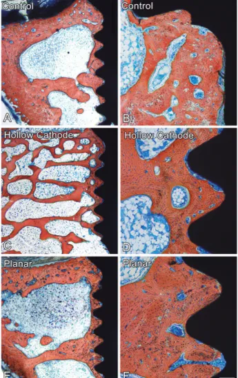
Bone formation was observed in close contact with all implants without relevant histological differences among the three evaluated surfaces (Figures 2A, C, and E). Implant surfaces were surrounded by lamellar bone and, at higher magnification, connective tissue was noticed between bone tissue and implant surfaces (Figures 2B, D, and F). The percentage of BIC was 24.5+14.9, 29.8+17.4, and 24.1+13.2 for control, hollow cathode, and planar surfaces, respectively, without
statistically signiicant difference (p=0.737) among
them (Figure 3A). The percentage of BABT was
Figure 2- Longitudinal sections of control (A-B); hollow cathode (C-D) and planar (E-F) Ti implant surfaces surrounded by bone and connective tissue, at 6 weeks. Stevenel’s blue and Alizarin red S. Scale bar: A, C and
E=500 μm and B, D and F=125 μm
34.8+14.2, 26.8+15.5, and 33.5+14.4 for control, hollow cathode, and planar surfaces, respectively,
without statistically signiicant difference (p=0.445)
among them (Figure 3B). The percentage of BAMA was 22.6+4.3, 26.9+9.9, and 22.1+6.2 for control, hollow cathode, and planar surfaces, respectively,
without statistically signiicant difference (p=0.637)
among them (Figure 3C).
DISCUSSION
Several Ti surface modifications have been proposed in order to improve the process of implant osseointegration1,2,6,14,15,17,20. As plasma-nitrided Ti
surfaces favor osteoblast differentiation, here, we have investigated bone tissue response to these surfaces and compared with machined ones. The results showed bone formation in close contact with all implant surfaces without relevant differences in terms of histological and histomorphometric parameters, indicating the lack of effect of plasma nitriding treatments on Ti implant osseointegration.
It has been reported that plasma nitriding treatments affect chemical, topographical, and roughness features4,10, improving surface hardness
without affecting Ti biocompatibility5,7,22. Compared
with conventional techniques, plasma treatment is inexpensive and environment friendly, needs low temperature and short time treatment and generates a uniform thickness layer3,23. In terms
of topography and roughness, it was previously observed that plasma-nitrided Ti discs exhibit less homogeneous and rougher discs compared with untreated surfaces, mainly those ones submitted to hollow cathode treatment10. Additionally, this
treatment results in higher percentage of Ti and contributes to the cleaning of surface as noticed by the reduction of C and O percentage10.
Distinct treatments generate Ti surfaces with different features, which affect bone cell/tissue response. It has been observed that Ti with nanotopography favors osteoblast differentiation in several culture models14,17,20. Also, biological
coatings such as bone apatite and type I collagen enhance bone formation in contact with Ti implants6,25. Regarding nitriding treatments,
previous studies demonstrated that Ti surfaces coated with nitride oxide increase cell growth rate and enhance osteoblast differentiation compared with machined surface8,10,19. In addition to in vitro studies that are useful to assess the inluence of Ti
surfaces on the osteoblast behavior in a controlled environment, in vivo experiments are of relevance as preclinical models. Thus, we have used an animal model to evaluate bone response to Ti
implants and no signiicant differences in terms
of histological and histomorphometric parameters were observed when plasma-nitrided surfaces were
compared with untreated one. In agreement with this, it has been shown that, despite modifying surface characteristics, bone formation in close contact with Ti implants is not deeply affected by nitriding treatments9,15,24. Bone contact, area, and
volume were not affected by nitride Ti produced by powder immersion reaction assisted coating when implanted in rat femora for 8 weeks24.
Nitrided Ti surfaces produced by glow-discharge plasma treatment had no effect on bone contact and area when implanted in rabbit tibia for 1, 3, and 6 weeks15. On the other hand, the increased
bone contact with nitrided Ti surface produced by plasma vapor observed 2 weeks post-implantation decreased after 1 and 3 months9. As our evaluation
was carried out at 6 weeks, a period in which the process of bone formation is completed in this animal model12, it is possible to suggest that some
effect of plama-nitrided Ti implant surfaces on bone formation, if any, could be noticed in early time-points.
CONCLUSION
In conclusion, our results showed that the plasma nitriding treatments used here create Ti implants that elicit similar bone tissue response to the untreated ones. Considering that these treatments improve the physico-chemical properties of Ti without affecting its biocompatibility, the
association with modiications generated by either
nanotechnology or functionalization with growth factors, which may favor bone formation, should be considered for developing new implant surfaces.
ACKNOWLEDGEMENTS
The authors would like to thank São Paulo Research Foundation (FAPESP) and National Council for Scientific and Technological Development
(CNPq) for inancial support. Adriana L. G. Almeida
and Sebastião C. Bianco are acknowledged for technical assistance during the experiments.
CONFLICTS OF INTEREST
The authors have no inancial interest in any
company or any of the products mentioned in this article.
REFERENCES
1- Albrektsson T. Hard tissue implant interface. Aust Dent J. 2008;53:S34-8.
2- Albrektsson T, Wennerberg A. Oral implant surfaces: Part 1 - review focusing on topographic and chemical properties of different surfaces and in vivo responses to them. Int J Prosthodont.
3- Alves C Jr, Araújo FO, Ribeiro KJ, Costa JA, Sousa RR, Sousa RS. Use of cathodic cage in plasma nitriding. Surf Coat Technol. 2006;201:2450-4.
4- Alves C Jr, Guerra-Neto CB, Morais GH, Silva CF, Hajek V. Nitriding of titanium disks and industrial dental implants using hollow cathode discharge. Surf Coat Technol. 2005;194:196-202. 5- Bordji K, Jouzeau JY, Mainard D, Payan E, Netter P, Rie KT, et al. Cytocompatibility of Ti-6Al-4V and Ti-5Al-2.5Fe alloys according to
three surface treatments, using human ibroblasts and osteoblasts.
Biomaterials. 1996;17:929-40.
6- Cecconi S, Mattioli-Belmonte M, Manzotti S, Orciani M, Piccioli A, Gigante A. Bone-derived titanium coating improves in vivo implant osseointegration in an experimental animal model. J Biomed Mater Res B Appl Biomater. 2014;102:303-10.
7- Clem WC, Konovalov VV, Chowdhury S, Vohra YK, Catledge SA, Bellis SL. Mesenchymal stem cell adhesion and spreading on microwave plasma-nitrided titanium alloy. J Biomed Mater Res A. 2006;76:279-87.
8- Durual S, Pernet F, Rieder P, Mekki M, Cattani-Lorente M, Wiskott HW. Titanium nitride oxide coating on rough titanium stimulates the proliferation of human primary osteoblasts. Clin Oral Implants Res. 2011;22:552-9.
9- Durual S, Rieder P, Garavaglia G, Filieri A, Cattani-Lorente M, Scherrer SS, et al. TiNOx coatings on roughened titanium and CoCr alloy accelerate early osseointegration of dental implants in minipigs. Bone. 2013;52:230-7.
10- Ferraz EP, Sá JC, Oliveira PT, Alves C Jr, Beloti MM, Rosa AL. The effect of plasma-nitrided titanium surfaces on osteoblastic cell adhesion, proliferation and differentiation. J Biomed Mater Res A. 2014;102:991-8.
11- Groessner-Schreiber B, Neubert A, Müller WD, Hopp M,
Griepentrog M, Lange KP. Fibroblast growth on surface-modiied
dental implants: an in vitro study. J Biomed Mater Res A.
2003;64:591-9.
12- Ivanoff CJ, Sennerby L, Lekholm U. Inluence of mono- and
bicortical anchorage on the integration of titanium implants. A study in the rabbit tibia. Int J Oral Maxillofac Surg. 1996;25:229-35.
13- Jafin RA, Berman CL. The excessive loss of Branemark ixtures
in type IV bone: a 5-year analysis. J Periodontol. 1991;62:2-4. 14- Kato RB, Roy B, Oliveira FS, Ferraz EP, Oliveira PT, Kemper AG, et al. Nanotopography directs mesenchymal stem cells to osteoblast lineage through regulation of microRNA-SMAD-BMP-2 circuit. J Cell Physiol. 2014;229:1690-6.
15- Larsson Wexell C, Thomsen P, Aronsson BO, Tengvall P,
Rodahl M, Lausmaa J, et al. Bone response to surface-modiied
titanium implants: studies on the early tissue response to implants with different surface characteristics. Int J Biomater. 2013;2013:412482.
16- Maniatopoulos C, Rodriguez A, Deporter DA, Melcher AH. An improved method for preparing histological sections of metallic implants. Int J Oral Maxillofac Implants. 1986;1:31-7.
17- Oliveira PT, Zalzal SF, Beloti MM, Rosa AL, Nanci A. Enhancement of in vitro osteogenesis on titanium by chemically
produced nanotopography. J Biomed Mater Res A. 2007;80:554-64.
18- Pjetursson BE, Karoussis I, Bürgin W, Brägger U, Lang NP. Patients' satisfaction following implant therapy. A 10-year prospective cohort study. Clin Oral Impl Res. 2005;16:185-93. 19- Rieder P, Scherrer SS, Filieri A, Wiskott HW, Durual S. TiNOx coatings increase human primary osteoblasts proliferation independently of the substrate: a short report. Biomed Mater Eng. 2012;22:277-81.
20- Rosa AL, Kato RB, Castro Raucci LM, Teixeira LN, Oliveira FS, Bellesini LS, et al. Nanotopography drives stem cell fate toward
osteoblast differentiation through α1β1 integrin signaling pathway.
J Cell Biochem. 2014;115:540-8.
21- Rosa AL, Oliveira CS, Beloti MM, Xavier SP, Oliveira PT. Effect of microcapsules containing TAK-778 on bone formation around osseointegrated implants: histomorphometric analysis in dogs. Implant Dent. 2006;15:97-103.
22- Sawase T, Yoshida K, Taira Y, Kamada K, Atsuta M, Baba K. Abrasion resistance of titanium nitride coatings formed on titanium by ion-beam-assisted deposition. J Oral Rehabil. 2005;32:151-7. 23- Silva JS, Amico SC, Rodrigues AO, Barboza CA, Alves C Jr, Croci
AT. Osteoblastlike cell adhesion on titanium surfaces modiied by
plasma nitriding. Int J Oral Maxillofac Implants. 2011;26:237-44. 24- Sovak G, Weiss A, Gotman I. Osseointegration of Ti6Al4V alloy implants coated with titanium nitride by a new method. J Bone Joint Surg Br. 2000;82:290-6.
25- Sverzut AT, Crippa GE, Morra M, Oliveira PT, Beloti MM, Rosa AL. Effects of type I collagen coating on titanium osseointegration: histomorphometric, cellular and molecular analyses. Biomed Mater. 2012;7:035007.