ABSTRACT
http://dx.doi.org/10.1590/1678-775720140124
TiF
4
and NaF varnishes as anti-erosive agents on
enamel and dentin erosion progression
in vitro
Livia Picchi COMAR, Cristiane de Almeida Baldini CARDOSO, Senda CHARONE, Larissa Tercilia GRIZZO, Marília Afonso Rabelo BUZALAF, Ana Carolina MAGALHÃES
Department of Biological Sciences, Bauru School of Dentistry, University of São Paulo, Bauru, SP, Brazil.
Corresponding address: Ana Carolina Magalhães - Departmento de Ciências Biológicas - Faculdade de Odontologia de Bauru - Universidade de São Paulo - Al. Octávio Pinheiro Brisolla, 9-75 - 17012-901 - Bauru - SP - Brazil - Phone/Fax: +55 (14) 3235-8497 - e-mail: [email protected]
Submitted: March 19, 2014 - Modiication: July 14, 2014 - Accepted: July 29, 2014
O
bjective: This study assessed the effect of luoride varnishes on the progression oftooth erosion in vitro. Material and Methods: Forty-eight enamel and 60 root dentin samples were previously demineralized (0.1% citric acid, pH 2.5, 30 min), leading to a baseline and erosive wear of 12.9 and 11.4 µm, respectively. The samples were randomly treated (6 h) with a 4% TiF4 varnish (2.45%F-, pH 1.0), a 5.42% NaF varnish (2.45%F-, pH 5.0), a placebo varnish and no varnish (control). The samples were then subjected to
erosive pH cycles (4x90 s/day in 0.1% citric acid, intercalated with artiicial saliva) for 5
days. The increment of the erosive tooth wear was calculated. In the case of dentin, this
inal measurement was done with and without the demineralized organic matrix (DOM).
Enamel and dentin data were analyzed using ANOVA/Tukey’s and Kruskal-Wallis/Dunn tests, respectively (p<0.05). Results: The TiF4 (mean±s.d: 1.5±1.1 µm) and NaF (2.1±1.7
µm) varnishes signiicantly reduced enamel wear progression compared to the placebo
varnish (3.9±1.1 µm) and control (4.5±0.9 µm). The same differences were found for dentin in the presence and absence of the DOM, respectively: TiF4 (average: 0.97/1.87 µm), NaF (1.03/2.13 µm), placebo varnish (3.53/4.47 µm) and control (3.53/4.36 µm). Conclusion: The TiF4 and NaF varnishes were equally effective in reducing the progression of tooth erosion in vitro.
Keywords: Sodium luoride. Titanium. Tooth erosion.
INTRODUCTION
Dental erosion is a clinical condition that is currently being studied since researchers have reported an increase in its prevalence17. Furthermore, dentists are becoming more aware about the etiology and early detection of this condition23.
A c i d e x p o s u r e c a u s e s e n a m e l s u r f a c e demineralization, and long-term demineralization leads to erosive enamel wear as a consequence of progressive mineral loss15. Dentin erosion presents a different histology lesion, since the progression of demineralization is modulated by the presence of the demineralized organic matrix (DOM)6,11. The DOM may be affected by enzymatic and chemical degradation, which may enhance the progression of dentin wear over time7,27. The primary preventive measure for dental erosion
is to reduce the frequency and duration of acid exposure22. As it is dificult to control the behavior of the patient, such as the frequency of acid intake or special drinking habits, other strategies have been proposed to control dental erosion that are rather less dependent on the compliance of the patient.
The most tested alternative is to increase the acid resistance of the teeth through the application of fluoride. Accordingly, highly concentrated
luoride applications such as oral rinses, gels and
varnishes have been tested5. The application of NaF at high concentration is able to promote a CaF2-like layerprecipitation on enamel26. The CaF
2 globules behave as a physical barrier, inhibiting the contact of the acid with enamel and/or acting as a
luoride reservoir. However, this layer presents low
On this basis, recent studies have focused on
other luorides, such as those containing polyvalent metals, which may have a higher eficacy than NaF
due to surface precipitation or incorporation of ions into sound and demineralized tissue.
Accordingly, the TiF4 has shown a promising erosion-inhibiting effect both in vitro and in situ
compared to other luorides12-14,19,30. The effect of TiF4 is related to both luoride and titanium3. It
is hypothesized that titanium may conlate with
phosphate groups, producing an acid-resistant surface coating25.
Most studies have applied luoride between the
erosive challenges, showing a positive effect of
different luoride formulations on the progression
of tooth erosion6,28. Recently, our research group has shown that an experimental TiF4 varnish can be more or at least as effective as a NaF varnish in reducing erosive tooth wear when applied only once on a previously sound surface18-21. However, there have been no data published regarding the effect of TiF4 and NaF varnishes on the reduction of tooth erosion progression when applied on pre-eroded tooth surfaces. Patients are usually treated
with luoride to prevent erosion when they have
signs of the lesion.
Therefore, the present study aimed to analyze and compare the effect of TiF4 and NaF varnishes on enamel and dentin erosion progression in vitro. The null hypothesis tested was that there
is no signiicant difference between TiF4 and NaF varnishes on the progression of erosive tooth wear when compared to the control.
MATERIAL AND METHODS
Sample preparation
Bovine teeth were prepared to obtain 48 enamel and 60 root dentin samples (4x4x3 mm). The teeth were stored in 0.1% buffered thymol solution (pH 7.0) at 4°C. The specimens were cut using an ISOMET low-speed saw cutting machine (Buehler Ltd., Lake Bluff, IL, USA) with two diamond
discs (Extec Corp., Enield, CT, USA), which were
separated by a 4 mm thick spacer. The surfaces
of the specimens were ground lat using
water-cooled silicon carbide discs (320, 600 and 1200 grades of Al2O3 papers; Buehler, Lake Bluff, IL,
USA), removing about 200 μm of the surface of the
tooth. Thereafter, the samples were cleaned in an ultrasonic device with deionized water for 5 min. The removal of the cement from the root dentin was
veriied using a microscope (x40 magniication).
Two-thirds of the outer surface (untreated area) of the specimens were covered with nail varnish in order to create sound areas on both sides of a central band of eroded enamel and dentin.
I n i t i a l e r o s i v e l e s i o n a n d p r o f i l e measurement
An initial erosive demineralization was created by immersion of the specimens in 0.1% citric acid (pH 2.5) for 30 min at room temperature under gentle agitation, using a shaking bath with horizontal movements (60 rpm). The acid solution was replaced every 5 min (30 ml/sample). After acid exposure, enamel and dentin wear (µm) were quantitatively determined using contact profilometry (Mahr Perthometer, Mahr Ltda, Göttingen, Germany). For the baseline profile measurement (initial erosive wear), the nail varnish was carefully removed using a scalpel and acetone solution (1:1 water). During the measurement, the dentin specimens were maintained in water (100% humidity) to avoid the DOM from shrinking.
The diamond stylus was moved from the irst
reference to the exposed area and then over the other reference area (2.5 mm long and 2 mm wide). Five profile measurements were performed at intervals of 0.5 mm. The vertical distance between the horizontal line drawn on the reference areas and the one drawn on the experimental (eroded)
area was deined as tooth wear using Mahr Surf
XT20 (Software Mahr Surf XT20, 2009, Mahr Ltda, Göttingen, Germany). The values were averaged
(μm) and used for the random allocation of the
samples into the groups (treatments).
Fluoride treatment and pH cycling
Twelve enamel and 15 dentin specimens were randomly distributed according to the baseline wear of each one of the 4 groups: a 4% TiF4 varnish (2.45% F-, pH 1.0), a 5.42% NaF varnish (2.45%
F-, pH 5.0), a placebo varnish (without luoride)
and no varnish (control). All varnishes contained colophonium, synthetic resin, thickening polymer,
essence, artiicial sweetener and ethanol.
The varnishes were applied in a thin layer using a microbrush, and the specimens were stored
in artiicial saliva. After 6 h, the varnishes were
carefully removed with acetone solution (1:1) and a scalpel blade, avoiding to touch the enamel surface19.
The samples were submitted to an erosive demineralization by immersion in 0.1% citric acid (pH 2.5) for 90 s 4 times a day for 5 d. After each demineralization, the specimens were rinsed with
deionized water (10 s) and transferred into artiicial
saliva (pH 6.8, 30 ml/specimen, unstirred, 25°C) for 2 h. After the last daily erosive treatment,
the specimens were also stored in artiicial saliva
overnight. The citric acid was renewed at each
erosive challenge, and the artiicial saliva was replaced daily. The artiicial saliva consisted of 0.2
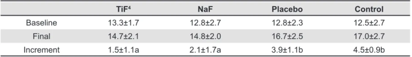
TiF4 NaF Placebo Control
Baseline 13.3±1.7 12.8±2.7 12.8±2.3 12.5±2.7 Final 14.7±2.1 14.8±2.0 16.7±2.5 17.0±2.7 Increment 1.5±1.1a 2.1±1.7a 3.9±1.1b 4.5±0.9b
Table 1- Mean±standard deviation of the baseline, inal and increment enamel erosive wear (µm) according to each
treatment
*Distinct lower case letters indicate signiicant differences among the treatment groups (ANOVA and Tukey’s test, p<0.0001,
n=12)
TiF4 NaF Placebo Control
Baseline 11.3 11.2 11.2 11.2
Final 12.3/13.1 12.0/13.2 14.6/16.0 14.8/15.4 Increment 0.97 (0.49)a
1.87 (0.95)a
1.03 (0.68)a
2.13 (0.73)a
3.53 (1.42)b
4.47 (1.91)b
3.53 (1.62)b
4.36 (2.18)b Table 2- Baseline, inal and increment dentin erosive wear means (µm), with and without DOM (interquartile range),
respectively, according to each treatment
*Distinct lower case letters indicate signiicant differences among the treatment groups (Kruskal-Wallis and Dunn, p<0.0001,
n=15)
K2HPO4, 3.3 mM urea, 2.4 mM NaH2PO4, and 11 μM ascorbic acid (pH 6.8)19.
Final proile measurement
After 5 days of pH cycling, the nail varnish was
removed, and 5 proiles were recorded as done at the baseline proile measurement. The values were averaged (μm), and the increment value (inal
wear – baseline wear) was calculated for enamel and dentin specimens (with and without the DOM in the case of dentin) and then submitted to statistical analysis.
After the profilometric analysis, the dentin samples were submitted to degradation of the
collagen ibrils in order to check the inluence of the DOM on the proile measurement and comparison
among the groups. A collagenase enzyme (Type VII from Clostridium histolyticum, product no. Co773, Sigma-Aldrich, St. Louis, MO, USA), with a collagen digestion activity of 1.98 U/µg, solid at 25°C, pH 7.5, in the presence of calcium ions, was used to remove the DOM. The collagen layer was removed
by adding 100 U/ml of enzyme into artiicial saliva
containing 20 mM HEPES, 0.70 mM CaCl2, 0.20 mM MgCl2.6H2O, 4 mM KH2PO4, 30 mM KCl, 0.30 mM NaN3, and EDTA-free protease inhibitor cocktail (Complete TM protease inhibitor of Cocktail, Santa Cruz Biotechnology, Santa Cruz, CA, USA) for 5 d at 37°C6.
Statistical analysis
The software GraphPad InStat (GraphPad Software, San Diego, CA, USA) was used. The assumptions of equality of variances and normal
distribution of data were veriied for all the variables
tested using Bartlett and Kolmogorov-Smirnov tests, respectively. As the equality of variances
was satisied for enamel, the differences among
treatments were analyzed using ANOVA followed by Tukey’s test. For dentin, the equality of variances was not satisfied, and the differences among treatments were analyzed using Kruskal-Wallis followed by Dunn’s test. The sample size was 12 for enamel and 15 for dentin, and the level of
signiicance was set at 5%.
RESULTS
The baseline erosive wear means were 12.9 and 11.4 µm for enamel and dentin, respectively. The mean increment of enamel wear (µm) was similar between TiF4 and NaF varnish. Both luoride
varnishes showed signiicantly less erosion (67%
and 53% decrease in enamel wear, respectively) compared to the placebo varnish and the control group (p<0.0001), which did not differ from each other. Table 1 shows the enamel erosive wear means.
DISCUSSION
Investigations of the potential of TiF4 to prevent tooth erosion have been performed since 19973. It has been speculated that its effect against erosion is
due to a high incorporation of luoride into enamel24 and a formation of an acid surface precipitation composed by TiO2 or hydrated titanium phosphate25.
Our group showed that the TiF4 incorporated in an experimental varnish presented a higher protective potential than the TiF4 solution on enamel erosion19, enamel erosion-abrasion24 and dentin erosion-abrasion in vitro21. We speculate that the better ability of the varnish to adhere to the tooth surface, allowing an increased contact time with the tooth, is responsible for its positive effect18,19,21. However, our previous studies have focused on the prevention of dental erosion rather than on the control of its progression. Accordingly, the varnishes were applied to previously sound surfaces in the previous studies. The question of the present study was whether TiF4 or NaF varnishes were also able to reduce the progression of tooth wear when applied on previously eroded surfaces.
In our study, we produced a baseline enamel and dentin erosive lesion by aggressive acid exposure done under agitation. Fast stirring can
increase the ive-fold of the enamel wear compared
to constant conditions. Clinically, this procedure simulates drinking habits such as rinsing, which may increase the risk of erosive wear8. The idea behind the procedure was also to simulate a patient with
a history of erosive lesions, for whom a luoride
application would be indicated, according to the Basic Erosive Wear Examination (BEWE) guidelines2. The varnishes were then applied on the eroded surface only once and removed after 6 h in order to simulate the clinical professional application19,20.
In the present study, all luoridated varnishes had a similar signiicant potential to reduce enamel and
dentin progression, therefore, the null hypothesis
can be rejected. However, there was no signiicant difference between both luoride varnishes.
The results observed for dentin agree with
recent data from our group, in which all luoridated
varnishes tested (experimental TiF4 and NaF,
Duraphat and Duoluorid varnishes) were similarly able to signiicantly reduce dentin wear compared to the placebo varnish, the luoridated solutions
(TiF4 and NaF)21 and the control group.
A limitation of analyzing erosion in dentin is the complexity of the lesion histology, which is characterized by centripetal mineral loss starting in the peritubular areas that progresses to the intertubular area, leading to a bulk tissue loss and the presence of a completely demineralized dentin surface zone that is stable while hydrated16.
Therefore, erosive wear is dificult to quantify since
the quality of the remaining organic layer may interfere in the measurement7. The limitation of the wear measurement is due to the shrinkage of the DOM that may occur under different environments.
Therefore, to generate reliable data, the proiles
must be measured with the samples immersed in water1. On the other hand, Schlueter, et al.29 (2011) advise that the DOM should be removed before the
proilometric measurement to prevent this problem
from occurring.
We found that the thickness of the DOM of our samples was about 1 µm, regardless of the treatment. It is important to highlight that, in the present study, the results were maintained when the wear was measured on dentin without the DOM.
Some studies have shown that the preventive effect of fluorides on dentin erosion is lower compared to enamel5,10 and highly dependent on the presence of the organic matrix9,27. Only a very
intensive luoridation was effective in the prevention
of dentin erosion5. Dentin erosion is much more complex than enamel erosion because the organic matrix plays an important role in the progression of wear. The DOM has a buffering capacity that prevents further demineralization, especially in the
presence of luoride6. At least for NaF, the enzymatic removal of the organic matrix substantially decreases its effect6,11,29. Considering that it is still unclear if what extent the organic matrix is retained under clinical conditions27, it would be interesting to analyze the effect of TiF4 varnish applied on eroded dentin with or without DOM.
In respect to the results of enamel, the present data are not in accordance with a previous study from our group19, in which we found signiicantly less erosion for the enamel samples treated with a TiF4 varnish than those treated with a NaF (Duraphat) or NaF/CaF2 (Duofluorid) varnish. Based on the experimental conditions, we speculate that the glaze-like surface layer produced by TiF4 may be thinner and with lower amounts of Ti on demineralized enamel surfaces compared to sound ones, as previously shown by Chevitarese, et al.4 (2004).
Based on these results, we suggest that the effect of other preventive measures (professional products) should be tested on previously eroded
tooth surfaces in future studies, since luoride
application is indicated only for patients with a high risk of erosion, according to BEWE guidelines2. The effect of the TiF4 varnish on eroded dentin surfaces in the absence of the DOM is another important topic to be further considered. Finally,
the interaction of this polyvalent metal luoride in
using in situ models.
CONCLUSIONS
Under the conditions of the present study, it can be concluded that the TiF4 varnish was as effective as the NaF varnish to reduce the enamel and dentin erosion progression in vitro.
Acknowledgments
The authors thank the high school students Andressa Alves and Gabriel Scarpim, who participated in some parts of this study through the Pre-Undergraduate Research Program of the University of São Paulo.
REFERENCES
1- Attin T, Becker K, Roos M, Attin R, Paqué F. Impact of storage
conditions on proilometry of eroded dental hard tissue. Clin Oral
Investig. 2009;13:473-8.
2- Bartlett D, Ganss C, Lussi A. Basic Erosive Wear Examination
(BEWE): a new scoring system for scientiic and clinical needs.
Clin Oral Investig. 2008;12(Suppl 1):S65-8.
3- Büyükyilmaz T, Ogaard B, Rølla G. The resistance of titanium
tetraluoride-treated human enamel to strong hydrochloric acid.
Eur J Oral Sci. 1997;105:473-7.
4- Chevitarese AB, Chevitarese O, Chevitarese LM, Dutra PB. Titanium penetration in human enamel after TiF4 application. J Clin Pediatr Dent. 2004;28:253-6.
5- Ganss C, Klimek J, Brune V, Schürmann A. Effects of two
luoridation measures on erosion progression in human enamel
and dentine in situ. Caries Res. 2004;38:561-6.
6- Ganss C, Klimek J, Starck C. Quantitative analysis of the impact
of the organic matrix on the luoride effect on erosion progression
in human dentine using longitudinal microradiography. Arch Oral Biol. 2004;49:931-5.
7- Ganss C, Lussi A, Scharmann I, Weigelt T, Hardt M, Klimek J, et al. Comparison of calcium analysis, longitudinal microradiography
and proilometry for the quantitative assessment of erosion in
dentine. Caries Res. 2009;43:422-9.
8- Ganss C, Lussi A, Schlueter N. Dental erosion as oral disease. Insights in etiological factors and pathomechanisms, and current strategies for prevention and therapy. Am J Dent. 2012;25:351-64.
9- Ganss C, Lussi A, Sommer N, Klimek J, Schlueter N. Eficacy of luoride compounds and stannous chloride as erosion inhibitors
in dentine. Caries Res. 2010;44:248-52.
10- Ganss C, Schlueter N, Klimek J. Retention of KOH-soluble
luoride on enamel and dentine under erosive conditions - a
comparison of in vitro and in situ results. Arch Oral Biol.
2007;52:9-14.
11- Hara AT, Ando M, Cury JA, Serra MC, González-Cabezas C,
Zero DT. Inluence of the organic matrix on root dentine erosion
by citric acid. Caries Res. 2005;39:134-8.
12- Hove L, Holme B, Øgaard B, Willumsen T, Tveit AB. The protective effect of TiF4, SnF2 and NaF on erosion of enamel by
hydrochloric acid in vitro measured by white light interferometry.
Caries Res. 2006;40:440-3.
13- Hove LH, Holme B, Stenhagen KR, Tveit AB. Protective effect of TiF(4) solutions with different concentrations and pH on development of erosion-like lesions. Caries Res. 2011;45:64-8. 14- Hove LH, Holme B, Young A, Tveit AB. The protective effect
of TiF4, SnF2 and NaF against erosion-like lesions in situ. Caries
Res. 2008;42:68-72.
15- Huysmans MC, Chew HP, Ellwood RP. Clinical studies of dental erosion and erosive wear. Caries Res. 2011;45(Suppl 1):60-8. 16- Kinney JH, Balooch M, Haupt DL Jr, Marshall SJ, Marshall GW Jr. Mineral distribution and dimensional changes in human dentin during demineralization. J Dent Res. 1995;74:1179-84.
17- Kreulen CM, Van't Spijker A, Rodriguez JM, Bronkhorst EM, Creugers NH, Bartlett DW. Systematic review of the prevalence of tooth wear in children and adolescents. Caries Res. 2010;44:151-9.
18- Levy FM, Magalhães AC, Gomes MF, Comar LP, Rios D, Buzalaf MA. The erosion and abrasion-inhibiting effect of TiF(4) and NaF
varnishes and solutions on enamel in vitro. Int J Paediatr Dent.
2012;22:11-6.
19- Magalhães AC, Kato MT, Rios D, Wiegand A, Attin T, Buzalaf MA. The effect of an experimental 4% TiF4 varnish compared to
NaF varnishes and 4% TiF4 solution on dental erosion in vitro.
Caries Res. 2008;42:269-74.
20- Magalhães AC, Levy FM, Rios D, Buzalaf MA. Effect of a single application of TiF(4) and NaF varnishes and solutions on dentin
erosion in vitro. J Dent. 2010;38:153-7.
21- Magalhães AC, Levy FM, Rizzante FA, Rios D, Buzalaf MA. Effect of NaF and TiF(4) varnish and solution on bovine dentin
erosion plus abrasion in vitro. Acta Odontol Scand. 2012;70:160-4.
22- Magalhães AC, Wiegand A, Rios D, Honório HM, Buzalaf MA. Insights into preventive measures for dental erosion. J Appl Oral Sci. 2009;17:75-86.
23- Mulic A, Vidnes-Kopperud S, Skaare AB, Tveit AB, Young A. Opinions on dental erosive lesions, knowledge of diagnosis, and treatment strategies among Norwegian dentists: a questionnaire survey. Int J Dent. 2012;2012:716396.
24- Mundorff SA, Little MF, Bibby BG. Enamel dissolution. II. Action
of titanium tetraluoride. J Dent Res. 1972;51:1567-71.
25- Ribeiro CC, Gibson I, Barbosa MA. The uptake of titanium ions by hydroxyapatite particles-structural changes and possible mechanisms. Biomaterials. 2006;27:1749-61.
26- Saxegaard E, Rölla G. Fluoride acquisition on and in human
enamel during topical application in vitro. Scand J Dent Res.
1988;96:523-35.
27- Schlueter N, Ganss C, Hardt M, Schegietz D, Klimek J. Effect
of pepsin on erosive tissue loss and the eficacy of luoridation
measures in dentine in vitro. Acta Odontol Scand.
2007;65:298-305.
28- Schlueter N, Ganss C, Mueller U, Klimek J. Effect of titanium
tetraluoride and sodium luoride on erosion progression in enamel
and dentine in vitro. Caries Res. 2007;41:141-5.
29- Schlueter N, Hara A, Shellis RP, Ganss C. Methods for the measurement and characterization of erosion in enamel and dentine. Caries Res. 2011;45(Suppl 1):13-23.
30- Wiegand A, Hiestand B, Sener B, Magalhães AC, Roos M, Attin T. Effect of TiF4, ZrF4, HfF4 and AmF on erosion and
erosion/abrasion of enamel and dentin in situ. Arch Oral Biol.