ABSTRACT
http://dx.doi.org/10.1590/1678-775720140122
Osteopontin expression in reactive lesions of
gingiva
Rathinam ELANAGAI, Veeran VEERAVARMAL, Ramdas Madhavan NIRMAL
Division of Oral and Maxillofacial Pathology, Rajah Muthaiah Dental College and Hospital, Annamalai University, Annamalai Nagar, India.
Corresponding address: Veeran Veeravarmal - Division of Oral and Maxillofacial Pathology, Rajah Muthaiah Dental College and Hospital, Annamalai University - Annamalai Nagar - Tamil Nadu - India - Phone: 9443659412 - e-mail: [email protected]
Submitted: April 2, 2014 - Modiication: September 28, 2014 - Accepted: September 30, 2014
R
eactive proliferations of the gingiva comprise lesions such as pyogenic granuloma (PG), inlammatory ibroepithelial hyperplasia (IFH), peripheral ossifying ibroma (POF), and peripheral giant cell lesion. Osteopontin (OPN) has a dual role, it promotes mineralization when it is bound to solid substrate, and on the other hand, it inhibits mineralization when it is seen in association with solution. Objectives: The study aimed to evaluate the expression of osteopontin in normal gingival tissue and different types of focal reactive proliferations of gingival tissue, and its role in the development of calciication within it. Material and Methods: The presence and distribution of osteopontin was assessed using immunohistochemistry in ive cases of normal gingival tissue and 30 cases of focal reactive proliferations of gingiva. Results: There was no expression of osteopontin in normal subjects. Few cases of pyogenic granuloma, inlammatory ibroepithelial hyperplasia, and all the cases of peripheral ossifying ibroma showed positivity for osteopontin in the inlammatory cells, stromal cells, extracellular matrix, and in the calciications. Conclusion: The expression of osteopontin in all the cases of peripheral ossifying ibroma speculates that the majority of the cases of peripheral ossifying ibroma originate from the periodontal ligament cells. The treatment modalities for peripheral ossifying ibroma should differ from other focal reactive proliferations of gingiva.Keywords: Osteopontin. Immunohistochemistry.
INTRODUCTION
The oral mucosa is under constant barrage of irritation due to various external and internal stimuli culminating in a wide spectrum of developmental,
inlammatory, reactive, and neoplastic diseases9.
The reactive proliferations manifest both clinically and histologically as non-neoplastic nodular swellings. They occur as the aftermath of chronic and repeated tissue injury and present as an exuberant or excessive tissue response while regressing on elimination of the initiating factor9,21.
Reactive lesions often present diagnostic challenges because of their overlapping and deceptive clinical presentation.
Reactive proliferations of the gingiva comprise lesions such as pyogenic granuloma (PG), inflammatory fibroepithelial hyperplasia (IFH),
peripheral ossifying ibroma (POF), and peripheral
giant cell lesion. Recently, localized juvenile spongiotic gingival hyperplasia has been added to this category4,20. These lesions develop in response
to local irritants such as masticatory force, defective restorations, dental plaque, calculus, trauma, and iatrogenic factor. These irritational factors in conjunction with the peculiar anatomy of the gingiva attribute to the high incidence of reactive proliferation at this site. The most common reactive
lesion is peripheral ibroma (56–61%), followed in
descending order by the pyogenic granuloma (19–
27%), the peripheral ossifying ibroma (10–18%),
and peripheral giant cell lesion (1.5–7%).
Long standing PG exhibits maturation and dystrophic calcifications that may mimic the histopathology of POF, which may be due to
metaplastic changes observed in the ibroblasts18,22.
Inlammatory ibroepithelial hyperplasia (IFH) is a hyperplastic ibrous tissue mass, usually located
irritation of periodontal ligament ibers. According
to some investigators, it represents the immature
variant of peripheral ossifying ibroma9.
Mineralization in POF can vary among
cementum-like material, bone, and dystrophic calciication2,10,11.
Whether all these reactive proliferations are separate entities or represent different stages in maturation of a single lesion has been a moot point for many years. Generally, the mineralization can
be inluenced by collagenous and non-collagenous
proteins present in the extracellular matrix. Osteopontin (OPN) is one such non-collagenous protein which is a highly phosphorylated sialoprotein and has high calcium binding potential. OPN is produced by osteoblasts at various stages of differentiation and by differentiated osteoblasts, osteocytes15,23, and osteoclasts7. The OPN in the
cemental lines may be utilized for osteoblasts cell
adhesion or to promote early calciication events
at this junction24. The OPN contains several serine
phosphorylated sites and stretching of negatively charged aspartic acid residues responsible for the attraction of calcium, and also influence mineralization.
The persistence of focal reactive growth of gingiva for prolonged period may result in the formation of
calciied structure within it. The initiating factor(s) inluencing the dystrophic calciication or cementum
or bone formation in these reactive lesions are poorly understood. The aim of the present study is to analyze the expression of osteopontin in normal gingival tissue and different types of focal reactive proliferations of gingiva in order to understand the possible role of osteopontin in the histogenesis of
calciications and ossiications in these lesions.
MATERIAL AND METHODS
This laboratory study consisted of 30 cases of histological variants of focal reactive proliferations that were subjected to histological examination in the Department of Oral and Maxillofacial Pathology, RMDC&H, Annamalai University, Tamilnadu, India. These archival tissues and normal oral mucosa were fixed in 10% buffered formalin
and embedded in parafin wax, sections of 4 µm
thickness were prepared from each case for routine hematoxylin and eosin and immunohistochemical staining. Histopathological sections of the focal reactive proliferations were reviewed by the
oral pathologists to conirm the diagnosis and to
ascertain representative areas. Five cases of normal oral mucosa were deemed as a control group. All the 30 cases of focal reactive proliferations (10 cases each of PG, IFH, and POF) and the control group were subjected to immunohistochemistry to evaluate the expression of osteopontin.
Two to three serial sections of 3 µm thickness
were made and taken on APES pre-coated slides.
The sections were deparafinized and rehydrated
in graded alcohol (100%, 90%, 70%, and 50%). The unmasking of antigen was done by the heat retrieval method in citrate buffer (pH 6.5). All the incubations were performed at room temperature using a humidifying chamber. At no time the tissue sections were allowed to dry during the staining procedure. After tapping off the excess buffer from the slides, the sections were sequentially treated with 3% hydrogen peroxide (DAKO REAL En Vision, Glostrup, Denmark) and protein block (power block, DAKO REAL En Vision, Glostrup, Denmark) 10 minutes each, to suppress endogenous peroxidase activity and prevent unwanted protein binding. After thorough washing in PBS, the sections were incubated in Polyclonal rabbit anti-human
osteopontin antibody (Thermoscientiic, Marietta,
Ohio, USA) for 30 minutes. The slides were washed and treated with Poly Horseradish peroxidase secondary antibody (DAKO REAL En Vision, Denmark) for 30 minutes. After thorough washing in PBS, the immunostaining was developed by treating with freshly constituted 3,3-diaminobenzidene
tetra hydrochloride (DAB) solution for ive minutes.
After thorough washing in distilled water, the slides were counterstained with Mayer’s hematoxylin and mounted with DPX.
The immunostained slides were evaluated by two blinded and calibrated pathologists independently. The positive areas were indicated by brown color precipitates and it was evaluated in stromal cells, extracellular matrix, calcifications, and
inlammatory cells. All areas in each section were
examined, and, in the areas where the intensity
was predominant, these ields were taken into
consideration. The intensity of staining (viewed at
magniication of 10x and 40x) was scored according
to the following scale: 0 (no staining); 1 (mild staining); 2 (moderate staining); and 3 (intense staining). The data were analyzed using SPSS 21.0 software. The chi-square test was used to
ind the difference between the intensity levels and
percentage positivity among the various groups. Differences with a probability value of <0.05 were
considered statistically signiicant.
RESULTS
The histopathological findings of all the 30 cases of focal reactive proliferations are given in
Figure 1. The ive normal subjects did not show any area of inlammation, calciication, or ulceration. PG showed surface ulceration in ive cases, and
vascularity was a predominant feature in all the cases. In IFH, one case showed small globules of
calciication in focal areas. The surface was not
cases exhibited calcifications which resembled trabecular bone in the majority of the cases, and few cases showed numerous basophilic
cementum-like calciication. The collagen distribution is similar
to IFH and showed equal number of cases with
moderate to densely ibrous stroma.
Osteopontin expression
All the cases of normal oral mucosa showed
no expression of OPN in the lamina propria. The expressions of OPN in all the 30 cases of focal reactive proliferations of gingiva are mentioned in the Figure
2. In PG, the stromal ibroblasts distributed along with inlammatory cells showed expression of OPN.
Interestingly, the cells with cuboidal morphology and the matrix adjacent to them showed OPN expression in PG (Figure 3a). A focal collection of stromal cells,
seen adjacent to blood vessel within the ibrovascular
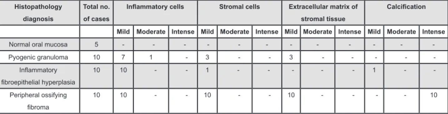
Histopathology diagnosis Total no. of cases
Inlammation Vascularity Collagen Calciication
Mild Moderate Intense Mild Moderate Intense Mild Moderate Intense Present Absent
Pyogenic granuloma 10 - 1 9 - - 10 6 4 - - 10
Inlammatory ibroepithelial
hyperplasia
10 - 4 6 3 3 4 - 5 5 1 9
Peripheral ossifying ibroma 10 - 4 6 3 5 2 - 5 5 10
-Figure 1- Histopathological indings of pyogenic granuloma, inlammatory ibroepithelial hyperplasia, and peripheral ossifying ibroma
Histopathology diagnosis
Total no. of cases
Inlammatory cells Stromal cells Extracellular matrix of stromal tissue
Calciication
Mild Moderate Intense Mild Moderate Intense Mild Moderate Intense Mild Moderate Intense
Normal oral mucosa 5 - - - - - - -
-Pyogenic granuloma 10 7 1 - 3 - - 3 - - - -
-Inlammatory ibroepithelial hyperplasia
10 10 - - 1 - - - - - 1 -
-Peripheral ossifying
ibroma
10 10 - - 10 - - 10 - - - - 10
Figure 2- Osteopontin expression in pyogenic granuloma, inlammatory ibroepithelial hyperplasia, and peripheral ossifying ibroma
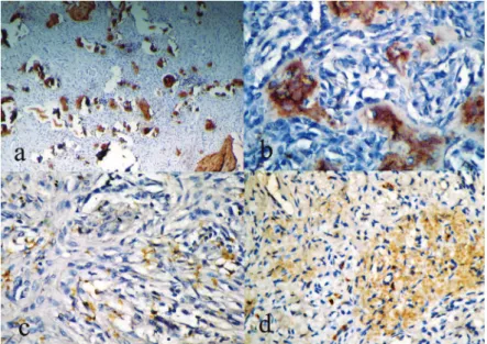
Figure 3- a) Pyogenic granuloma showing osteopontin (OPN) expression in stromal cells and extracellular matrix (40x); b)
component of PG, showed OPN expression. In PG, the extracellular matrix expression of OPN was observed
in areas of inlammatory iniltrate. Lymphocytes, mast
cells, macrophages, plasma cells, and osteoclast-like giant cells also exhibited positivity (Figure 3d). One case showed an area with osteoid appearance surrounded by stellate and cuboidal cells with few
inlammatory cells that showed OPN expression
in the cells with cuboidal and stellate morphology (Figure 3b).
The IFH showed OPN expression in the
inlammatory component, stromal cells, and also in the calciication. None of the cases showed positivity in the extracellular matrix. The inlammatory cells
such as lymphocytes, plasma cells, macrophages, and mast cells showed mild expression for OPN
in focal areas in all cases of IFH. The calciication resembling dystrophic calciication was observed in
close relation to chronic inlammatory cells in one
area in IFH (Figure 3c).
The POF demonstrated OPN expression in the stromal cells with cuboidal or stellate morphology
which were seen close to the calciied structures
(Figure 4c). In all the cases of POF, the expression of OPN was observed in calcifications which resembled bone, cementum, and/or dystrophic
calciication (Figures 4a, b). All the cases of POF
exhibited OPN expression in the extracellular matrix and the distribution was focal in nature (Figure 4d). The extracellular matrix positivity was
observed predominantly in areas of inlammation
and in areas of early bone formation (Figure 4d). Histopathologically, these later areas appeared as eosinophilic, extracellular matrix with less cellularity. There was focal and mild expression of
OPN in the inlammatory cells of all the cases.
Figure 4- a) Peripheral ossifying ibroma (POF) showing osteopontin (OPN) expression in calciication resembling bone and cementum under lower magniication; b) The bony trabeculae expressing OPN in 40x; c) POF showing OPN expression
in stromal cells (40x); d) The extracellular matrix of POF showing OPN expression under 40x
Groups Inlammatory cells Stromal cells Extracellular matrix Calciication
Normal mucosa Vs. PG
0.014 0.171 0.171 NIL
Normal mucosa Vs. POF
0 0 0 NIL
Normal mucosa Vs. IFH
0 No difference 0.464 NIL
PG Vs. POF 0.171 0.003 0.003 NIL
PG Vs. IFH 0.171 0.582 0.211 NIL
IFH Vs. POF No difference among
the group
0 0 0
PG=pyogenic granuloma; POF=peripheral ossifying ibroma; IFH=inlammatory ibroepithelial hyperplasia
The comparison of OPN expression in the
inlammatory component among normal and the
PG, IFH, and POF was found to be statistically
signiicant (Table 1). The statistical analysis showed no difference in OPN expression in the inlammatory iniltrate seen in IFH and POF. The expression of
OPN was observed in all the cases of POF in the stromal tissue, while there was a total absence of
expression in the ibroblast of the lamina propria of normal tissue. The statistical comparison between
normal tissue and POF was found to be signiicant
(p<0.05) (Table 1). The expression of OPN in the stromal cells is compared between PG and POF, on statistical evaluation the results were found
to be signiicant (p=0.0003).There was a marked
difference in the expression of stromal cells for the OPN marker between IFH and POF (p<0.05) (Table
1). There was no signiicance on comparing the
expression of OPN in the stromal tissue of control tissue, PG, and IFH.
The expression of OPN in the extracellular matrix of normal oral mucosa, PG, IFH showed no significance. On comparing the results of extracellular matrix expression of OPN in normal tissue and POF, it was observed to be highly
signiicant (p<0.05). The extracellular matrix of
POF exhibited positive expression for OPN antibody in all the cases. Thus, the statistical comparison
of the expression was found to be signiicant at p
value (p=0.003). POF demonstrated expression in the extracellular matrix, on comparing the expression between IFH and POF; results were
shown to be highly signiicant (p<0.05) (Table
1). Histopathologically, there was total absence
of calciication in oral mucosal tissue and PG. Furthermore, the expression of OPN in calciied
tissue, when compared between IFH and POF,
showed highly signiicant results (p<0.05).
DISCUSSION
In the present study, 10 cases of PG exhibited high vascularity and inflammatory component distributed in loose fibrous connective tissues which ascertain its immature nature (Figure 1). Microscopically, IFH showed abundant collagen
ibers with few to many blood vessels. Therefore,
this group is composed of lesions in various stages
of progression. The calciication was observed in one case of IFH, and was conined to a single site (Figure 1). The morphology of this calciication was akin to the dystrophic calciication described in chronic inlammatory conditions or the calciications due to
degenerating and senescent cells5.
The histogenesis of POF remains controversial. According to one view, it is believed to arise from the periodontal ligament tissue/periostium due to its increased occurrence in the gingiva, a tissue
which is in proximity to the periodontal ligament18.
Contrarily, it is also believed to be a mature PG and some features are attributed to the secondary changes due to repeated trauma1,18,20.
POF demonstrated ibrous proliferation with plenty of ibroblasts in association with mineralized
tissue in the form of bone, cementum-like material,
dystrophic calciication, or a combination of all20.
Moderate to intense inflammatory cells were noted in most of the cases and vascularity was a predominant feature in cases which exhibited
intense inlammation. Further it was observed that
dense, mature collagen was seen only in cases with
minimum vascularity. All cases showed calciication and ibrous connective tissue in equal proportion. The calciied structures resembled bony trabeculae
with osteoid material in the periphery. Some mineralized structures presented as small globules
mimicking dystrophic calciication or cementum
(Figure 1).
The soft tissue possesses certain inhibitory factors/signals that prevent it from undergoing calcification. During disease process, there may be a deviation from the normal process or elimination of inhibitory factors leading to
calciication of soft tissue5. In the present study,
the lamina propria of the normal subjects showed no expression of OPN. When the normal tissue undergoes pathological changes, the OPN may be expressed in the stromal tissue14. This view
is supported by the results obtained from the current study. OPN is expressed in activated
chronic inlammatory cells such as lymphocytes,
mast cells, and macrophages3,6,13,17. The PG, IFH,
and POF expressed OPN in inlammatory iniltrate
which included lymphocytes, macrophages, plasma cells, and mast cells morphology. The results of
OPN expression in the inlammatory component
between normal oral mucosa, PG, IFH, POF was
found signiicant. This could be attributed to the reduction in the number of inlammatory component
in normal oral mucosa.
Few giant cells seen in fibrous background of PG also showed OPN expression7,15,23,28. Two
possible explanations could be drawn for stromal
cell positivity for OPN in PG. The irst mechanism is that macrophages in the chronically inlamed
tissue release pro-osteogenic cytokine, which stimulates the vascular smooth muscle cells to undergo osteogenic differentiation (or) release of
calcifying matrix vesicle and initiate calciication26;
the second mechanism may represent the immature POF without calcification. Thereby, this study speculates, based on the observations above, that the OPN expressing stromal cells may be osteoblast cells derived from PDL.
Nevertheless, it is not possible to state clearly
or osteoblastic differentiation pertaining to PDL origin as a result of consistent association of these
lesions with inlammation. Expression of OPN in extracellular matrix was observed in 3 cases of PG and all the cases of POF, whereas IFH failed to show any expression. Presumably, such areas could be the site of initiation of mineralization in the future.
In one case of PG, OPN expression was conined
to the extracellular matrix near the OPN positive stromal cells. In the other two cases, extracellular matrix positivity was seen in areas admixed with
ibroblast and inlammatory cells. However, absence
of OPN expression in the stromal cells in these two cases could be due to the release of OPN into the extracellular matrix. In POF, the areas of osteoid and the areas where the cells exhibited stellate morphology and separated apart, representing the initial phase of osteoid formation expressed OPN.
These were the sites where the calciications are
initiated.
One case of IFH and all the 10 cases of POF
showed OPN expression in the calciied structures
resembling bone, cementum, and dystrophic calcification. According to Giachelli12 (1999),
dystrophic calciication possesses several features
of bone mineralization, including the presence of non-collagenous proteins such as OPN matrix GLA protein, Osteocalcin, SPARAC, and bone morphogenic protein. The data from the present
study for OPN expression in calciied structure which resembled dystrophic calciication validates
the view that the non-collagenous proteins have a role in mineralization8,12.
All the cases of POF showed expression of
OPN in the calciications resembling bone and/ or cementum. There was a signiicant difference
in OPN expression in the mineralized component between POF and IFH. The mineralization seen
in IFH was closely associated with inlammatory component. The inlammation induced dystrophic calciication is purportedly the mechanism involved in the formation of calciied structure in this group. The IFH having exhibited dystrophic ossiication in
one area may presumably represent maturing PG
that passes through the stages of ibrous epulis and matures into ibroepithelial polyp27.
The information available in the literature and the results obtained from the present study insinuate
that the calciied structures in POF resembled bone, cementum, and/or dystrophic calciication. The reason for the presence of calciied structures
with morphology of bone and cementum in POF may be its tissue of origin. Alternatively, either
ibroblastic metaplasia occurring in longstanding
PG (or) osteogenic differentiation of smooth muscle
cells are inluenced by underlying inlammatory
mechanism19,25. The combination of dystrophic
calciication, bone and cementum in POF may be
due to the occurrence of both the mechanisms simultaneously.
Except for a few cases, most of the cases of PG showed no expression of OPN in stromal cells and extracellular matrix, which contradicts the concept that the PG on maturation develops into POF. However, all the cases of POF subjected for OPN expression exhibited positivity in stromal
cells, extracellular matrix, and calciication. From
the results, we opine that the majority of POF are probably de novo in origin. The indings of the
present study with regard to OPN expression in POF contradicted the results of Ono, et al.16 (2008), who
observed OPN expression only in 50% of the cases of POF. The stromal cells exhibited stellate and/or cuboidal morphology, and cells in the periphery of
ossiication showed OPN expression, while such inding was not reported16.
The hypothesis given by many investigators for the varied histopathological presentation of focal reactive lesions implies different stages in maturation of a single entity. The results from the current study are in part concurrent with this concept. The epithelium present both in normal oral mucosa and in focal reactive proliferation is totally negative for OPN expression. The odontogenic cyst and tumors showed expression of OPN in the epithelium. The epithelium in reactive proliferation is not altered genetically, and that could be the reason for its absence.
After due cogitation, we reckon that the present study observed OPN expression in the stromal cells of PG, IFH, and POF. The results suggest that there is an osteoblastic differentiation of stromal cells in focal reactive proliferations of gingiva. The pyogenic granuloma showed OPN expression in the stromal cells and extracellular matrix in few cases, which favors the concept that pyogenic granuloma may mature into POF. However, POF showed OPN expression in stromal cells, extracellular matrix,
and in areas of ossiication in all the cases. This inding leads us to conclude that there is a deinite
veracity to the idea of co-definite osteoblast/ cementoblast differentiation and OPN plays a role in
the development of ossiication. The current study
also theorizes that the majority of POF arises from the periodontal ligament cells. Hence, the treatment modalities for POF should differ from other focal reactive lesions of gingiva.
The data collected from the current study also upholds the view that PG may mature into IFH and POF on certain occasions. To ponder on the issue whether these lesions are separate entities or different phases during maturation of single entity, studies need to be carried out using
speciic markers involved in the differentiation of
osteoblast, cementoblast, and in the development
ACKNOWLEDGEMENT
We would like to thank Dr. V. Srinivas Prasad, Post-graduate student in the Division of Oral and Maxillofacial Pathology, Rajah Muthaih Dental College and Hospital, Annamalai University, Annamalai Nagar, for language editing of the manuscript.
REFERENCES
1- Bhaskar SN, Jacoway JR. Peripheral ibroma and peripheral ibroma with calciication: report of 376 cases. J Am Dent Assoc.
1966;73(6):1312-20.
2- Buchner A, Hansen LS. The histomorphologic spectrum of
peripheral ossifying ibroma. Oral Surg Oral Med Oral Pathol.
1987;63(4):452-61.
3- Cantor H. The role of Eta-1/osteopontin in the pathogenesis of immunological disorders. Ann N Y Acad Sci. 1995;760:143-50. 4- Chang JY, Kessler HP, Wright JM. Localized juvenile spongiotic gingival hyperplasia. Oral Surg Oral Med Oral Pathol Oral Radiol Endod. 2008;106(3):411-8.
5- Cotran RS, Kumar V, Collins T, Robbins SL. Robbins & Cotran
pathologic basis of disease. 6th ed. London: Saunders; 1999.
1425 p.
6- Denhardt DT, Mistretta D, Chambers AF, Krishna S, Porter JF, Raghuram S, et al. Transcriptional regulation of osteopontin and the metastatic phenotype: evidence for a Ras-activated enhancer in the human OPN promoter. Clin Exp Metastasis. 2003;20(1):77-84.
7- Dodds RA, Connor JR, James IE, Rykaczewski EL, Appelbaum E, Dul E, et al. Human osteoclasts, not osteoblasts, deposit
osteopontin onto resorption surfaces: an in vitro and ex vivo study
of remodeling bone. J Bone Miner Res. 1995;10(11):1666-80. 8- Donley GE, Fitzpatrick LA. Noncollagenous matrix proteins
controlling mineralization; possible role in pathologic calciication
of vascular tissue. Trends Cardiovasc Med. 1998;8(5):199-206.
9- Efiom OA, Adeyemo WL, Soyele OO. Focal reactive lesions of
the gingiva: an analysis of 314 cases at a tertiary health institution in Nigeria. Niger Med J. 2011;52(1):35-40.
10- Farquhar T, Maclellan J, Dyment H, Anderson RD. Peripheral
ossifying ibroma: a case report. J Can Dent Assoc.
2008;74(9):809-12.
11- Garća de Marcos JA, Garća de Marcos MJ, Arroyo Rodŕguez
S, Chiarri Rodrigo J, Poblet E. Peripheral ossifying ibroma: a
clinical and immunohistochemical study of four cases. J Oral Sci. 2010;52(1):95-9.
12- Giachelli CM. Ectopic calciication: gathering hard facts about
soft tissue mineralization. Am J Pathol. 1999;154(3):671-5. 13- Giachelli CM, Liaw L, Murry CE, Schwartz SM, Almeida M. Osteopontin expression in cardiovascular diseases. Ann N Y Acad Sci. 1995;760:109-26.
14- Liu SJ, Hu GF, Liu YJ, Liu SG, Gao H, Zhang CS, et al. Effect of human osteopontin on proliferation, transmigration and expression of MMP-2 and MMP-9 in osteosarcoma cells. Chin Med J (Engl). 2004;117(2):235-40.
15- McKee MD, Nanci A. Postembedding colloidal-gold immunocytochemistry of noncollagenous extracellular matrix proteins in mineralized tissues. Microsc Res Tech. 1995;31(1):44-62.
16- Ono N, Nakashima K, Rittling SR, Schipani E, Hayata T, Soma K, et al. Osteopontin negatively regulates parathyroid hormone receptor signaling in osteoblasts. J Biol Chem. 2008;283(28):19400-9.
17- Patarca R, Freeman GJ, Singh RP, Wei FY, Durfee T, Blattner F, et al. Structural and functional studies of the early T lymphocyte
activation 1 (Eta-1) gene. Deinition of a novel T cell-dependent
response associated with genetic resistance to bacterial infection. J Exp Med. 1989;170(1):145-61.
18- Patil K, Mahima VG, Lahari K. Extragingival pyogenic granuloma. Indian J Dent Res. 2006;17(4):199-202.
19- Peacock JD, Levay AK, Gillaspie DB, Tao G, Lincoln J. Reduced
sox9 function promotes heart valve calciication phenotypes in
vivo. Circ Res. 2010;106(4):712-9.
20- Rossmann JA. Reactive lesions of the gingiva: diagnosis and treatment options. Open Pathol J. 2011;5:23-32.
21- Shenoy SS, Dinkar AD. Pyogenic granuloma associated with bone loss in an eight year old child: a case report. J Indian Soc Pedod Prev Dent. 2006;24(4):201-3.
22- Shetty DC, Urs AB, Ahuja P, Hai HC, Sikka S, Sahu A, et al.
A polarizing microscopic analysis of the calciied masses based on their collagen ibre orientation in peripheral ossifying ibroma.
Pathol Lab Med Int. 2010;2:79-84.
23- Sodek J, Chen J, Nagata T, Kasugai S, Todescan R Jr, Li IW, et al. Regulation of osteopontin expression in osteoblasts. Ann N Y Acad Sci. 1995;760:223-41.
24- Standal T, Borset M, Sundan A. Role of osteopontin in adhesion, migration, cell survival and bone remodeling. Exp Oncol. 2004;26(3):179-84.
25- Steitz SA, Speer MY, Curinga G, Yang HY, Haynes P, Aebersold R, et al. Smooth muscle cell phenotypic transition associated with
calciication: upregulation of Cbfa1 and downregulation of smooth
muscle lineage markers. Circ Res. 2001;89(12):1147-54. 26- Ten Cate AR. Oral histology: development, structure, and
function. 4th ed. St. Louis: Mosby; 1994. 532 p.
27- Zain RB, Fei YJ. Fibrous lesions of the gingiva: a histopathologic analysis of 204 cases. Oral Surg Oral Med Oral Pathol. 1990;70(4):466-70.